
Abstract
Primary pulmonary neoplasms in children are very rare, and because of their rarity, delays in diagnosis and treatment are common. Bronchial typical carcinoid accounts for 80% of primary malignant tumors, but, there are less than 40 proven cases in children reported in literature. Atypical carcinoids (AC) are the least common type of pulmonary carcinoids among children and to the best of our knowledge less than 10 cases have been reported in the English literature so far. Herein we present an extremely rare case of AC in a 15-year-old child and review the previously reported and published cases of pulmonary AC in pediatric age group.
Introduction
Pulmonary carcinoid tumors are malignant neuroendocrine tumors, representing 1% to 2% of all lung tumors. 1 According to histopathologic criteria (WHO 2004), carcinoids are divided into four groups i.e. typical and atypical carcinoids, large cell neuroendocrine carcinoma and small cell lung carcinoma. 2 The typical age range for carcinoid tumors is 40-50 years. 2 These tumors are very rare in pediatric population and to the best of our knowledge less than 10 cases of atypical carcinoids (AC) have been reported in the English literature. 3
Herein we report our experience with an extremely rare case of AC in a 15 year-old girl presented with cough and sign and symptoms of pneumonia.
Case Report
A 15-year-old healthy girl presented with cough and dyspnea for 3 days duration. Her past medical history showed reactive airway and asthma since last year. Physical examination showed blood pressure: 125/85; pulse rate:
124/min; respiratory rate: 20/min; and temperature: 36.7°C. Heart examination was normal. There were decreased breathing sounds over the left lung. There was no wheezing or rales. Abdominal examination was normal with no organomegaly. Extremities were normal with no clubbing, edema or cyanosis.
Laboratory examination showed leuckocytosis (WBC=16.4×10³/mL), hemoglobin was normal (Hb=12 gr/L) and erythrocyte sedimentation rate was 14.
With the clinical impression of pneumonia, a chest X-ray was performed; it showed lung infiltration and consolidation (Figure 1). Spiral computed tomography scan of the chest showed a small hypodensity in left main bronchus (Figure 2). According to this finding and the patient's vague history of foreign body ingestion, rigid bronchoscopy was performed which showed a small polyp like mucosal projection in left main bronchus, measuring 1×0.5 cm. The mass was excised by bronchoscopy. The patient had an uneventful postoperative course. The specimen in the pathology department received as a small nodule of about 1 cm with grey color and soft consistency. Microscopic findings showed bland looking epithelial to spindle shaped cells with mild atypia and nesting pattern separated by delicate fibrovascular stroma (Figure 3A,B). The cells were rich in mitosis, i.e. there was 4 mitosis/0 HPF. The cells were strongly positive with Synaptophysin. According to the WHO criteria (2004) the tumor was diagnosed as AC. The patient was scheduled for bronchial sleeve operation, but unfortunately she didn't accept to perform any additional procedure, but now after about 6 months, she is doing well and symptom free.
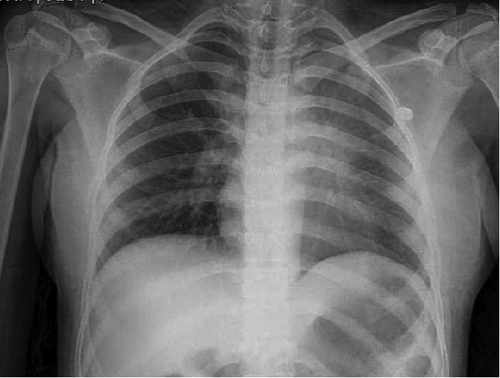
Chest X-ray shows left lung infiltration.

Spiral computed tomography scan shows left lung opacity and lesion.

A, B) Sections from lung tumor show nests of bland looking cells separated by delicate fibrovascular cores with high mitotic counts (Hematoxylin and Eosin ×250, ×400).
Discussion
Pulmonary tumors in children are rare; the most common lesions seen in clinical practice are metastatic disease. Primary lung tumors in pediatric age group are extremely rare, and approximately 75% are malignant. This group consists of carcinoid tumors (40%), bronchogenic carcinoma (17%), and pleuropulmonary blastoma (15%).4,5 Bronchial carcinoid accounts for 80-85% of primary malignant tumors. However, there are fewer than 40 proven cases in children in the literature. 5 Among the four types of carcinoid tumors of lung in children, AC are the least common and to the best of our knowledge less than 10 cases have been reported in the English literature.6–11 The details of the previous cases have been summarized in Table 1.
Characteristics of the reported cases of pulmonary atypical carcinoid in pediatric population.

The difference in histological criteria between TC and AC were first described by of Arrigoni et al. and later modified by Travis et al. and finally was fixed in 1999 by World Health Organization. 2
The tumor is more common in girls and the most common presenting symptoms have been cough, fever and hemoptysis in the previous cases. All of them have been primarily diagnosed as pneumonia. 10 The previous tumors have been in left, right and main bronchus. 11 The method of diagnosis has been different imaging studies with or without preoperative diagnosis by biopsy.10,11
The surgical procedures in the seven previously reported cases have been pneumonectomy, lobectomy and sleeve operation.9,10 With an average size of 2-4 cm, these tumors rarely were entirely exophytic, and may infiltrate the bronchial wall and surrounding lung parenchyma in an iceberg fashion. 12
There was a case of pulmonary A in a 19 year old male who presented with skull metastasis as the primary sign of disease. He has not been operated and just received chemo therapy. 9 This patient died in one year because of liver and lung metastasis. 9 There was another patient with subcarinal lymphnode metastasis at the time of diagnosis, that also died in less than one year. 5 In this 13-year old female left pneumonectomy and chemotherapy has not been successful. 6 Another patient who has received chemoradiation with surgery was a 21 year old female. She has been alive and symptom free after 18 months post surgery. 8
Our case is doing well after 6 months post resection, however her family haven't agreed to proceed for the sleeve operation yet. Whole body scan and all the abdominal imaging were normal. Prognosis in pulmonary carcinoids of children is generally good, but careful longterm follow-up is needed for potential recurrences or associated tumors. 12
Conclusions
As a conclusion, AC is a rare pulmonary tumor with potential of malignancy. Although AC can very rarely be seen in pediatric population, however it should be kept in mind whenever looking at a mass with carcinoid features in a child.