
Abstract
Pleomorphic adenoma is the most common benign salivary gland tumour. It can occur in any salivary gland, but is most frequently found in the parotid gland. Chondroid metaplasia is a frequent finding in pleomorphic adenoma. Other forms of metaplasia have been described, but are encountered less frequently. We report a rare case of unusual pleomorphic adenoma of the parotid gland with schwannoma-like feature.
Introduction
Salivary gland tumors account for about 3% of all head and neck neoplasia. The parotid gland is the main site for these tumors and about 95% of them are of epithelial origin. 1 Benign tumors represent 54% to 79% and 21% to 46% are malignant. The proportion of benign versus malignant tumor varies greatly by site. In the major salivary glands, such as the parotid and the submandibular gland, the majority of the tumors are benign contrary to the minor salivary glands, such as the sublingual and the floor of the mouth, where most of them are malignant.
Case Report
A 47-year-old woman was referred to us for a pain localized in the right retromandibular area, since about two months. Careful head and neck palpation revealed only a slight swelling in the right retromandibular region, without any well delineated palpable tumor. Head and neck MRI revealed a well defined tumor of 3.2 cm of size in the deep lobe of the right parotid gland with parapharyngeal extension, consistent with a pleomorphic adenoma (Figure 1). A right total parotidectomy with identification and preservation of the facial nerve was performed to remove the tumor appropriately. Macroscopic examination showed an intra-glandular, well delimited, white and firm, homogeneous nodule of 3.5cm of size. Histopathologically the tumor was completely circumscribed by a thin fibrous capsule. The major part of the tumor consisted of spindle cells with nuclear palisading resembling Verocay bodies (Figure 2B). At the periphery, some foci were composed of myxoid stroma with epithelial and myoepithelial-lined tubules (Figure 2A). Immunohistochemistry demonstrated the expression of p63 (Figure 3A) and CD10 (Figure 3B) in both tumour components and cytokeratin was positive in epithelial and myoepithelial cells. Histopathology diagnosis was a schwannoma-like pleomorphic adenoma of the parotid gland.
Magnetic resonance imaging of the patient with a radio opac lesion in the right parotid gland.
(A) Low magnification of schwannoma-like pleomorphic adenoma with both components (hematoxylin-eosin, magnification 100×); (B) Pathologic features of the schwannoma-like pleomorphic adenoma with palisading spindle-shaped cells (hematoxylin-eosin, magnification 200×).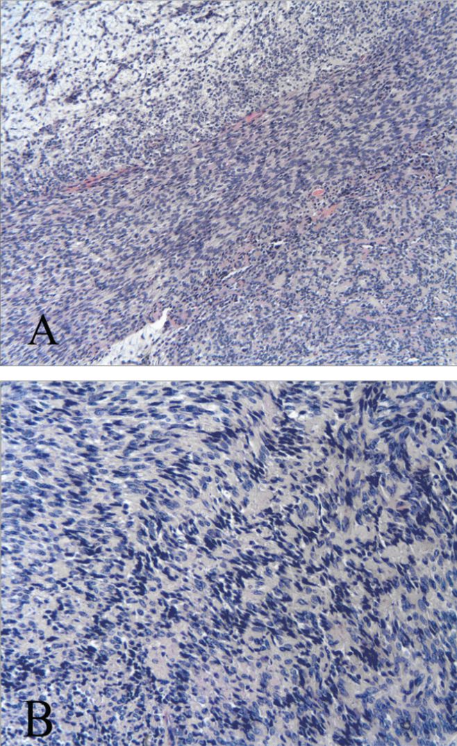
Immunohistochemical staining with (A) p63 and (B) CD10 (magnification for A: 400×, and B: 200×).
Discussion
Benign mixed tumour, also referred to as pleomorphic adenoma, is the most common benign salivary gland tumour, of which approximately 80% occur in the parotid gland, 10% in the submandibular glands and 10% in the minor salivary gland of the oral cavity. This tumour is most often observed in 30 to 60 years old patients and is more frequent in women than in men. 2 It is usually a solitary slow growing painless mass.
Pleomorphic adenomas are known for their morphologic and architectural variability. They share common features of epithelial, myoepithelial and mesenchymal components. The proportion of each of these elements can vary widely.
1
The stromal component of these tumours is most often predominantly myxoid with focal chondroid or fibrous aspects. Modified myoepithelial cells are thought to play an important role in the histopathological changes of the stroma. Sometimes squamous or osseous metaplasia is found in pleomorphic adenoma but is encountered less frequently. On a retrospective study on 83 pleiomorphic adenomas over a period of five years, we found an overall prevalence of 5% of metaplasia (
Squamous cell metaplasia in pleomorphic adenoma is an uncommon and most often accidental finding.3–6 Squamous cell metaplasia has been reported as a potential pitfall in fine-needle aspiration cytology (FNAC) of pleomorphic adenoma, which can be confused with a squamous cell or mucoepidermoid carcinoma. Different hypotheses have been advanced for its etiopathology. Firstly, it may be a repair process following FNAC, as reported by Li
Osseous metaplasia can be found in all salivary gland locations such as the parotid, submandibular and minor salivary glands. In some cases an important chondroid matrix was present with the formation of enchondral ossification at the borders. 9 Others described the formation of osteoid indicating a possible direct differentiation from myoepithelial cells10,11 Osseous metaplasia can also be found in carcinoma ex pleomorphic adenoma12,13 and mixed tumour of the skin.14,15
To our knowledge only five cases of schwannoma-like pleomorphic adenoma were reported in the English literature.16–18 Previous cases included four women and one man, aged from 39 to 75 years (Table 1). The majority of the lesions, as ours, were located in the parotid gland with one exception in the hard palate. All cases including this one had palisading areas of spindle-shaped cells in an otherwise classical pleomorphic adenoma.
Schwannoma-like features in pleomorphic addenoma. Review of the literature.

Spindle cell tumours are rare in the salivary glands, representing from 1.9% to 5% of parotid neoplasms.1,19,20 Differential diagnoses of benign spindle cell tumours in salivary glands include neurogenic tumours: schwan-noma, composed of areas with Verocay bodies and neurofibroma, as well as smooth muscle proliferations: leiomyoma.
Immunohistochemistry (IHC) may solve this dilemma. Leiomyomas are negative for cytokeratin positive for smooth muscle markers including alpha-smooth muscle actin, desmin and caldesmon, whereas benign neurogenic tumours, also negative for cytokeratine, express neurogenic marker (S100-Protein, CD57 and neurofilament).
Shwannoma-like pleomorphic adenomas are composed of modified myoepithelial cells expressing p63, CD10 and cytokeratin by IHC.
16
Merino
Fine needle aspiration cytology is an important diagnostic procedure used to evaluate salivary gland lesions and to help in their pre-operative management. Typically, FNAC of pleomorphic adenoma shows a combination of cohesive epithelial cells in a pale myxoid matrix. When the cellularity is abundant and no matrix is identified this diagnosis is challenging. In the presence of spindle-shaped cells immunohistochemical analysis may be used to identify the myoepithelial phenotype.
Pleomorphic adenoma with schwanoma-like feature is a rare variant that could be confused with a schwannoma or a leiomyoma, but immunohistochemical study is helpful to differentiate these entities.