
Abstract
Gliosarcomas (GS) are highly malignant and rare tumors of the central nervous system with a poor prognosis. We report here on four patients with GS, the median survival for whom was 9.25 months. Prognosis of GS remains poor, and a multidisciplinary approach (surgery, radiation therapy, and chemotherapy) seems to be associated with slightly more prolonged survival times.
Introduction
Gliosarcomas (GS) are highly malignant and rare tumors of the central nervous system with a poor prognosis. They account for 2–8% of all glioblastoma multiforme (GBM), and 0.48% of all intracranial tumors.1–4 They are primary brain tumors characterized by a biphasic pattern displaying both glial and mesenchymal components. 5 GS are predominant in males, and usually affect patients in the fifth to sixth decades of life. The clinical presentation, natural history, and radiologic profile are similar to those of primary glioblastoma.2,6,7
The clinical presentation varies according to location and tumor size. The most common symptoms are seizures, focal neurological deficits, headache, and other symptoms related to increased intracranial pressure.7,8 Treatment includes tumor resection, postoperative radiation therapy, and sometimes chemotherapy.4,8 At present, radiation therapy (60–66 Gy in 30 fractions) and chemotherapy are the standard of care for GB and, consequently, for GS.9,10
Between January 1999 and October 2004, 66 patients underwent radiation therapy for newly diagnosed GBM at the Department of Radiation Therapy, University of Ankara. We report here on four patients with GS, retrieved from this series. The aim of this retrospective study is to analyze clinical features, treatment, and survival.
Materials and Methods
Patients
In the GS group, two patients were male and two were female. The mean age was 56.5±4.65 years (range, 50–61 years). Three of the patients had headache, one had motor deficit, and all were on steroid medication during treatment. Preoperative computed tomography (CT) scans or magnetic resonance imaging (MRI) was performed in all patients (Figure 1). The locations, all supratentorial, included left temporal in one, left frontoparietal in two, and right frontoparietal areas and corpus callosum in one patient (Figure 2). Midline shift was not observed.
Cranial magnetic resonance image of Case #1.
Cranial magnetic resonance image of Case #3.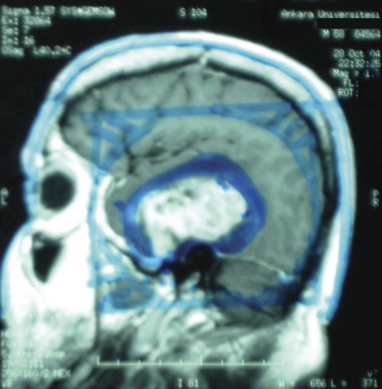
Treatment
All of the patients underwent tumor resection, which was classified as gross total. The median interval between surgery and the initiation of radiation therapy was 29.5 days (range, 19–38 days). All patients were treated with Cobalt.60 Clinical Target Volume (CTV) was established as Gross Tumor Volume (GTV) plus 2–2.5 cm, and 0.5 cm was added as a safety margin for Planning Target Volume (PTV). All of the patients in the GS group received 60 Gy in 30 fractions. Three patients had chemotherapy with temozolomide. All cases tolerated radiation therapy well, without interruption of their treatment because of side effects.
Statistics
Survival was calculated from the first day of radiation therapy, using the method of Kaplan and Meier. 11
Results
Clinical features
Patient characteristics are shown in Table 1.
Characteristics, treatment, and survival of patients in the gliosarcoma group.
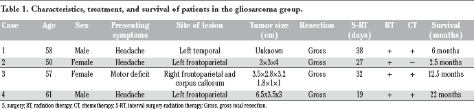
S, surgery; RT, radiation therapy; CT, chemotherapy; S-RT, interval surgery-radiation therapy; Gross, gross total resection.
Survival
All patients in the GS group had died at the time of analysis. The median survival was 9.25 months.
Discussion
In our present report, GS accounted for 5.5% of all GBM, with a mean age of 56.5±4.65 years. The age distribution was similar to that of GBM patients. Other reported series determined that GS account for 2–8% of all GBM with ages ranging from the fifth to the sixth decades.1–4,12,13 This is in accordance with our findings (Table 1). GS reportedly affect males more frequently than females.2,4 However, the male to female ratio was 2:2 in our GS group; we failed to observe a male preponderance. Furthermore, although a temporal lobe predilection has been reported by other authors,2,13 the majority of our GS patients presented with frontoparietal lesions, but the small numbers in our study make this difficult to interpret. In spite of some reports of metastatic spread,13,14 none of the tumors of our patients evolved with systemic dissemination.
Several authors reported significant biological similarities in the behavior of GS and GBM,2,4,6,13 suggesting that the same treatment should be applied for these two kinds of tumor. On the basis of this observation, we have treated patients with GS as those with GBM. All of our GS patients underwent gross total resection and all had postoperative radiation therapy. The median interval between surgery and the initiation of radiation therapy was 29.5 days (range, 19–38 days). Three of the four patients received chemotherapy with temozolomide, and one had no chemotherapy because of her poor clinical condition (Karnofsky Performance Status 40).
Morantz et al 3 commented on the effect of chemotherapy on the outcome. They found a mild increase in survival in GS patients with additional chemotherapy (36 weeks) compared with radiation therapy alone (33 weeks). In our study, median survival was 9.25 months for our patients. The median survival in GS patients ranges from 6 to 14.8 months in the reported studies.1,3,4,13,15,16 It has to be noted that survival times were calculated from different points of time: date of onset, diagnosis, surgery, or first day of radiation therapy. Heesters et al 17 reported a median survival of seven months for their six GS patients. Lutterbach et al 4 analyzed 12 GS patients and 410 GBM patients and found that the median survival was 11.5 months for the GS patients compared with 8.1 months for the entire GBM group. Perry et al 13 analyzed 25 GS patients and reported a median survival of six months. Meis et al. 2 analyzed 26 GS patients and 1453 GBM patients and report a median survival of 8.3 months for GS patients compared with 9.6 months for the GBM patients; their findings were contrary to the other reports.
In the reported studies,3,4,13 all patients died within 20 months. In the study of Galanis et al, 12 only one of 18 patients survived for 29 months. Winkler et al 18 presented a rare case of GS in a 61-year-old woman who was stable over 22 years. We concluded that despite the fact that our cases were very few, the presenting clinical features, treatment, and survival of GS are similar to that of GBM. Although the prognosis of GS remains poor, a multidisciplinary approach (surgery, radiation therapy, and chemotherapy) seems to be associated with slightly more prolonged survival times.