
Abstract
Transplantation of hormone-producing cells is an experimental endocrine dysfunction treatment. The present study investigated the effects of orchidectomy (OE) and transplantation of interstitial cell suspension (ICS) on rat sexual behavior. Adult experimental animals were divided into two populations. One of these populations had sexual experience before the experiment and the other did not. Each population was divided into three groups: control group and two orchidectomized groups. One of the orchidectomized groups was treated with ICS, and the other was treated with the vehicle. The changes in the sexual behavior were investigated on the following parameters: mount latency (ML), intromission latency (IL), ejaculation latency (EL), mount frequency (MF), intromission frequency (IF), copulatory efficacy (CE), and IF/EL ratio. The investigation of these changes lasted 4 weeks after ICS transplantation. The parameters of sexual behavior reflected a decrease in sexual function after OE at the beginning of the observation, especially for the animals that did not have a sexual experience. However, it was shown that sexual activity increased in the following 4 weeks. We have indicated that the loss of gonads attenuated the capacity to acquire sexual experience; nonetheless, it did not mean that the animals completely lost this capacity. Transplantation of ICS facilitated the maintenance of male sexual behavior after OE, fractionally enlarged the size of regressed seminal vesicles of the animals, and increased the free testosterone (T) level. These findings suggest the ICS can be considered as a temporal source of androgens, which can facilitate a restoration of sexual activity.
Introduction
Male hypogonadism occurs commonly in clinical practice and has significant effects on the well-being of patients. It can be a result of primary gonadal failure, such as Klinefelter's syndrome or normal aging, as well as a result of gonad loss after orchidectomy (OE). OE is often prescribed to patients with testicular and prostate cancer or after traumatization of the testes 1 3 . Clinical signs of testicular function after the treatment involve a decrease in serum levels of total testosterone (T) and free T. Thus, the patients may complain about decreased libido, impotence, low energy, and a decline in a general sense of well-being. An appropriate hormonal therapy for men with these conditions can partly eliminate these symptoms 4 . However, it requires an understanding of normal physiological regulation of testes and pathophysiology underlying testicular dysfunction. Moreover, it is important to mention that there are several adverse effects of hormone replacement. Serum concentrations of T are often greater than a physiological range during the first few days after injection of T enanthate and T cypionate, but less than the physiological range at the end of the dosing period before the next injection 5 . These fluctuating T levels may lead to undesirable swings in libido and sexual performance and side effects on the cardiovascular system and prostate 4 . Therefore, more research is needed to investigate these issues.
The development of transplantation methods in andrology was associated with the creation and experimental approbation of different variants of testis transplantation. Currently, the experimental transplantations of various cells and tissues from the testes are being used to find ways of coping with testicular dysfunction 6 11 . Hence, it is unclear whether there are enough resources in these transplants to compensate for the hormone insufficiency and to what extent T is important in some cases.
The effects of castration and androgen replacement on sexual behavior are known to be highly variable in men and experimental animals. Undoubtedly, T replacement therapy improves some aspects of male sexual function, including libido, ejaculation, and erectile function. However, it has been shown in a previous study that the extent of the sexual behavior reduction varies widely among species as well as among individuals within given species or strain 12 . Regardless of whether sexual behavior was retained, the level of plasma T was reduced in these animals by castration. It is furthermore arguable that the circulating levels of T and estradiol are prerequisites for sexual activity in castrated male rats 13 . Finally, previous studies14,15 have demonstrated that some men who were subjected to castration due to testicular cancer, prostate cancer, or convicted of sex crimes retained the capacity to develop an erection in response to sexual stimulation for a long time. Additionally, it was shown 16 that dehydroepiandrosterone (DHEA) and androstenedione (4-A) supplements failed to support male rat sexual motivation (time and urinary marks near an inaccessible receptive female). Moreover, 4-A reduced motivation similar to the suppressive effects of corticosteroids. The results suggest that DHEA and 4-A are not merely precursors of sex hormones but also provide support for these steroids in influencing the brain and behavior in a unique fashion that is dissimilar from the effects of T on male sexual behavior.
As far as the animals are concerned, their sexual activity is a complex behavioral process. It is controlled by various nervous system structures as well as by interactions between the hypophysis, the hypothalamus, and the sexual glands. Sexual activity is connected with sexual contacts between individuals of different sex for reproduction. The effectiveness of this process for animals depends on the male capacity for behaviors such as pursuit, anogenital sniffing, and implementation of intromissions, ejaculations, and female activity in behavioral estrus.
It has been shown that of the several types of testis transplantations, some can be more efficient than others 7 . Successful testicular interstitial cell transplantation for the recovery of T level of rats has been shown in a previous study 17 . However, it is important to find out whether T is crucial for sexual behavior in some cases. In the present study, we hypothesized that the transplantation of steroidogenic cells can have an impact on sexual behavior. However, there are still not enough data about different aspects of the sexual behavior after transplantation. For example, there is not enough information about the ability to have sexual experiences after castration and testis interstitial cell transplantation. Considering the actuality of these problems, the main goal of this study was to examine the sexual behavior of hypogonadal male rats after allotransplantation of interstitial cell suspension (ICS). The data may be useful for understanding the reasons underlying copulatory endocrine dysfunction and for the improvement of a general sense of well-being in patients who are suffering from these disorders.
Materials and Methods
Animals and Experimental Design
Male and female Wistar and female Wistar rats were obtained from Bio ModelService (Kiev, Ukraine) and were kept in the animal house of the Institute for Problems of Cryobiology and Cryomedicine of NAS of Ukraine (Kharkiv, Ukraine). The animals were housed six per cage, with a 12-h light— dark cycle. The female rats were housed in separate cages. All rats were supplied with standard laboratory food and water. The experimental protocol followed the Bioethical Standards of Preclinical Research carried out on laboratory animals in the Scientific Departments of the Institute for Problems of Cryobiology and Cryomedicine NAS of Ukraine and was approved by the Committee for Bioethics in Animal Experimentation at the Institute for Problems of Cryobiology and Cryomedicine NAS Ukraine. These standards are subject to the legislative documents of Ukraine such as the General Ethical Principles of Animal Experimentation and the Bioethical Expertise of Preclinical and Other Research Carried Out on Animals. All these documents are subject to the European Convention for the Protection of Vertebrate Animals Used for Experimental and Other Scientific Purposes.
The male rats were divided into six groups with six animals in each group. The groups were divided depending on their sexual experience. Groups 1—3 had sexual experience before the experiment. The male rats were trained four times with sexually receptive females for sexual experience before the experiment. Groups 4—6 did not have any experience. Groups 1 and 4 served as the controls of groups 2 and 3 and groups 5 and 6, respectively. Groups 2, 3, 5, and 6 were orchidectomized.
The surgical procedures were carried out under ketamine—xylazine anesthesia {ketamine [80 mg/kg body weight (BW); Brovapharma, Brovary, Ukraine]; xylazine (20 mg/kg BW; Interchemie werken De Adelaar Eesti AS, Viimsi, Estonia)}. In the case of OE, a ventral mid-line incision of about 1 cm in length was made, and both testes were exposed. The vas deferens and vessels were ligated and cut, and the testes were then removed. Afterward, the muscles as well as skin were sutured layer by layer. In the case of the control animals, their testes were exposed through the midline incision, and then they were returned to the abdominal cavity.
Groups 3 and 6 were transplanted with ICS under ketamine—xylazine anesthesia 1 week after the OE. ICS isolation and transplantation were carried out as outlined below: the donor Wistar rats were sacrificed by cervical dislocation and immersed in 70% ethanol (Vishpha, Zhitomir, Ukraine) for 5 min. The testes were decapsulated, trimmed of blood vessels, and placed in 15-ml centrifuge tubes with 4 ml of Dulbecco's modified Eagle's medium (DMEM; PAA, Pasching, Austria) per testis with 0.2 mg/ml collagenase (type I) (Sigma-Aldrich, St. Louis, MO, USA) and 0.1 mg/ml DNase I (Sigma-Aldrich) for 10 min in a thermostatic shaking water bath (90 cycles/ min at 34°C). Ten milliliters of collagenase-free DMEM was added to each tube, and the seminiferous tubule mass was removed by filtration through doubled, 100-um nylon mesh. The filtrates were centrifuged at 325 × g for 3 min at room temperature. The supernatants were discarded. The residues were resuspended in 10 ml of DMEM and supplemented with 100 IU/ml penicillin (Arterium Corporation, Kiev, Ukraine) and 100 ug/ml streptomycin (Arterium Corporation). The procedure of sedimentation was repeated. The cell concentration in the ICS was adjusted to 10 8 cells/ml by DMEM with the antibiotics. Viability of the cells was measured by trypan blue dye (Sigma-Aldrich) exclusion test, and the amount of viable cells accounted for 90% of the total amount of cells in ICS. The histochemical staining of 3β-hydroxysteroid dehydrogenase (3β-HSD) was carried out to detect the steroidogenic enzyme in Leydig cells. The staining was carried out as previously described 18 . The percentage of positively stained cells accounted for 10% of the total amount of cells in ICS.
The recipient animals of groups 3 and 6 got a single subcutaneous injection of 20 ul of ICS into the scrotum (2 × 10⁷ cells per body) 1 week after OE. The animals of groups 2 and 5 received a subcutaneous injection of the DMEM/antibiotic vehicle into the scrotum. Moreover, the animals of groups 3 and 6 got a single dose of cyclosporine (20 mg/kg BW, orally; Ivax Pharmaceuticals Inc., Miami, FL, USA) 1 day before the ICS transplantation by catheter. After the ISC transplantation, the dose was reduced to 10 mg/kg/day. The animals of groups 3 and 6 got the cyclosporine doses every day, except Sundays, while the animals of groups 1, 2, 4, and 5 received phosphate-buffered saline (PBS). The volume of orally administered solution was 1 ml. One week after transplantation (2 weeks after OE), the animals fully recovered after these operations in terms of their behavior and appearance, that is, they were actively moving and had a good appetite.
Four weeks after the transplantation, the animals were sacrificed by cervical dislocation. Separated sera samples of all animals were collected and stored at the same time until assayed. Free serum T concentrations were determined without dilution using a competitive immuno-enzymatic colorimetric method (Free T Kit; Xema Co. Ltd., Moscow, Russia) in duplicate samples within a single assay, with intra-assay coefficient of variation of 8%. Moreover, the seminal vesicles from all the transplanted rats were weighed to assess the bioactive circulating T. The weight of the seminal vesicles was defined as the ratio of the weight of the animal's organ per the animal's weight. Additionally, they were fixed in 10% neutral-buffered formalin, dehydrated in graded ethanol, cleared in xylene, and embedded in paraffin wax. The sections were cut at 5-um thickness, stained with hematoxylin and eosin (H&E; Leica Microsystems, Wetzlar, Germany), and observed under a light microscope (MEIJI Techno, Saitama Japan).
Mating Behavior Test Procedure and Parameters
In the present study, all female rats were ovariectomized before behavioral assessments in order to simulate menopause. The oviducts were ligated and removed, and the abdominal wall and skin were sutured.
Two weeks after ovariectomy, four behavioral tests were conducted after the transplantation. The first was carried out 1 week after the transplantation. The following tests were conducted within a period of 1 week after the first test. Each male rat was allowed a 10-min exposure to a female rat in behavioral estrous for copulatory behavior. The estrous in the ovariectomized females was induced by subcutaneous administration of estrone (25 ug/rat; Zdorovye, Kharkiv, Ukraine) 48 h before the test and progesterone (500 ug/rat; Biofarma, Brovary, Ukraine) 4 h before the test. After the preparations, the female rats performed characteristic estrous behavior (lordosis and the desire for copulation). During the sexual behavior test, a male rat was placed in the mating cage 5 min prior to the introduction of a sexually receptive female rat. The test was carried out between 5:00 pm and 7:00 pm under dim light.
The following parameters were used as described previously 19 : the mount latency (ML) or the interval between placing a female animal near a male animal and the first copulation attempt. They were registered by real-time monitoring. The copulation is the behavior in which the male animal mounts on the back of the female. The intromission latency (IL) is the interval between placing a female animal near a male animal and the first male intromission. Intromission is a copulatory behavior in which the male animal genital organ enters the female vagina. The ejaculatory latency (EL) is the interval between placing a female animal near a male and the first ejaculation. The mount frequency (MF) is the number of times that the male animal copulates with the female animal without intromissions. MF is calculated in the copulation series until the first ejaculation. The intromission frequency (IF) refers to the number of times the male copulates with intromissions and performs this action in a copulation series until the first ejaculation.
Copulatory efficacy (CE) is measured by the formula CE=IF/(MF + IF). The IF/EL ratio was also measured. It shows to what extent the copulation was successful. The IL, IF, and CE of the groups were not counted and compared when at least one of their animals did not have intromissions and/or ejaculations. These cases will be discussed below.
Statistical Analysis
All data were processed by Statistica 10 package (StatSoft Inc., Tulsa, OK, USA) for Windows (Microsoft, Armonk, NY, USA), and the results were expressed as median with 25th and 75th interquartile range. The statistical differences between groups were determined using nonparametric Kruskal-Wallis test. Multiple comparisons were tested using Mann-Whitney U-test with Bonferroni correction. Values of p < 0.05 were considered statistically significant.
Results
The effects of the OE and ICS transplantation on the free T serum level and the weight of seminal vesicles are presented in Table 1. The free T serum and the weight of the seminal vesicles decreased considerably in orchidectomized animals of groups 2-5 when compared with corresponding control animals (p≤0.05) (Table 1). Moreover, the free T level of orchidectomized rats of groups 2 and 5 was lower than the method sensitivity. It is important to note that the free T level and the weight of the seminal vesicles did not depend on sexual experience of the experimental animals (p ≤ 0.05). The free T serum level and the weight of the seminal vesicles of orchidectomized animals with transplantation of ICS (groups 3 and 6) were higher (p ≤ 0.05) (Table 1) than in the corresponding orchidectomized animals without the transplantation (groups 2 and 5). However, it was clearly shown that these parameters of all the orchidectomized animals were significantly lower than in control groups 1 and 4 (p ≤ 0.05) (Table 1). After OE, the mucosal folds of the seminal vesicles were less prominent, and degeneration of glandular cells was observed in groups 2, 3, 5, and 6 (Fig. 1).

The histology of seminal vesicles. (A) Control animals. They have a typical pseudostratified columnar epithelium (arrows) that consists of columnar and basal cells. The lumen of the seminal vesicles is full of secretory fluid (SF). (B, C) Orchidectomized animals without and with interstitial cell suspension (ICS) transplantation, respectively. Thinning and flattening of the epithelium were observed in both cases. The secretory fluid of the vesicles was weakly expressed. There was thickening of the lamina propria. Scale bars: 50 μm.
The Effects of the Orchidectomy and Transplantation of Interstitial Cell Suspension (ICS) on the Free Testosterone Level (pg/ml) and the Weight (mg/g) of Seminal Vesicles
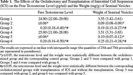
The results are expressed as median with interquartile range (the quantities of 25th and 75th percentiles are represented in parentheses).
p≤0.05, the testosterone level and the weight were statistically different between the orchidectomized group and the corresponding control group. Groups 2 and 3 were compared with group 1. Groups 5 and 6 were compared with group 4.
p≤0.05, the testosterone level and the weight were statistically different between the corresponding orchidectomized groups with transplantation of ICS and without the transplantation. Group 3 was compared with group 2, and group 6 was compared with group 5.
The investigation into the effects of the OE and ICS transplantation on ML indicated that in the groups that were orchidectomized, ML increased (Table 2) by week 1 compared with the control groups, especially in groups 5 and 6 (p ≤ 0.05) (Table 2). However, ML of orchidectomized animals had a tendency to be lower in the course of observation.
The Effects of the Orchidectomy and Transplantation of Interstitial Cell Suspension (ICS) on Mount Latency (ML) (s)

The results are expressed as median with interquartile range (the quantities of 25th and 75th percentiles are represented in parentheses).
p ≤ 0.05, the ML was statistically different between the orchidectomized group and the corresponding control group. Groups 2 and 3 were compared with group 1. Groups 5 and 6 were compared with group 4.
p ≤ 0.05, the ML was statistically different between the corresponding orchidectomized groups with transplantation of ICS and without the transplantation. Group 3 was compared with group 2, and group 6 was compared with group 5.
p ≤ 0.05, the ML was statistically different between the corresponding groups without sexual experience and with the experience. Group 4 was compared with group 1, group 5 with group 2, and group 6 with group 3.
The effects of the OE and ICS transplantation on IL showed that in groups 2 and 3, IL significantly increased (p ≤0.05) (Table 3) by week 1 when compared with the control group 1 (Table 3). Intromissions were not observed for groups 5 and 6 by week 1. Only two of six animals from group 5 and half of group 6 had a few intromissions by week 2, and the IL lasted more than 350 s. It is important to note that IL of all orchidectomized rats dropped significantly during the experiment, especially by week 4, with the exception of group 5. Additionally, for group 3, it remained much lower than for group 2 over a period of 4 weeks, and for group 6, it remained lower than for group 5 by week 4 (p ≤0.05) (Table 3).
The Effects of the Orchidectomy and Transplantation of Interstitial Cell Suspension (ICS) on Intromission Latency (IL) (s)

The results are expressed as median with interquartile range (the quantities of 25th and 75th percentiles are represented in parentheses). ILs of groups 5 and 6 by weeks 1 and 2 were not presented because not all animals of the groups had intromissions. These cases are discussed in the Results.
p ≤ 0.05, the IL was statistically different between the orchidectomized group and the corresponding control group. Groups 2 and 3 were compared with group 1. Groups 5 and 6 were compared with group 4.
p ≤ 0.05, the IL was statistically different between the corresponding orchidectomized groups with transplantation of ICS and without the transplantation. Group 3 was compared with group 2, and group 6 was compared with group 5.
p ≤ 0.05, the IL was statistically different between the corresponding groups without sexual experience and with the experience. Group 4 was compared with group 1, group 5 with group 2, and group 6 with group 3.
The investigation into the effects of OE and ICS transplantation on EL indicated that orchidectomized animals of groups 2, 3, 5, and 6 did not ejaculate; hence, the EL was not obtained for these groups. However, the EL of group 1 came to 471 s by week 1. With the exception of a slight rise by week 2, the EL kept decreasing until it reached a low of 352 s in the final week. Only two of six animals of group 4 had ejaculations by week 2, and half of them had ejaculations by week 3. Their EL was more than 500 s by weeks 2 and 3 and then decreased to 487 s by week 4.
The effects of the OE and ICS transplantation on MF indicated that the MF of groups 1 and 2 steadily decreased, whereas the MF of groups 3-6 demonstrated an upward trend by week 4 (Table 4). It is important to note that the MF of orchidectomized animals with transplants of ICS (groups 3 and 6) became higher (Table 4) than the MF of the corresponding groups without the transplants (p≤0.05) (groups 2 and 5).
The Effects of the Orchidectomy and Transplantation of Interstitial Cell Suspension (ICS) on Mount Frequency (MF)

The results are expressed as median with interquartile range (the quantities of 25th and 75th percentiles are represented in parentheses).
p ≤ 0.05, the MF was statistically different between the orchidectomized group and the corresponding control group. Groups 2 and 3 were compared with group 1. Groups 5 and 6 were compared with group 4.
p ≤ 0.05, the MF was statistically different between the corresponding orchidectomized groups with transplantation of ICS and without the transplantation. Group 3 was compared with group 2, and group 6 was compared with group 5.
p ≤ 0.05, the MF was statistically different between the corresponding groups without sexual experience and with the experience. Group 4 was compared with group 1, group 5 with group 2, and group 6 with group 3.
The effect of the OE and ICS transplantation on IF showed that OE itself drastically decreased (Table 5) the IF of groups 2 and 3 at the beginning when compared with control group 1 (p≤0.05) (Table 5). The IF of group 3 was higher when compared with group 2 (p≤0.05), and the IF of group 6 was higher than that of group 5 (p≤0.05) (Table 5) by weeks 3 and 4.
The Effects of the Orchidectomy and Transplantation of Interstitial Cell Suspension (ICS) on Intromission Frequency (IF)

The results are expressed as median with interquartile range (the quantities of 25th and 75th percentiles are represented in parentheses). IFs of groups 5 and 6 by weeks 1 and 2 were not presented because not all animals of the groups had intromissions. These cases are discussed in the Results.
p≤0.05, the IF was statistically different between the orchidectomized group and the corresponding control group. Groups 2 and 3 were compared with group 1. Groups 5 and 6 were compared with group 4.
p≤0.05, the IF was statistically different between the corresponding orchidectomized groups with transplantation of ICS and without the transplantation. Group 3 was compared with group 2, and group 6 was compared with group 5.
p≤0.05, the IF was statistically different between the corresponding groups without sexual experience and with the experience. Group 4 was compared with group 1, group 5 with group 2, and group 6 with group 3.
The investigation into the effects of OE and ICS transplantation on CE indicated that in the groups that were orchidectomized, the CE was significantly lower than in the corresponding control groups (p≤0.05) (Table 6), especially in groups 5 and 6 (Table 6), with the exception of group 3 during the first 3 weeks of observation. The CE of group 3 generally had a tendency to be higher when compared with group 2. Also, the CE of group 6 had the same tendency when compared with group 5 (Table 6) by weeks 3 and 4. It should be noted that the CE of each group had a gradual increase over the period of the observation.
The Effects of the Orchidectomy and Transplantation of Interstitial Cell Suspension (ICS) on Copulatory Efficacy (CE)

The results are expressed as median with interquartile range (the quantities of 25th and 75th percentiles are represented in parentheses). CEs of groups 5 and 6 by weeks 1 and 2 were not presented because not all animals of the groups had intromissions and ejaculations. These cases are discussed in the Results.
p≤0.05, the CE was statistically different between the orchidectomized group and the corresponding control group. Groups 2 and 3 were compared with group 1. Groups 5 and 6 were compared with group 4.
p≤0.05, the CE was statistically different between the corresponding orchidectomized groups with transplantation of ICS and without the transplantation. Group 3 was compared with group 2, and group 6 was compared with group 5.
p≤0.05, the CE was statistically different between the corresponding groups without sexual experience and with the experience. Group 4 was compared with group 1, group 5 with group 2, and group 6 with group 3.
The IF/EL ratio of group 1 had an upward trend from the first to the fourth week and accounted for 0.050, 0.056, 0.076, and 0.071 s−1, respectively. The proportion of group 4 by week 4 accounted for 0.047 s−1.
Discussion
Sexual behavior is a complex process that is dependent on sex, age, sexual experience, and other factors. This study examined how OE, sexual experience, and transplantation of ICS influence the free T level, the parameters of sexual behavior, the secretory epithelium, and the weight of seminal vesicles of orchidectomized rats.
The present study showed that the OE led to the withdrawal of testicular androgens, resulting in a decrease in free T levels and seminal vesicle degradation. Although the level of free T dramatically decreased after OE, it was maintained at low but detectable levels in the transplanted animals. This could be one of the reasons we observed sexual behavior enhancement in these groups compared with orchidectomized animals without ISC transplantation. However, this level of T was not sufficient to completely reconstitute seminal vesicles after OE.
When it comes to sexual behavior, we can see that the presence of sexual experience gave certain advantages for animals in groups 1-3 in terms of manifestation of male capacity for copulation compared to animals in groups 4-6, which did not have the experience. It is clear that the animals of group 1 had a very low ML and IL, especially by week 1 when compared with the animals of group 4. Their CE had a tendency to be higher than that of animals in group 4 with little sexual experience. Moreover, all the animals of group 1 had ejaculations during the first week, whereas the animals of group 4 had them only by week 4, and their IF/EL increased mostly at the expense of EL, which decreased during the experiment. The IF/EL of group 4 by week 4 was the same as in animals of group 1 by week 1. Therefore, it can be concluded that all the animals of groups 4-6 gained sexual experience over the period of the experiment.
The OE drastically altered male sexual performance in all experimental groups and the capacity to gain sexual experience for the rats in groups 5 and 6 as well. All the parameters of sexual behavior declined. However, it is important to note that the orchidectomized animals did not totally lose male sexual behavior and the capacity to gain sexual experience. Accordingly, we registered that the animals with sexual experience (groups 1-3) had a higher CE after OE compared with the animals that did not have one (groups 4-6). Additionally, we indicated that a loss of gonads attenuated the capacity to acquire sexual experience.
The possibility of in vivo transplantation of Leydig cells as a new biologic androgen replacement therapy was investigated in the 1980s and 1990s. It was shown that levels of T were correlated with the number of autotransplanted rat Leydig cells 20 . Moreover, according to van Dam et al. 21 , isolated Leydig cells could secrete steroids after subcutaneous transplantation. It was shown in another study that the number of Leydig cells increased after allotransplantation of newborn rat testes under the kidney capsule 22 . The secretion of T by these cells was demonstrated by increased seminal vesicle weight.
Notwithstanding the distrust of such free transplantation for the treatment of androgen deficiency 7 , it can be suggested that transplantation can be useful for the maintenance of male sexual behavior of rodents after OE. In this study, it was observed that the orchidectomized animals of groups 3 and 6, which were treated with the transplantation of ICS, had generally higher means of MF, IF, and CE compared to the animals of groups 2 and 5, which did not get the treatment. Animals of group 6 developed copulatory skills more quickly than the animals in group 5.
It was demonstrated that transplanted Leydig cells helped to increase T levels in the blood 17 . Similarly, it was observed that the level of free T as well as the weight of the seminal vesicles in the orchidectomized animals with the transplantation of ICS (groups 3 and 6) were higher when compared with the animals without the transplantation (groups 2 and 5). These findings show that the secretion of androgens was higher in the groups transplanted with ICS.
Previous studies showed an almost complete abrogation of androgens after castration23,24. However, it is not clear why the orchidectomized animals, especially of groups 2 and 5 without ISC transplantation, mounted and had intromissions over the course of the study.
It is generally known that dopamine (DA) is released during copulation due to stimulus from a receptive female 25 . There are three main integrative targets for the DA: the nigrostriatal system, the mesolimbic system, and the medial preoptic area (MPOA)25,26. The MPOA focuses on the motivation onto the specific sexual targets, increases copulatory rate and efficiency, and coordinates genital reflexes 26 . According to Hull et al. 25 , the main paradigm can be outlined as follows: T increases the activity of nitric oxide synthase (NOS) in the MPOA, which produces more nitric oxide (NO), which in turn promotes DA release in both basal and sexual situations. DA promotes sexual motivation, genital reflexes, and copulation 27 . Thus, further research is necessary to determine whether there are factors that increase the DA release or whether the reduced extracellular DA level is sufficient to activate sexual behavior of orchidectomized rats by weeks 3 and 4.
Taking this information into account, we considered some additional mechanisms by which androgen levels can be increased in an organism, thus facilitating DA release and manifestation of sexual behavior. It was shown that the levels of dihydrotestosterone, T, DHEA, and 4-A in the ventral prostate of rats increased gradually from the second week after the castration 28 as well as the DHEA and 4-A levels in the adrenal glands. This finding supports the concept that adrenal glands could partly compensate for the loss of testicular androgen. On the other hand, it was shown 29 that OE of middle-aged rats (a model of andropause) led to the reliable decrease in serum T levels and DHEA, indicating an immediate impact of T on DHEA secretion, while exogenous T administration results in both T and DHEA increase. Thus, it has been suggested that the increase in free T levels after ISC transplantation could be a reason that sexual behavior improved in groups 3 and 6 when compared with orchidectomized animals without the transplantation (groups 2 and 5).
Apart from DHEA and its derivatives, the 11-beta-hydroxyandrostenedione level remained unchanged 30 after castration. It was suggested 31 that the steroid can be metabolized and yield androgens in steroid-responsive peripheral tissues, as in the case of androgen precursor metabolites of adrenal origin. It is possible that this steroid alone and in combination with T was able to maintain sexual behavior in animals of groups 2, 3, 5, and 6. Other steroid-producing sites, for example, skin and nervous system, may possibly contribute to this compensatory activity32,33, which in turn promotes DA release and stimulates sexual behavior.
It is important to note that there are data 34 about an adverse effect of cyclosporine on T synthesis. We failed to get a satisfactory result in terms of T secretion when we used ICS allotransplantation without immunosuppression (data not shown). Moreover, we used nearly maximal doses of cyclosporine to elucidate ICS functioning after OE, allotransplantation, and immunosuppression, which could provide better insight into the feasibility of using it in the clinic.
Thus, ICS transplantation could be considered as a possible source of androgens for castrated individuals, which can facilitate the recovery of sexual activity and/or realize the shift of the sexual activity to the steroid-independent way.
The scope of this work cannot show the overall potential of the free transplantation. In some cases, the outcome of cell transplantations can be improved by applying different models (e.g., intratesticular injection of cells under various models of hypogonadism 35 ). It is now imperative that fertility preservation is considered as part of the care offered to patients who are suffering from hypogonadism and decreased libido 36 . This can only be approached within a multidisciplinary setting, including research in the areas of tissue retrieval, cryopreservation, and transplantation.
Conclusion
Based on the results of this study, it can be concluded that the loss of gonads drastically altered sexual behavior and the rate of sexual experience acquisition. However, this loss did not totally inhibit these processes. It can be concluded that the transplantation of ICS can have a positive effect on the recovery of the sexual behavior after OE and enhance the rate of sexual experience acquisition. Given declining libido in men after castration or due to other disorders, this study could lead to discoveries with possible implications for the improvement of the general sense of men's well-being.
Footnotes
Acknowledgment
The authors declare no conflicts of interest.