
Abstract
Alzheimer's disease (AD) is characterized by a progressive loss of memory and other cognitive disturbances. The neuropathology of AD includes the major hallmarks of toxic amyloid-β oligomer accumulation and neurofibrillary tangles, as well as increased oxidative stress, cholinergic dysfunction, synapse loss, changes in endogenous neurotrophic factors, and overall degeneration of the brain. Adult mesenchymal stem cells (MSCs) offer the potential for a readily available treatment that would be long lasting, have low likelihood of rejection, and could target a variety of pathological deficits. MSCs have been shown to be effective in alleviating symptoms in some transgenic models of AD, but the optimal location for transplanting MSCs has yet to be determined. In the present study, the behavioral effects of transplantation of MSCs into the lateral ventricles, the hippocampus, or both of these regions were compared in the 5xFAD mouse model of AD. The results indicate that MSC transplants effectively reduce learning deficits in the 5xFAD mouse model and demonstrate a clear impact of MSCs on the levels of Aβ42 in the brains of 5xFAD mice. Overall, these findings support the hypothesis that MSCs may be a viable treatment for AD, especially when injected into the lateral ventricles.
Keywords
Introduction
The major hallmarks of Alzheimer's disease (AD) include extracellular amyloid-β (Aβ) plaques and intracellular neurofibrillary tangles (NFT) throughout the brain (7). Other neurophysiological changes include synapse loss, oxidative stress, inflammation, and cholinergic dysfunction (7), as well as alterations in neurotrophins (15,29). Combinations of these factors lead to extensive neuronal dystrophy throughout the brain and, in particular, the hippocampus, the basal forebrain, and large cortical neurons (7).
Current treatments for AD are, at best, only moderately effective and are targeted specifically to only one or two (such as cholinergic or glutamatergic) aspects of the multifaceted dysfunction observed in AD. An alternative strategy for treating AD is to use bone marrow-derived mesenchymal stem cells (MSCs), which have been shown to have pleiotropic effects, including reduction in oxidative stress, as well as upregulating production of critical neurotrophic factors, such as brain-derived neurotrophic factor (BDNF) and nerve growth factor (NGF) (27). Direct injections of BDNF (3) and NGF (33) have been shown to produce beneficial effects in rodent models of AD, but unfortunately these effects are short-lived, and NGF can cause painful side effects in humans (33).
MSCs as Treatment for Alzheimer's Disease
MSCs offer an excellent source of the type of neurotrophic factors that are expected to be both effective and long lasting in the treatment of AD. MSCs are multipotent cells that make up about one in every 100,000 cells of the bone marrow and have the ability to differentiate into blood cells, cartilage, bone, muscle, or adipose tissue (25), as well as into glia and neurons (18). MSCs appear to be hypoimmunogenic (27), do not proliferate as readily as other stem cell alternatives (10), and release a variety of beneficial cytokines and trophic factors that appear to improve the health of synapses and decrease apoptosis and inflammation (17). Additionally, MSCs can provide long-term functional benefits without harmful side effects in rodent models of Huntington's disease (HD) (9,28).
MSCs have been shown to reduce neuropathological dysfunction in rodent models of AD (2,19–21,31,35), but the optimal location for transplantation in the treating of neuropathology in the AD brain has not been adequately addressed. The goal of the present study was to further investigate the efficacy of MSC transplants as a potential treatment for AD by comparing the behavioral and pathological effects following their transplantation into the lateral ventricles, hippocampus, or into both of these two regions.
Materials and Methods
Animals
The effects of injecting MSCs into various locations in the brain were tested in mice bred from 5xFAD (AD) male mice and female wild-type (WT) F1 littermates (006554; Jackson Laboratories, Bar Harbor, ME, USA). Mice used for behavioral and histological analyses were randomly assigned to one of the following groups: (1) LV (n = 8), where AD mice received bilateral implantations of MSCs into the lateral ventricles; (2) Hipp (n = 8), where AD mice received bilateral implantations of MSCs into the hippocampus; (3) LV-Hipp (n = 8), where AD mice received bilateral implantations of MSCs in both the hippocampus and lateral ventricles; (4) WT Surgery Control (n = 6), where WT mice were put under anesthesia and received an incision, but not injections or burr holes; (5) WT Sham (n = 6), where WT mice received bilateral injections of vehicle, Hank's balanced salt solution (HBSS; Invitrogen, Carlsbad, CA, USA), into either the lateral ventricles, hippocampus, or both regions; (6) AD Surgery Control (n = 6), where AD mice were put under anesthesia and received an incision, but not injections or burr holes; and (7) AD Sham (n = 6), where AD mice were injected with HBSS into either the LV, hippocampus, or both regions. All MSCs injected into the hippocampus were labeled with Hoechst (bisBenzimide, H33258; Sigma-Aldrich, St. Louis, MO, USA), while all MSCs injected into the LV were positive for green fluorescent protein (GFP), with the exception of a MSC Control group (n = 2), which contained mice that received bilateral transplantations of MSCs labeled with Hoechst into the LV to ensure that GFP, itself, or the difference in cell passaging did not alter the experimental outcomes. All groups contained equal numbers of male and female mice.
Another group of mice that was used for protein analysis was given the same treatment as groups described above and included (1) LV (n = 6), (2) Hipp (n = 6), (3) LV-Hipp (n = 6), (4) WT Sham (n = 6), and (5) AD Sham (n = 6). Each protein analysis group contained three male and three female mice.
Mice were housed in groups of one to five with same-sexed animals. Food and water were available ad libitum. The mice were kept on a 12-h light/dark cycle, lights on at 900 h, and housed in clear plastic cages. In order to reduce the stress of the mice, they were individually handled prior to surgery. Gentling included 2-min sessions where the experimenter picked up the mouse three times and then carried the mouse around a room in order to simulate moving a mouse from a cage to a testing apparatus. This training was conducted three times in the week prior to surgery. All care of the mice followed the guidelines of the National Institute of Health and was approved by Central Michigan University Institutional Animal Care and Use Committee (IACUC; approval #10-14).
Genotyping
At 2–3 weeks of age, tail snips were collected for standard polymerase chain reaction (PCR) analysis to test for the presence of the APP gene. The PCR amplification was conducted using the MyiQ cycler (Bio-Rad, Hercules, CA, USA) with primers for APP, including transgenic of P1, oIMR 3610 (AGG ACT GAC CAC TCG ACC AG) and P2, oIMR 3611 (CGG GGG TCT AGT TCT GCA T) as well as internal positive controls of P3, oIMR 8744 (CTA GGC CAC AGA ATT GAA AGA TCT), and P4, oIMR 8745 (GTA GGT GGA AAT TCT AGC ATC ATC C). DNA was analyzed using gel electrophoresis.
Isolation and Culture of MSCs
Isolation and culture of MSCs were conducted as previously published (22). C57BJL/6 or GFP-positive mice were used as donor mice. Following euthanasia by cervical dislocation, the femur and tibia were removed, cleaned, and cut in half. The bone marrow was then extracted via aspiration with a 25-gauge needle and 1-ml syringe, primed with bone marrow media [α-modified Eagle medium (α-MEM) (Invitrogen) with 10% fetal bovine serum (FBS) (Invitrogen), 10% horse serum (Invitrogen), 1% streptomycin, and penicillin (Sigma-Aldrich)]. Cells were then centrifuged at 200 ° g for 7 min at 4°C, the supernatant was suctioned off, and the remaining pellet was resuspended in 1 ml of bone marrow serum. Cells suspended in 10 μl of bone marrow serum were then counted using a hemocytometer, and the remaining cells were then divided into one to three flasks (75 cm2) in 15 ml of medium stored at 37°C in 5% CO2. Cells were passaged when they reached 75% confluency. During passaging in a sterile biosafety cabinet, excess media was removed, at which point the plastic adhesion of MSCs to the flask was interrupted using 5 ml 0.25% trypsin-ethylenediaminetetraacetic acid (EDTA) (Sigma-Aldrich). The trypsin then was counteracted using equal levels of bone marrow serum. The solution was then centrifuged at 200 ° g for 7 min at 4°C. The excess bone marrow serum and 0.25% trypsin in EDTA was removed, and the cells were resuspended in 1 ml, counted using a hemocytometer, then redistributed into 1 million cells per flask with fresh bone marrow serum. GFP cells (which may lose their labeling if passaged too many times) were used on passages one to three, while Hoechst-labeled cells were passaged up to 11 times. All GFP cells were at least 85% positive for GFP expression. GFP expression was measured using a BD LSR-II flow cytometer (BD Biosciences, San Jose, CA, USA), utilizing FACSDiva Version 6.1.3 software (BD Biosciences). In order to detect GFP, fluorescein isothiocyanate (FITC) was used, which has an excitation of 490 nm and emission of 525 nm wavelength, with the long pass dichotic mirror being set at 505 nm and bandpass filter being set at 530/30 nm.
On the day of the surgery, all cells went through their final passage, at which point the cells to be injected into the hippocampus were labeled with Hoechst (1:1,000; Sigma-Aldrich) for 5 min, while GFP cells to be injected into the lateral ventricles were processed through a normal passaging procedures. Cells were suspended in a concentration of 200,000 cells/μl in HBSS vehicle (Invitrogen). Cells were kept on ice until the time of injection.
Surgery
Mice at 6 months of age were anesthetized with 2–4% isoflurane (Piramal, Bethleham, PA, USA) in 500 ml/L oxygen. Heads were then shaven and disinfected with 4% chlorhexidine gluconate (Walgreens, Deerfield, IL, USA), with their eyes being protected with Artificial Tears Ophthalmic Ointment (Walgreens). The mice were then secured into a rodent stereotax via tooth and ear bars set so that lamba and bregma were at the same level. During surgery, anesthesia was maintained with a mixture of 0.5–2% isoflurane (Piramal Healthcare, Andhra Pradesh, India) and 500 ml/L oxygen. Body temperature was monitored using a rectal probe and maintained with a TCAT-2CU temperature controller (Physitemp, Clifton, NJ, USA). A subdural injection of 0.1 ml of 1–2% lidocaine injectable local anesthetic (Sparhawk Laboratories, Lenexa, KS, USA) was made over the skull prior to making the incision. A 2-cm incision was made, and the skin and underlying fascia was retracted. A burr hole was drilled on each side of the skull, directly over the site of injection at -0.2 anterior/posterior from bregma (A/P) and ±1.0 medial/lateral from bregma (M/L) into the ventricle, -1.2 A/P and ±1.0 M/L into the hippocampus, or all four locations. An injection of 2 μl of MSCs or vehicle, or two injections of 1 μl of MSCs, per side, were made at a rate of 0.2 μl/min at -2.4 and -2.2 dorsal/ventral from bregma (D/V), with all mice receiving a total of 4 μl of MSCs or vehicle. After a 3-min wait period, the needle was slowly withdrawn. The burr holes were then covered with bone wax, and the scalp was sutured with stainless steel wound clips. Topical analgesic (1–2% topical lidocaine: viscous solution; Hi-tech Pharmacal Co., Inc., Amityville, NY, USA) and topical antibiotic (Neomycin-based triple antibiotic ointment; Walgreens) was applied to the wound. Finally, the mice were placed on a supplementary heat source and monitored as they regained consciousness. Daily follow-up monitoring was continued for 7 days after surgery, and the wound clips were removed by day 10.
Behavior
Mice were tested for alterations in tests of memory, anxiety, activity, as well as motor and sensory function. Behavioral testing commenced after 10 days of rest (Fig. 1). The mice were weighed the day of surgery (designated as week 0), in order to obtain their initial weight, and the day of perfusion (week 10) to obtain their final weight. Their percentage change in weight was calculated.

Transplantation locations and behavioral time line. Time line of behavioral testing beginning at 6 months of age. EPM (elevated plus maze); D (dark condition); L (light condition); PPI (prepulse inhibition); RAWM (radial-arm water maze).
Radial-Arm Water Maze 8-Choice Memory Task
The radial-arm water maze (RAWM) was used to measure spatial working and reference memory, beginning at week 3 postsurgery. The protocol (14,30) allowed for simultaneous testing of working memory and reference memory. The RAWM task consisted of 25 days of testing, with a 3-day rest period after the first 10 days of training. Mice were tested once in the morning and once 4 h later. The apparatus consisted of a pool (134 cm in diameter) containing eight evenly spaced arms (45 cm in length, 10 cm in width, and 45 cm deep) with collapsible escape platforms at the ends of arms 1, 3, 4, and 7 (clockwise from due North), hidden just below the surface of the water [for detailed description of the RAWM, see Dunbar et al. (11)]. The water was made opaque with nontoxic white tempura paint kept at room temperature, approximately 20–21°C. Mice were placed into the center of the maze facing north and given a total of 3 min to locate all four platforms. When a platform was located, the mice were allowed to rest for 10 s, at which point the platform was lowered by an experimenter using a pulley system outside the room. The number, order, and pattern of entries into the arms were determined. After each trial, the mice were towel dried and placed on a supplementary heat source.
Water T-Maze Two-Choice Learning Task
In order to assess learning in a less complicated task, the water T-maze test was conducted on week 10 postsurgery. The apparatus included a T-shaped water tank of Plexiglas, with a stem and two arms each measuring 45 cm long ° 15 cm wide, and stem measuring 15 ° 37 cm. The water was made opaque with nontoxic white tempura paint and filled the maze to a depth of 20 cm and was maintained at room temperature. A clear, Plexiglas escape platform, 10 ° 15 cm in area, was placed into each end of the arms, 1 cm below the water surface.
Mice were acclimated to the T-maze prior to testing in three phases. First, the mice were placed directly on a platform located in the center of the stem. Second, the mice were placed within 1 cm of the platform and allowed to climb onto it. Third, the mice were required to swim the length the stem to reach the escape platform. The mice were given a maximum of 1 min to complete the third phase, after which they were guided to the platform. Mice had 2 min to rest between each acclimation phase.
The test of learning on the T-maze was adapted from that used by Deacon (6). The mice were placed into the stem of the tank facing the wall directly opposite the choice point, with platforms raised in both arms of the maze, and allowed to swim to the arm of their choice. The platform opposite to the initial choice of the mouse was kept in the tank while the preferred platform was removed. The mouse was then given 20 trials to learn the new location of the platform. In each trial, the mouse had a maximum of 1 min to find the platform, after which it was guided by hand to it. After each trial, the mice were then towel dried and given 2 min of rest before the next trial began. The number of errors (i.e., entries into an arm that did not contain the platform) was recorded as the dependent measure.
Spontaneous Motor Activity
On week 2 postsurgery, activity levels of the mice were assessed using an open-field maze. Mice were placed in a transparent, 45 cm ° 23 cm ° 40 cm chamber for 1 h. Total movement, as well as time in the periphery and time in the center, was calculated using Motor Monitor Software (Kinder Scientific, Poway, CA, USA).
Motor Coordination
In order to assess motor coordination in the mice, a rotor-rod (ROTOR-RODTM; San Diego Instruments, San Diego, CA, USA) task was administered on week 2 postsurgery. Prior to surgery, the mice were trained to balance on a 3.175-cm rotating rod over a padded surface that was placed 45.72 cm below the rod. The mice were then trained at 5 rpm for 60 s or for up to 10 trials. On week 2 postsurgery, latency to fall from a rotating rod was measured during three trials at speeds of 5, 10, and 15 rpm, each 60 s/trial as described by Dey and colleagues (9). Mice were allowed to rest for 5 min between trials.
Clasping
Clasping, the withdrawing of the forelimbs or hind limbs into the torso for 1 s or more, was measured on week 2 postsurgery (9). Clasping has been observed in 5xFAD mice at 12 months of age (16), but not at 8 months. Mice were suspended from their tails approximately 50 cm above the table surface for 30 s. The maximum number of limbs simultaneously withdrawn to the torso during the 30 s was counted (giving a maximum score of four per trial). Mice were tested for three trials with 3-min rest periods between trials.
Activity in Elevated-Plus Maze
The elevated-plus maze was used to measure anxiety of the mice on weeks 2 and 10 postsurgery. The apparatus included an elevated plus maze (Model EPM2001; Hamilton-Kinder, Poway, CA, USA), with two open arms and two enclosed arms, measuring 34 ° 6 cm, elevated to 57 cm above the floor. Laser beams positioned around the maze were used to monitor movement of the mice, and the information was transformed into time spent in closed arms and time spent in the open arms, using Hamilton-Kinder Motor Monitor software. For each day of testing, mice were placed into the center of the maze facing an open arm, and their activity was monitored for each single 5-min trial. In a given testing session, male mice were tested prior to female mice, and all equipment was cleaned with LabSan-256Q (Sanitation Strategies, Williamston, MI, USA).
Prepulse Inhibition
On weeks 2, 9, and 10, sensory gating was measured using a test of prepulse inhibition (PPI). PPI determines the ability of a subject to inhibit the startle response to an unforeseen loud tone, called a pulse, paired with a low-volume tone, called a prepulse (4), which precedes the loud tone during some of the trials. PPI percentage is the percentage change in startle response to the pulse alone, relative to the startle that occurs when the prepulse precedes the pulse (4). A decrease in PPI percentage has been reported for in dementia patients of the AD type, compared to normal controls (34).
The procedure for the PPI task followed that of Dey and colleagues, (8), where a mouse was placed into a 9-cm ° 2.5-cm Plexiglas compartment within a soundproof cabinet, measuring 35.56 ° 27.62 ° 49.53 cm (Model SM-100; Hamilton-Kinder). A speaker, placed approximately 24 cm above the mouse, emitted startle, prepulse, or white noise, and the level of startle was measured in Newtons (N), using a piezo-transducer, which is connected to Hamilton-Kinder Motor Monitor software. The test included a 5-min acclimation period, followed by 30 trials, in which the mice were exposed to either a 120-decibel (db) pulse for 50 ms (pattern A), a 16-db prepulse for 30 ms (pattern B), or an 8-db prepulse for 30 ms (pattern C) in an ABCBACBAC order. Percentage of PPI was calculated using the formula PPI% = (PO - PW/PO), where PO is the startle to the original pulse, and PW is the startle to the pulse, preceded by a prepulse (26).
Immunohistochemistry Euthanasia
Mice assigned to the immunohistochemistry groups were injected with an overdose of Fatal-Plus solution (Med-Vet International, Mettawa, IL, USA) and perfused transcardially with 0.1 M phosphate-buffered saline (PBS) (Sigma-Aldrich), followed by 4% paraformaldehyde (Sigma-Aldrich). The brains were stored in a solution of 4% paraformaldehyde in 0.1 M PBS overnight. The following day, brains were moved to a solution of 30% sucrose until they sank. The brains were then frozen in methylbutane at -30°C and stored at -80°C. The brains were subsequently sliced into 30-μm-thick sections on a cryostat (Ultrapro 500C; Leica, Buffalo Grove, IL, USA) and stored in nine wells of PBS at 4°C. Each section was approximately 270 μm apart.
Fluorescent Labeling
Free-floating coronal brain sections were fluorescently labeled against glial fibrillary acidic protein (GFAP) to locate activated astrocytes or neuronal nuclei (NeuN) to label adult neurons. The brain sections were rinsed three times for 5 min in 0.1 M PBS. Nonspecific binding was blocked using a solution that contained 10% normal goat serum (NGS; Jackson Immunoresearch Laboratories, West Grove, PA, USA), 0.1% Triton X-100 (Sigma-Aldrich), and 0.1 M PBS for 1 h at 20°C. The brain sections were then exposed to the primary antibody, polyclonal rabbit to GFAP (1:1,000; Abcam, Cambridge, MA, USA) or monoclonal mouse to NeuN (1:1,000; Millipore, Billerica, MA, USA) accordingly, in a solution of 0.1 M PBS, and allowed to incubate overnight at 4°C. The following morning, the brain sections were rinsed three times in 0.1 M PBS for 5 min. The secondary antibodies included goat anti-rabbit Alexa Fluor 594 (1:500; Invitrogen) or goat anti-mouse Alexa Fluor 594 (1:500; Invitrogen) accordingly, in a solution of 0.1 M PBS, which was incubated for 1 h at 20°C. After three rinses of 5 min in 0.1 M PBS, the tissue was mounted onto microscope slides in 0.1 M phosphate buffer (PB) and air dried. Slides were then coverslipped using fluoromount (Sigma-Aldrich).
Fluorescent Microscopy
The presence of GFP- or Hoechst-labeled cells was confirmed using the Zeiss Axiovert 200/Axiovert 200M inverted fluorescent microscope (Carl Zeiss Inc., Thornwood, NY, USA), and imaged using Axio Vision 4.6.3 software (Carl Zeiss Inc.). Evidence for migration and colabeling with NeuN or GFAP was assessed.
DAB Labeling
The coronal brain sections were labeled against Aβ42, to determine plaque load; choline acetyl transferase (ChAT), to determine number of cholinergic neurons; NeuN, to determine the number of neurons; and CD11b, to determine the levels of activated microglia. The brain sections were rinsed three times in 0.1 M PBS, and then placed in 0.3% hydrogen peroxide in order to deactivate endogenous peroxidases. The brain sections labeled for NeuN and CD11b remained in the 0.3% hydrogen peroxide for 5 min, while the tissue labeled for Aβ42 and ChAT remained for only 30 s. The brain sections were then rinsed one time in distilled water (dH2O), then two times in 0.1 M PBS. Nonspecific binding was blocked by incubating sections for 1 h at 20°C in a solution of 10% NGS and 0.1% Triton X-100 (Sigma-Aldrich) in 0.1 M PBS. For sections labeled for ChAT, 0.3% Triton X-100, rather than 0.1% Triton X-100, was used. The brain sections were then incubated overnight at 4°C (except those used for ChAT, which were incubated at 20°C), in a solution of 0.1 M PBS containing primary antibodies, rabbit monoclonal to Aβ42 (1:500; Invitrogen), rabbit monoclonal to ChAT (1:500; Millipore), mouse monoclonal to NeuN (1:1,000; Millipore), or rat monoclonal to CD11b (1:1,000; Abcam). The following day, the brain sections were rinsed three times in 0.1 M PBS. The brain sections were then incubated for 1 h at 20°C in a 0.1 M PBS solution containing their corresponding secondary of biotinylated goat anti-rabbit secondary antibody (1:500; Vector Laboratories, Burlingame, CA, USA), biotinylated goat anti-mouse secondary antibody (1:500; Vector Laboratories), or biotinylated goat anti-rat secondary antibody (1:500; Vector Laboratories). After three rinses in 0.1 M PBS, the brain sections were incubated for 45 min at 20°C in an avidin-biotin peroxidase (ABC) mixture containing 1 ml 0.1 M PBS, and 4 μl of reagent A and 4 μl reagent B from an ABC Elite kit (Vector Laboratories) and rinsed in 0.1 M PBS three times. Finally, the brain sections were incubated for 2.5 min at 20°C in a fresh 3,3'-diaminobenzidine (DAB) solution using a DAB kit (Vector Laboratories), then rinsed one time in dH2O, followed by three rinses in 0.1 M PBS. Brain sections were mounted onto charged slides in 0.1 M PB. Once dry, the brain sections were further dehydrated using 30-s ethanol rinses (at 70%, 95%, and 100%, respectively) followed by two immersions, for 1 min each, in xylenes (Sigma-Aldrich). The brain sections were then coverslipped using Depex Mounting Medium (Electron Microscopy Sciences, Hatfield, PA, USA).
Cytochrome Oxidase
Additional sections were stained for cytochrome oxidase (CYO), a marker for cellular metabolism. A cytochrome solution of 19.2 ml of 0.1 M PB, 0.8 g of sucrose, 4 mg cytochrome C (Sigma-Aldrich), and one tablet of 10 mg of DAB (Sigma-Aldrich) were combined. Tissue sections were mounted onto charged slides. The brain sections were then rinsed 3 ° 5 min in 0.1 M PBS then moved into the cytochrome solution for a period of exactly 3 h. The brain sections were moved into dH2O to stop the reaction and then allowed to dry. The slides were then further dehydrated in three baths of ethanol solutions for 30 s (at 70%, 95%, 100%, respectively), followed by two immersions in xylenes (Sigma-Aldrich) for 2 min each, and finally coverslipped using Depex Mounting Medium (Electron Microscopy Sciences).
Histological Analyses
For histological analyses, the areas of interest included the medial septum (MS), lateral septum (LS), layers 5 and 6 of the cortex, the fornix, and the subiculum and were delineated in all cases by AP coordinates (in mm) from bregma (24) (Fig. 2).
Optical Densitometry
Brain sections labeled for Aβ42, CD11b, and CYO were scanned using Nikon Coolscan IV (Nikon, Tokyo, Japan). The average density was measured using SigmaScanPro Image Analysis Version 5.0 (Jandel Scientific, San Rafael, CA, USA) (Fig. 2). Accumulations of Aβ42 were measured in the following structures and A-P coordinates (from bregma): (1) LS, at 1.18, 0.91, 0.64; (2) layers 5 and 6 of the cortex at 0.91, 0.64, 0.37, 0.10, -0.17, and -0.44; (3) the fornix at -0.17, -0.44, and -0.71; (4) the CA3 region of the hippocampus at -1.25, -1.53, -1.79; (5) the dentate gyrus (DG) at -1.22, -1.52, and -1.19; and (6) the early subiculum at -2.60, -2.87, and -3.14. Similarly, CD11b was measured in LS, at 1.12, 0.85, and 0.61, in the fornix at -0.23, -0.50, and -0.77, and in the CA3 region of the hippocampus at -1.31, -1.58, and -1.85. Finally, CYO staining was measured in the MS and LS, at 1.21, 0.94, and 0.67, in layers 5 and 6 of the cortex at 0.40, 0.12, -0.14, -0.41, -0.68, -0.95, -1.22, -1.49, and in the CA3 region of the hippocampus at -1.22, -1.49, and -1.76. In order to control for background, a small portion of the cortex, immediately adjacent to the midsagittal fissure that was consistently free of plaques and microglia, was used as a control for both CD11b and Aβ42 for density measures of all structures, except that of the fornix. In assessments of the fornix, a carefully chosen small portion of the CC, free of plaques or activated microglia, was used as a control. All analysis of CYO used the CC as a control for background.

Areas selected for densitometry/stereology analyses. (A) Medial septum (striped) and lateral septum (white) boundaries: 1, midsagittal fissure; 2, a connective line between the anterior commissures; 3, most lateral edge of medial septum; 4, corpus callosum (CC); 5, lateral ventricle; and 6, a line perpendicular to the most ventral portion of the lateral ventricle. (B) Cortex (white) and fornix (striped) boundaries: 1, vertical line 2 mm from the midsagittal fissure; 2, CC; 3, layer 4 of cortex; 4, line perpendicular to the most ventral–lateral portion of the CC; 5, a diagonal line from the most ventral–medial coordinate of the fornix to the most dorsal edge of portion of the fornix adjacent to the ventricle; and 6, the outer edges of the fornix. (C) CA3 region boundaries: 1, the pyramidal cell layer; 2, line perpendicular to the hippocampal fissure (hf); and 3, line parallel to hf. (D) Subiculum boundaries: 1, CC, 2, thickness determined by location of pyramidal band.
Cell Counting
ChAT-positive cells in the MS were counted at 100° in sections at approximately +1.15, +0.88, +0.61 using stereological techniques. The ventral boundary was a connecting line between the anterior commissures, the medial boundary the midsagittal fissure, and the third diagonal boundary was the visible distinction between the MS and the surrounding areas (Fig. 2A). An automated stage (Applied Scientific Instruments, Eugene, OR, USA) and Stereologer 2000 version 2 (Systems Planning and Analysis, Inc., Alexandria, VA, USA) software were used. The program was based on region point counting, guards were set at 10 μm, and frames were set approximately 100 μm or until an acceptable coefficient of error (CE) was obtained.
Cells positive for NeuN were counted in layers 5 and 6 of the cortex in sections at approximately 0.01, -0.26, -0.53, -0.8, -1.07, -1.34, -1.61, and -1.88. The boundaries for layer 6 included a medial boundary of a vertical line 2 mm from the midsagittal fissure, ventral boundary of the corpus callosum, dorsal boundary of layer 5 of the cortex, and ending perpendicular to the most ventral lateral portion of the corpus callosum (Fig. 2B). Boundaries for layer 5 included the same medial and lateral boundaries of layer 6, but included dorsal boundary of layer 4 of the cortex, and ventral boundary of layer 6 of the cortex. The apparatus and setting in NeuN stereology were the same as that used for the analyses the number of ChAT-positive cells, except that the frame size was set at 250 μm, or until an acceptable CE was obtained.
Protein Assay Euthanasia
A separate cohort of mice was utilized for ELISAs. Mice were euthanized via cervical dislocation. The brains were rapidly removed, weighed, and flash-frozen in liquid nitrogen. The brains were stored at -80°C, prior to analysis. The WT group (n = 5) contained two males and three females. All other groups, LV (n = 6), Hipp (n = 6), LV-Hipp (n = 6), and AD (n = 6), contained an equal number of male and female mice.
Synapatophysin and Aβ Protein Assays
The left hemisphere was used to measure synaptophysin (SYN), protein found in synaptic vesicles. In preparation of the ELISA for SYN, 800 μl of cold 5 M guanidine HCl (Sigma-Aldrich), 50 mM Tris HCl (Sigma-Aldrich), 0.03% Tween-20 (Sigma-Aldrich) in Dulbecco's PBS (Sigma-Aldrich) with 1° protease inhibitor (539131; Calbiochem, San Diego, CA, USA) was added to the hemisphere (approximately 150 mg wet weight), and the tissue was homogenized for 5 min. The homogenate was then transferred to a 1.5-ml tube and mixed using a drill with a plastic pit at low speed for 60 s. Next, the tube was placed on ice, and sonicated in three increments of 10 s, with 10-s wait periods between sonications. The homogenate was then mixed at room temperature for 3–4 h. The final solution then diluted at 1:50 in the reaction buffer either containing 5% bovine serum albumin (BSA; Sigma-Aldrich) if assigned to be used in ELISA, or PBS, alone, to be used in the BCA (bicinchoninic acid) protein assay. The tubes were centrifuged at 13,000 ° g for 20 min at 4°C. The supernatant was then decanted and stored on ice until used in the BCA protein assay (Thermo Scientific, Pierce Protein Biology Products, Rockford, IL, USA) or SYN ELISA (Life Sciences Advanced Technologies, St. Petersburg, FL, USA) kits.
The right hemisphere was used for Aβ42 ELISA. For this analysis, 150 mg/ml of wet weight of the hemisphere was homogenized in Dulbecco's PBS and protease inhibitor cocktail (539131; Calbiochem) to reveal the soluble portion, which was then frozen until ready for use. The remaining pellet was resuspended as the insoluble portion, and extraction was continued, following the same procedures as that used for analysis of SYN.
A BCA kit (Thermo Fisher Scientific, Waltham, MA, USA) was used in order to determine the total protein extracted. Solutions contained 1:40 of soluble protein from Aβ42 extractions or 1:500 of that extracted for insoluble Aβ42 or SYN suspended in dH2O. The standard curve solutions contained an equal amount of buffer as the protein solutions. Solutions were loaded into 96-well plates in triplicate. A microplate photometer (Spectra Max, M2; Molecular Devices, Sunnyvale, CA, USA) and software (Softmax Pro, version 5.3; Molecular Devices) were used to determine the optical density of the final solution.
The ELISAs were conducted following the instructions of the manufacturers of the polyclonal competing SYN (Life Sciences Advance Technologies) and monoclonal sandwich Aβ42 (Invitrogen) ELISA kits. The amount of protein put into each well was based on the results of the BCA protein assay. SYN was loaded in duplicate, while soluble Aβ42 and insoluble Aβ42 were loaded in triplicate. The ELISAs were also conducted with their respective standard curves made with the solutions provided by the manufacturer.
Data Analysis
A one-way analysis of variance (ANOVA) was used to assess the effect of group on the following dependent variables: (1) stereotypic search strategy in the RAWM; (2) the number of errors in the T-maze during the probe test; (3) the percentage of time spent in the peripheral portion of the chamber and total distance traveled in the open-field task; (4) the percentage of time spent in the open arms and total distance traveled in the elevated plus maze; (5) the average number of arms withdrawn to the torso during the three trials of the clasping test; (6) the latency to fall at 5, 10, and 15 rpm on the rotarod task; (7) the density of Aβ42 in the LS, cortex, fornix, CA3 region of the hippocampus, DG, and subiculum; (8) the density of CD11b in the LS, fornix, and CA3 region of the hippocampus; (9) the density of CYO in the MS, LS, cortex, CA3 region of the hippocampus, and the DG; (10) the number of ChAT-positive neurons in the MS; (11) the number of NeuN-positive neurons counted in layers 5 and 6 of the cortex; and (12) the levels of SYN and both insoluble and soluble Aβ42, as measured by ELISA. In tests with multiple trials, two-way repeated measures ANOVAs were conducted, assessing effects of group on weight, the number of errors in the T-maze on trials 1–10 and 11–20, reference memory errors, working memory errors correct, and working memory errors incorrect in the RAWM, and the percentage of response decrement across three trials in the PPI tasks. Partial correlations, using Pearson's partial correlation coefficient analyses controlled by group, were conducted in order to determine relationships between all dependent variables. Alpha levels were set at 0.05 for all analyses. Fisher protected least significant difference post hoc tests (PLSD) were used when the omnibus ANOVA was significant. All analyses were conducted using Statistical Package for the Social Sciences (SPSS) version 22.0 (IBM Corporation, Armonk, NY, USA).
Results
There were no significant differences on any measure between WT Surgery Control and WT Sham groups, so all WT mice were combined to form one WT group. Similarly, no differences on any measures were found between the 5xFAD-positive (AD) Surgery Control and AD Sham groups, and so these were combined to form the AD control group. There were also no significant differences between male and female mice on any measure. Mice injected with GFP-positive labeled cells into the lateral ventricles did not significantly differ from the MSC Control group (n = 2) that had been injected with Hoescht-labeled cells into the lateral ventricles, so these were combined into a single lateral ventricles (LV) group. Thus, the final groups included WT (n = 13), LV-Hipp (lateral ventricles and hippocampus; n = 8), LV (n = 10), Hipp (n = 9), and AD (n = 13).
Weight Was Not Affected in 5xFAD Mice
Initial weights on the day of surgery and final weight on week 11 postsurgery were compared between groups. The two-way repeated measures ANOVA did not reveal a significant group ° time interaction [F(4, 44) = 1.615, p = 0.187], overall effect of group [F(4, 44) = 1.133, p = 0.354], or effect of time [F(4, 44) = 0.685, p = 0.412] on weight. Final weight data from four mice, including those from LV (n = 1), Hipp (n = 2), and AD control (n = 1), were missing and so were not included in the analysis.
Learning Deficits of 5xFAD Mice Were Improved by MSC Treatment
The RAWM was used in order to assess both working and reference memory at weeks 3–7 postsurgery (when the mice were approximately 7–8 months of age). One mouse (LV-Hipp) failed the initial swim pretest so these data were not included in the analysis. The two-way repeated measures ANOVAs did not reveal a significant group ° trial interaction or difference between groups in the number of working memory errors correct (WEC; in which mice reentered arms that previously contained a platform) [F(4, 47) = 1.031, p = 0.407 and F(4, 47) = 2.182, p = 0.086], working memory errors incorrect (WEI; in which mice reentered arms that never contained a platform) [F(4, 47) = 1.081, p = 0.300 and F(4, 47) = 1.268 p = 0.296], or reference errors (RE; in which mice initially entered an arm that did not contain a platform) [F(4, 47) = 1.054, p = 0.350 and F(4, 47) = 0.962, p = 0.437], respectively. There were overall effects of trial on RE [F(4, 47) = 3.242, p < 0.001] and WEI [F(4, 47) = 2.198, p = 0.002] that indicate that RE and WEI errors decreased over time in all mice regardless of group. However, the effect of trials on WEC did not reach significance [F(4, 47) = 1.352, p = 0.131].
Stereotypic search strategies, as measured by the percentage of entries into an immediately adjacent arm, was significantly different between groups [F(4, 47) = 4.029, p = 0.007]. Fischer's PLSD indicated that WT mice were less likely to utilize a stereotypic search strategy compared to AD, Hipp, and LV-Hipp groups, and only the mice given MSCs into the LV did not differ significantly from the WT mice on this measure (Fig. 3).

5xFAD mice were more likely to utilize a stereotypic search strategy, as measured by the number of entries into the adjacent arm in the radial-arm water maze task. Only the wild-type (WT) and the 5xFAD mice bilaterally injected with mesenchymal stem cells (MSCs) into the lateral ventricles (LV) made significantly fewer adjacent arm entries than did AD control mice. The 5xFAD mice with bilateral injections of MSCs into the hippocampus (Hipp) or both the lateral ventricles and the hippocampus (LV-Hipp), and 5xFAD mice (AD) all utilized a stereotypic search pattern significantly more often than did WT mice.
The water T-maze, conducted on week 9 postsurgery, was used to assess learning deficits across two trial blocks including trials 1–10 and 11–20. The two-way repeated measures ANOVA revealed a significant group ° trial block interaction [F(4, 48) = 3.687, p = 0.011], which indicated that AD mice made significantly more errors compared to WT [p = 0.001], LV [p = 0.033], and Hipp-LV [p = 0.028] groups in trial block 1–10, but did not significantly differ from the Hipp group [p = 0.076] (Fig. 4). AD [p < 0.001], LV [p = 0.015], and Hipp [p = 0.023] groups significantly improved on trial block 11–20 compared to trial block 1–10.

The learning deficits of 5xFAD mice in trials 1–10 were attenuated when mesenchymal stem cells (MSCs) were injected bilaterally into the lateral ventricles and hippocampus (LV-Hipp) or lateral ventricles alone (LV). The 5xFAD mice that received bilateral injections of MSCs into the hippocampus (Hipp), as well as the 5xFAD (AD) control mice, took significantly longer to learn the test than did the wild-type (WT) controls.
Activity, Balance, Clasping, and PPI Were Not Affected in 5xFAD Mice
One-way ANOVAs did not reveal significance between groups on (1) total distance traveled [F(4, 48) = 0.090, p = 0.985] or the percentage of time in the center of the open-field maze [F(4, 48) = 1.383, p = 0.254]; (2) in clasping [F(4, 48) = 1.151, p = 0.344]; or (3) in the latency to fall from the rotorod at 5 rpm [F(4, 48) = 1.298, p = 0.284], 10 rpm [F(4, 48) = 0.697, p = 0.598], or 15 rpm [F(4, 48) = 1.523, p = 0.210]. A two-way repeated measures ANOVA did not reveal significant group ° test interaction [F(4, 46) = 1.465, p = 0.205], differences between groups [F(4, 46) = 2.132, p = 0.092], or effect of repeated test [F(4, 46) = 0.457, p = 0.422], on sensory gating functioning in 8-db trials, as measured by percentage of attenuation in the force of the startle response in the PPI task on postoperative weeks 2, 9, and 10. Similarly, a two-way repeated measures ANOVA did not reveal a significant group ° test interaction [F(4, 46) = 1.465, p = 0.197], between-group differences [F(4, 46) = 2.216, p = 0.082], or effect of repeated test [F(4, 46) = 1.114, p = 0.324], on sensory gating functioning in 16-db trials of the PPI task on those same weeks.
Time in the Open Arms of the Elevated-Plus Maze Was Increased
On week 2 postsurgery, measures of activity in the elevated-plus maze in a dark room were taken. A oneway ANOVA revealed significant between-group differences in the percentage of time spent in the open arms compared to the closed arms [F(4, 48) = 4.998, p = 0.002]. The PLSD indicated that WT mice spent significantly less time in the open arms compared to all other groups, suggesting a decrease in this measure of anxiety in the 5xFAD mice (Fig. 5). On week 10 post-surgery, measures of activity in the elevated-plus maze in a well-lit room were taken. The one-way ANOVA revealed significant between-group differences [F(4, 47) = 5.806, p = 0.001] and the PLSD indicated that LV-Hipp mice spent more time in the open arms compared to all other groups, and Hipp mice spent a greater percentage of time in the open arms compared to WT mice (Fig. 5). These results suggest that while all 5xFAD mice demonstrated less than normal anxiety levels in the elevated-plus maze in the dark room, only the LV-Hipp and Hipp groups maintained this level when retested in a well-lit room.

Results of the elevated-plus maze differed in dark (A) and light settings (B). (A) The 5xFAD mice that received bilateral injections of mesenchymal stem cell (MSCs) into the lateral ventricles (LV), hippocampus (Hipp), or both locations (LV-Hipp), as well as 5xFAD controls (AD) spent more time in the open arms than wild-type (WT) controls in the dark setting. (B) However, in the light setting only LV-Hipp and Hipp mice differed from WT, and the LV-Hipp mice spent significantly more time on the open arm compared to all other groups.
Immunohistochemistry
All mice used in the behavioral analysis were perfused and their brains were analyzed using immunohistochemistry (IHC). The brain of one mouse in the LV-Hipp group retained a high amount of blood after postfixation and was excluded from the majority of IHC analyses due to nonspecific staining/labeling.
Labeled MSCs Migration and Did Not Colabel with GFAP and NeuN
Inspection of tissue under a fluorescent microscope indicated that GFP and Hoescht-labeled MSCs had very limited migration from the injection site. Most MSCs injected into the hippocampus tended to remain clustered near the injection site, while those injected into the lateral ventricles were sparsely found in the tissue around the lateral ventricles. No evidence was found to indicate colabeling of transplanted MSCs with glial fibrillary acetic protein (GFAP) or neuronal nuclei (NeuN).
Amyloid-β42 Was Significantly Reduced by MSCs in the Fornix and Subiculum
A significant increase in density of Aβ42-labeling was observed in the fornix [F(4, 48) = 10.051, p < 0.001], CA3 region of the hippocampus [F(4, 48) = 2.161, p = 0.047], and the subiculum [F(4, 48) = 11.362, p < 0.001]. Transplanted MSCs significantly attenuated Aβ42 density in the fornix and subiculum of all treatment groups, and the Aβ42 density in the CA3 region of the hippocampus of Hipp and LV-Hipp treatment groups did not significantly differ from the WT group (Fig. 6). Density of Aβ42 was also increased in the lateral septum (LS) [F(4, 48) = 8.676, p < 0.001], and cortex [F(4, 48) = 4.709, p = 0.003], but none of the MSC transplants had any effect on the Aβ42 density in those structures. Density of Aβ42-labeling in the DG did not reach statistical significance [F(4, 48) = 2.261, p = 0.076].

(A) MSCs attenuated the increase in the density of Aβ42 labeling in the fornix and of 5xFAD transgenic mice (AD) when injected bilaterally into the lateral ventricles (LV), the hippocampus (Hipp), or both locations. (B) However, this protective effect was not observed in the CA3 region of LV mice, which, like the untreated AD controls, had significantly more Aβ42 labeling than WT controls. (C) MSCs also attenuated increased density of Aβ42 labeling in the subiculum of all treatment groups. Images collected with Nikon scanner are random samples from each group. Scale bar: 100 μm.
Pearson's partial correlations revealed significant relationships between Aβ42 and T-maze errors and between Aβ42 and CD11b labeling (Tables 1 and 2). These data suggest that as Aβ42 increased, both errors in the water T-maze and CD11b in specific regions tended to increase.
Correlations Between Aβ42 and T-Maze Errors
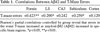
Pearson's partial correlations controlled by group reveal that errors in the water T-maze increased as amyloid-β42 (Aβ42) increased in specific brain regions.
p < 0.05
p < 0.01.
Correlations Between Aβ42 and CD11b

Pearson's partial correlations controlled by group reveal that inflammation as measured by CD11b (activated microglia) increased as Aβ42 increased in specific brain regions.
p < 0.05
p < 0.01.
Inflammation Was Not Increased in the Fornix or MS of LV-Hipp Mice
Significant between-group differences were observed in the density of CD11b labeling in the CA3 region of the hippocampus [F(4, 48) = 4.741, p = 0.003], the LS [F(4, 47) = 2.775, p = 0.038], and the fornix [F(4, 48) = 3.314, p = 0.018]. Post hoc analyses revealed significantly increased inflammation in the CA3 region of the hippocampus of the AD group and all treatment groups. However, increased inflammation was observed in the fornix of the AD, LV, and Hipp groups and in the MS of the AD and LV groups, but no increases in LV-Hipp mice (Fig. 7).

Protective effect of mesenchymal stem cells (MSCs) against inflammation in 5xFAD mice as measured by CD11b in the lateral septum (LS) and fornix regions. Mice injected with MSCs into both the lateral ventricles and the hippocampus (LV-Hipp) or the hippocampus alone (Hipp) did not have a significant increase in CD11b labeling in the LS. Images collected with Nikon scanner are random samples from each group. LV, 5xFAD mice injected with MSC into the lateral ventricles. Scale bars: 100 μm.
There Were No Observable Changes in Cytochrome Oxidase Staining
No between-group differences were observed for CYO densitometry measures in the cortex [F(4, 48) = 0.680, p = 0.609], CA3 region of the hippocampus [F(4, 48) = 1.675, p = 0.171], MS [F(4, 48) = 1.018, p = 0.408], LS [F(4, 48) = 0.888, p = 0.478], or dentate gyrus [F(4, 48) = 0.766, p = 0.553]. These data indicate that the level of metabolic activity, as measured by CYO labeling, did not appear to be significantly decreased in any of the structures of 8-month-old 5xFAD mice.
Partial correlations controlled by group revealed negative correlations between the percentage of time in the open arms in the light condition of elevated-plus maze and density of CYO in the dentate gyrus [r(50) = -0.326, p = 0.020], with similar trends in the MS [r(50) = -0.248, p = 0.080], cortex [r(50) = -0.259, p = 0.066], and the CA3 region of the hippocampus [r(50) = -0.248, p = 0.080]. These data suggest that as mice with lower levels of anxiety (higher percentage of time in the open arms) tended to have higher levels of CYO activity. Negative correlations were also observed between clasping scores and density of CYO in the LS [r(50) = -0.394, p = 0.004], cortex [r(50) = -0.298, p = 0.032], and CA3 region of the hippocampus [r(50) = -0.330, p = 0.017], with similar trends in the dentate gyrus [r(50) = -0.244, p = 0.081], and the MS [r(50) = -0.259, p = 0.064], suggesting that as CYO decreased in various regions, the incidence of clasping in all mice increased.
NeuN Labeling in the Cortex Was Not Reduced in 8-Month-Old 5xFAD Mice
No significant differences were observed between groups for the number of neurons labeled with NeuN, in either layer 5 [F(4, 50) = 0.963, p = 0.437] or layer 6 [F(4, 48) = 0.816, p = 0.521] of the cortex. These data indicate that the 8-month-old 5xFAD mice do not have significant neuronal loss in layers of these cortical regions.
ChAT Labeling in the MS Was Related to Aβ42 and CYO Levels, but Unaffected by MSC Transplants
ChAT-labeled neurons were counted in the MS using nonbiased stereology. The one-way ANOVA did not reveal significant differences between groups [F(4, 48) = 0.594, p = 0.344], suggesting that there is not a significant loss of ChAT-labeled cells in the 5xFAD at 8.5 months of age. However, the number of ChAT-labeled cells negatively correlated with the density of Aβ42 labeling in the dentate gyrus [r(50) = -0.283, p = 0.039], suggesting that as density of Aβ42 in the dentate gyrus increased, the number of ChAT-labeled cells counted in the MS decreased. The number of ChAT-labeled cells was also positively correlated with the density of CYO in the MS [r(50) = 0.311, p = 0.025], indicating that as counts of ChAT-labeled cells in the MS increased, CYO in the MS also increased.
Soluble and Insoluble Aβ Was Increased in 5xFAD Mice, but Synaptophysin Levels Were Unaffected
Soluble protein was operationally defined as protein that was extracted in Dulbecco's PBS. The standard curve ranged from 1 to 500 pg/ml, with r2 = 1.0, and y = (0.0206 - 3.32)/(1 + (x/252)1.59) + 3.32. Soluble Aβ42 was significantly different between groups [F(4, 24) = 7.625, p < 0.001], and the PLSD analysis indicated that WT mice had less soluble Aβ42, when compared to all other groups.
Insoluble protein was operationally defined as protein that was extracted in a guanidine HCl-based solution. The standard curve ranged from 1 to 1,000 pg, with r2 = 0.999, and y = (0.0141 - 1.34)/(1 + (x/465)1.58) + 1.34. Significant differences in insoluble Aβ42 were observed, [F(4, 24) = 3.237, p = 0.029], and the PLSD analyses indicated that WT had significantly less insoluble Aβ42, when compared to all other groups. Insoluble Aβ42 positively correlated with errors in the T-maze, on trials 11–20 [r(26) = 0.420, p = 0.026], which suggests that as insoluble Aβ42 increased, errors in the T-maze tended to increase.
ELISA measures of SYN revealed a standard curve, OTD = (1.95 - 0.364)/[1 + (concentration/5.8)1.06] + 0.364, with levels ranging from 1 to 50 ng, and r2 = 0.996. One-way ANOVAs revealed no significant between-group differences for SYN, but WT mice tended to have greater amounts of SYN. However, partial correlations, controlled by group, revealed that insoluble SYN negatively correlated with insoluble Aβ42 [r(26) = -0.458, p = 0.014], which indicated that as insoluble Aβ42 increased, SYN decreased. Further analysis of this measure revealed a trend that suggested that SYN tended to be negatively correlated with errors on trials 1–10 [r(26) = -0.321, p = 0.096] and trials 11–20 [r(26) = -0.364, p = 0.057].
Discussion
The results of the present study indicate that MSC transplants into 5xFAD mice can (1) reduce cognitive deficits; (2) alter the levels of Aβ42 and signs of inflammation in the brains of 5xFAD mice; and (3) tended to be most effective when injected into the lateral ventricles.
MSCs Induced Selective Beneficial Effects on Cognitive Disruptions of 5xFAD Mice Related to Levels of Aβ42
MSCs significantly improved both search strategies and learning as measured in the RAWM (Fig. 3) and water T-maze (Fig. 4), respectively. Given a lack of evidence that MSCs differentiated into neurons, their protective effect on cognitive processing is likely due to their abilities to regulate inflammation, release neurotrophic factors, and improve Ab clearance (17,20). Errors in the water T-maze increased as Aβ42 labeling in numerous regions of the brain (Table 1), and total Aβ42 insoluble protein increased. The subiculum and the fornix were the only regions where significant attenuation was observed in all MSC treatment groups, and of these regions the fornix most strongly correlated with the behavioral results, suggesting the fornix as a region of interest in treating AD. Decreases in fornix volume are observed in those with AD and are thought to be a critical point in the transition from mild cognitive impairment to AD clinical diagnosis (5,23).
Results of open-field, rotor rod, and PPI support that the attenuation of learning deficits is not confounded by the influence of activity, motor, or sensory gating abilities, respectively. MSCs transplanted into the hippocampus or into both the hippocampus and the lateral ventricles tended to prolong the abnormal antianxiety effects as measured by the elevated-plus maze. However, the antianxiety effects of the MSCs observed in the elevated plus-maze did not significantly correlate with measures taken from the water T-maze or RAWM, supporting the notion that the effects of the MSC transplants are mnemonic in nature and are not activity or anxiety dependent.
Subtle Increases in Anxiety in 5xFAD Mice Are Amplified by MSCs
Alterations of anxiety as measured by the elevated-plus maze in 5xFAD mice are likely subtle. Measured fluctuations in anxiety in the elevated-plus maze indicate that 5xFAD mice display significantly decreased anxiety on week 2 postsurgery when tested in the dark condition, but not on week 10 postsurgery, when tested in the light condition (Fig. 5). The decrease in anxiety observed in the 5xFAD mice (6.5 months of age) in the dark condition corresponds to the aging study of Jawhar and colleagues (16). The dark condition only utilizes the openness stressor, while the light condition includes both the openness stressor and a brightness stressor that mice naturally avoid. It is likely that a ceiling effect is observed when using the light condition, and that a dark condition is necessary to detect the subtle change in anxiety in 5xFAD mice.
The observed reduction of anxiety in the elevated-plus maze light condition when MSCs were injected into the hippocampus suggests that MSCs may have clinical utility for the treatment of mood disorders. Improvements of depressive symptoms were observed when MSCs were injected into the hippocampus of a rat model of depression (32). These decreases in depressive symptoms were accompanied by increases in hippocampal neurogenesis and were thought to be related to the release of neurotrophic factors by the stem cells (32).
Differing Effects Based on Route of Administration of MSCs
This is the first study to examine injections of MSCs, or any type of stem cell treatment, into multiple regions of the brain in the 5xFAD mouse model of AD. There appear to be only modest cognitive improvements when the cells are injected directly into the hippocampus, while there are more definitive improvements in mice injected with MSCs into both the lateral ventricles and the hippocampus or the lateral ventricles alone. Only an intermediate effect is observed in the T-maze (Fig. 4) for mice injected with MSCs into the hippocampus, and there are no effects in the RAWM (Fig. 3). While there was an MSC-induced attenuation of Aβ42 labeling in several regions of the Hipp group (Figs. 7 and 8), the fact that this did not translate into behavioral improvements in the measures used in this study suggests that its potential clinical utility may be limited.

A significant genotype effect was observed in the amount of Aβ42 extracted with Dulbecco's PBS and guanidine as assessed by enzyme-linked immunosorbent assay. The 5xFAD mice that received bilateral injections of mesenchymal stem cells (MSCs) into the lateral ventricles (LV), hippocampus (Hipp), or both locations (LV-Hipp), as well as AD control, had significantly more Aβ42 than wild-type (WT) controls, regardless of extraction media. However, no treatment effect of the MSC transplants was observed.
Dividing the cells between the lateral ventricles and the hippocampus provided improvements in the water T-maze (Fig. 3), as well as attenuation of Aβ42 in the hippocampus and other regions (Fig. 7), but injections directly into the lateral ventricles appear to work as well or even increase the effectiveness of the MSCs. There was a similar attenuation in neuropathological measures, such the decrease of Aβ42 in multiple regions of the brain (Fig. 7). Behaviorally, the mice with MSC injections into the lateral ventricles were the only ones to demonstrate improvement in both learning-related tasks (Figs. 3 and 4). While it is not completely clear if injecting into the lateral ventricles is more effective than dividing the MSCs between the hippocampus and the lateral ventricles, it is certain that the simple division of the MSCs between the regions does not provide an additive treatment effect. An additive effect may be observed if the amounts of MSCs injected into the LV-Hipp group are doubled, which we plan to explore in future studies.
The lack of CD11b reduction in mice with MSC injections into the lateral ventricles is supportive of MSC efficacy. Recent reports suggest that while MSCs increase anti-inflammatory cytokines and reduce proinflammatory cytokines, they also, counterintuitively, increase microglial activation when in the presence of Ab (21,31). This increase in microglial activation is dependent on production of chemoattractive factor CCL5 by MSCs, which increases the migration of healthy microglia that are capable of Ab phagocytosis as a means to replace dysfunctional microglia found in the AD brain (21).
While improvements occur in multiple MSC transplant groups, the injection of MSCs into the lateral ventricles alone offers the most clinically applicable treatment. Except for the rare cases for familial AD where patients may be diagnosed in their late 20s, the majority of AD patients are over the age of 65, with a large percentage that are over the age of 75 (1). The major brain surgery required for hippocampal injection may be too great of a risk for older patients. In the present study, injection of MSCs into the lateral ventricles appears to be more effective than injecting into the hippocampus.
Given these results, injecting the cells into the cerebral spinal fluid may provide an excellent alternative to more invasive brain surgery. However, since the long-term effectiveness of MSCs is still unknown, multiple administrations of MSCs may prove necessary for effectively treating AD. Injections through the cerebral spinal fluid could be administered multiple times, and this would allow the sampling of the spinal fluid to monitor the patient's progress, in terms of Aβ42 and other markers of AD in the spinal fluid (13), and provide a more practical approach for titrating the amount of cells to be delivered at various time points.
However, there remain several major caveats that need to be addressed before the efficacy of this treatment and the preferred route of administration can be fully evaluated. For example, the long-term fate of the injected MSCs needs to be thoroughly studied to ensure this procedure does not produce toxic side effects or tumor formation. Although evidence of long-term survival of MSCs has not been consistently observed, nor has such survival proven to be necessary for their therapeutic efficacy (10,27,28), further studies aimed at tracking the fate of these cells are needed to ensure the safety of this procedure and mitigate possible adverse effects that may occur over time, especially if repeated administration is to be considered.
In addition, more research is needed to determine how to best prepare the cells prior to transplantation in order to optimize their efficacy. Recent work in our lab (12) suggests that the number of passages and the source of the MSCs can affect the efficacy following transplantation.
Conclusions
In summary, our work supports a growing body of knowledge suggesting that MSC transplants can provide ameliorative effects for the neuropathology observed in the 5xFAD mouse model of AD. The MSCs in this study attenuated learning deficits observed in 5xFAD mice and also led to a decrease of Aβ42 in regions surrounding the injection sites. When comparing the various routes of administration, those MSCs injected into the lateral ventricles seemed to provide the most consistent ameliorative effects, either independently of, or in conjunction with, the hippocampal transplants. Given that no detrimental side effects of MSC transplants have been reported in clinical testing, the application of MSCs for clinical use in AD appears feasible. If a safe and effective delivery method, such as intravascular injections or delivery via the cerebral spinal fluid, can be developed, then MSCs may provide a new therapeutic approach in the treatment of AD.