
Abstract
The role(s) of collagenase G (ColG) and collagenase H (ColH) during pancreatic islet isolation remains controversial, possibly due to the enzyme blends used in the previous studies. We herein examined the role of ColG and ColH using highly pure enzyme blends of recombinant collagenase of each subtype. Rat pancreases were digested using thermolysin, together with ColG, ColH, or ColG/ColH (n = 9, respectively). No tryptic-like activity was detected in any components of the enzyme blends. The efficiency of the collagenase subtypes was evaluated by islet yield and function. Immunohistochemical analysis, in vitro collagen digestion assay, and mass spectrometry were also performed to examine the target matrix components of the crucial collagenase subtype. The islet yield was highest in the ColG/ColH group (4,101 ± 460 islet equivalents). A substantial number of functional islets (2,811 ± 581 islet equivalents) was obtained in the ColH group, whereas no islets were retrieved in the ColG group. Mass spectrometry demonstrated that ColH reacts with collagen I and III. In the immunohistochemical analysis, both collagen I and III were located in exocrine tissues, although collagen III expression was more pronounced. The collagen digestion assay showed that collagen III was more effectively digested by ColH than by ColG. The present study reveals that ColH is crucial, while ColG plays only a supporting role, in rat islet isolation. In addition, collagen III appears to be one of the key targets of ColH.
Introduction
Pancreatic islet transplantation is a promising treatment for restoring normoglycemia in type 1 diabetic patients (24,27,28). However, there are still many issues to be resolved regarding this treatment. One is that two or more donor pancreases are needed to render one diabetic patient insulin independent. Considering the organ shortage, a more efficient isolation procedure is needed, wherein a large number of high-quality islets can be stably obtained from one donor pancreas. Although several factors are related to the outcome of islet isolation (12,19,21,23), the enzymes used for digestion are one of the crucial factors (12,17,21).
To isolate islets, the pancreatic tissue first has to be dissociated by enzymatic degradation of the extracellular matrix (ECM) without damaging the structural and functional integrity of the islets. Crude collagenase preparation from Clostridium histolyticum, which have been routinely used for this purpose, are complex mixtures of different collagenases, neutral protease, and various other enzymes that possess tryptic-like activity (TLA) (8,32). Although highly purified collagenases are readily available in recent clinical islet isolations, these enzymes still contain a certain level of TLA. The original crude collagenase preparations contain at least six different collagenases with molecular weights ranging from 68 to 125 kDa (4,5). On the basis of their activities toward native collagen and the synthetic peptide 2-furanacryloyl-l-leucylglycyl-l-prolyl-l-alanine (FALGPA), the six collagenases are divided into two classes. Collagenase G (ColG) (corresponding to class I collagenases) has high collagenase activity and moderate FALGPA activity, while collagenase H (ColH) (corresponding to class II collagenases) has moderate collagenase and high FALGPA activity (4–6).
The role of the collagenase subtypes has been examined in several studies, but still remains controversial. An early study of rat islet isolation suggested that ColH plays a more important role (32), while another study showed that equal amounts of ColG and ColH were required for efficient rat islet isolation (7). Antonioli et al. reported that, in human islet isolation, ColH was correlated with the islet yield and digestion time (1). Conversely, Kin et al. reported that a ColH/ColG ratio <0.204 resulted in a high islet yield (17).
A possible explanation for these discrepancies is the enzymes used in each study. In most of the previous studies, crude collagenases produced by C. histolyticum were purified and fractionated by anion exchange chromatography (1,9,32). Therefore, a risk of contamination with an unwanted collagenase subtype or unknown proteases derived from C. histolyticum could not be ruled out. The use of highly purified recombinant collagenase of each subtype could help to overcome this problem. In fact, Wolters et al. (32) reported that the pancreas dissociation was different between chromatographically purified ColG and recombinant ColG. Of particular interest, Brandhorst et al. (9) recently reported that TLA also facilitates islet isolation (8). This novel finding suggests that some unknown protease(s), other than collagenases and neutral protease, may affect pancreas dissociation, since collagenases and neutral protease are known to have no TLA. Hence, TLA-free enzyme preparations should be used to investigate the roles of specific collagenase subtypes.
Another possible explanation for the discrepancies among the previous studies is the difference in the pancreatic ECM among species (15,29). A broad variation of the ECM is also seen in individual human pancreases (3,16,25,30). Therefore, elucidating the target ECM of each collagenase subtype would be beneficial not only for investigating the role of the two types of collagenase but also for optimizing the outcome of islet isolation.
The present study examined the roles of ColG and ColH in islet isolation using TLA-free enzyme preparations of highly purified recombinant collagenase of each subtype. We also investigated the molecular composition of the target ECMin rat pancreases of the crucial collagenase subtype using an immunohistochemical analysis and mass spectrometry.
Materials and Methods
Animals
Rat pancreases were obtained from 10-week-old male inbred Lewis rats (Japan SLC, Inc., Shizuoka, Japan) weighing 265 to 300 g. Eleven- to 12-week-old male inbred athymic mice (nude; nu/nu BALB/c, Japan SLC, Inc.) were used as a streptozotocin model of diabetes and for the intraperitoneal glucose tolerance test (IPGTT). All animals used in this study were handled in accordance with the Guide for the Care and Use of Laboratory Animals published by the National Institutes of Health (2) and the guidelines for animal experiments and related activities at Tohoku University (approved protocol ID: 2012 NICHe-Animal-5). All surgeries were performed under anesthesia, and maximal efforts were made to minimize suffering.
Construction of ColG-His and ColH-His Expression Systems
The coding region of ColG was amplified with a colG-F primer and colG-R primer. The primers (Operon Biotechnology, Tokyo, Japan) used in the present study are shown in Table 1. The genomic deoxyribonucleic acid (DNA) from C. histolyticum was used as a template. As a result of the substitutions, the amino acid residues at 1007–1008 were changed from Asn–Lys to Ser–Arg. The promoter region of the β-galactosidase [lactose (lacZ)] gene was amplified with lac-F and lac-R primers, and the plasmid (pUC19; Takara Bio Inc., Kyoto, Japan) was used as a template. The polymerase chain reaction (PCR) fragment for ColG was digested with Bacillus amyloliquefaciens H (BamHI; Takara Bio Inc.), and the promoter fragment was digested with Haemophilus influenzae Rd (HindIII; Takara Bio Inc.). Both digested fragments were inserted into the plasmid, pBR322 (Takara Bio Inc.), which was digested with BamHI and HindIII (lacZ-ColG-pBR322). The histidine (His)-tag coding region and following multicloning site of pET24a (Novagen, Darmstadt, Germany) were amplified with a set of primers for His-F and His-R. The PCR fragments were digested with BamHI and Xanthomonas badrii (XbaI; Takara Bio Inc.). The digested DNA fragments were inserted into the plasmid described above (lacZ-ColG-His-tag-pBR322), digested with BamHI and XbaI. The expression plasmid for the collagenase H-His-tag fusion protein was constructed using the same method described above, with the exception of the PCR for the ColH coding sequence. The coding region of ColH was amplified with colH-F and colH-R primers. The amino acid residue 980Gly in ColH was substituted with 980Ala. The plasmids, pColG-His and pColH-His, were used for the transformation of Escherichia coli χ1176 (a stock strain of Meiji Seika Pharma Co., Ltd., Tokyo, Japan).
The PCR Primers Used in the Study

PCR, polymerase chain reaction; colG, collagenase G; lac, β-galactosidase (lactose); His, histidine.
Enzyme Production and Purification
The E. coli carrying pColG-His or pColH-His were cultured in 100 ml of Terrific Broth [1.2% tryptone (Becton, Dickinson and Company, Franklin Lakes, NJ, USA), 2.4% yeast extract (Becton, Dickinson and Company), 0.94% K2HPO4, 0.22% KH2PO4, 0.8% glycerol] containing 100 μg/ml 2,4-diaminopimeric acid, 20 μg/ml thymidine, 50 μg/ml ampicillin (Sigma-Aldrich, St. Louis, MO, USA), and 0.1 mM isopropyl β-d-1-thiogalactopyranoside (all from Wako Pure Chemical Industries, Ltd., Osaka, Japan unless specified otherwise) at 28°C for 16 h. Ten milliliters of POPculture reagent (Merck-Millipore, Darmstadt, Germany) was added to the culture broth and mixed well. The mixture was filtered with a 0.2-μm cellulose acetate filter (Nalge Nunc International, New York, N Y, USA) to remove any remaining bacteria. A sixfold volume of binding buffer (20 mM phosphate buffer pH 7.5 containing 0.5 M NaCl and 20 mM imidazole, all from Wako Pure Chemical Industries) was added to the filtrate. The mixture was applied to 100 ml of Ni-NTA agarose column (Qiagen, Duesseldorf, Germany). The column was washed adequately with binding buffer, and then the enzyme was eluted with the same buffer excluding 500 mM imidazole. The collected His-tagged ColG and ColH were dialyzed against 24.1 mM 4-(2-hydroxyethyl)-1-piperazineethanesulfonic acid (HEPES)– Hanks' Balanced Salt Solution (HBSS; both Gibco, Life Technologies, Grand Island, NY USA) at pH 7.6 and were concentrated with a Pellicon 3 cassette ultrafiltration module (Merck-Millipore).
Enzyme Activity and Blending
In the present study, recombinant ColG, ColH, and thermolysin (TL) (as a neutral protease) (Peptide Institution, Inc., Osaka, Japan) were used to prepare highly pure, TLA-free enzyme blends. The enzyme blend activity was adjusted to be equal to that of the crude collagenase from C. histolyticum (Sigma collagenase type V; Sigma-Aldrich) using Azocoll (Calbiochem®, Merck Millipore), 4-phenylazobenzyloxycarbonyl-Pro-Leu-Gly-Pro-D-Arg (Pz-PLGPR) and Azocasein (both Sigma-Aldrich Japan, Tokyo, Japan) as substrates (Table 2). The crude collagenase was just used as one example to decide the effective amount of ColG and ColH. The TLA activity was measured by the cleavage of Na-benzoyl-l-arginine p-nitroanilide (Bz-Arg-pNA; Wako Pure Chemical Industries, Ltd., Osaka, Japan) at 37°C and pH 7.5. Notably, no TLA was detected in any components of the enzyme blends. In all the experimental groups, 0.3 mg of TL was added. The GH group contained ColG and ColH. The G and H groups consisted of ColG or ColH, respectively.
Characterization of Blended Enzyme Components

Pz-PLGPR, 4-phenylazobenzyloxycarbonyl-Pro-Leu-Gly-Pro-D-Arg; Bz-Arg-pNA, Na-benzoyl-l-arginine p-nitroanilide; ColG, collagenase G; ColH, collagenase H; ND, not detected.
Islet Isolation
Rat islet isolation was performed as described previously (26). In brief, before the removal of the pancreas, the cannulated bile duct was injected with 10 ml of cold HBSS containing enzyme blends. After digestion at 37°C for 14 min, density-gradient centrifugation was performed using Histopaque-1119 (Sigma Diagnostics, St. Louis, MO, USA) and Lymphoprep™ (Nycomed Pharma AS, Oslo, Norway) to isolate the pancreatic islets. The islet count was performed as islet equivalents (IEQs) under a scaled microscope using diphenylthiocarbazone (Wako Pure Chemical Industries) staining. The islets were cultured in Roswell Park Memorial Institute (RPMI)-1640 containing 5.5 mmol/L glucose (Gibco, Life Technologies) and 10% fetal bovine serum (Equitech-Bio, Inc., Kerrville, TX, USA) at 37°C in 5% CO2 and humidified air for 3 h before examination.
In Vitro and In Vivo Evaluation of Islet Function
The adenosine diphosphate (ADP)/adenosine triphosphate (ATP) ratio and the ATP/DNA ratio were measured to evaluate the energy status of the cultured islets. After picking up 80 IEQs, the ApoGlow™ kit (Lonza Rockland, Inc., Rockland, ME, USA) was used for the ADP and ATP measurements, as described previously (13). Using the same sample, the DNA content was measured using a DNA Quantify kit (Primary cell, Ishikari, Japan) as described (31). The insulin/DNA ratio was measured as previously described (31). The in vivo islet function was assessed in nude mice rendered diabetic by an intravenous injection of 237.5 mg/kg streptozotocin (Sigma-Aldrich) 6 days before transplantation of 700 IEQs under the left kidney capsule (n = 6). Blood glucose levels were measured in blood obtained from tail veins using a Precision Xceed (Abbott Japan Co., Ltd., Chiba, Japan). Mice whose nonfasting blood glucose levels exceeded 400 mg/dl in two consecutive measurements were considered diabetic. After transplantation, nonfasting blood glucose levels less than 200 mg/dl on two consecutive measurements were defined as normoglycemic and were considered to indicate graft function. The IPGTT was performed 30 days after islet transplantation. After a 14-h fast, d-glucose (2.0 g/kg; Otsuka Pharmaceutical Factory, Inc., Tokushima, Japan) was infused intraperitoneally as a single bolus, and the blood glucose concentrations were determined before and at 1, 3, 5, 10, 15, 20, 25, 30, 45, 60, 90, and 120 min after the glucose injection. The results of the IPGTT were evaluated by the area under the curve (AUC) and Kg values. Nephrectomy of the left kidneys was performed 35 days after transplantation to demonstrate the immediate return of hyperglycemia.
Sequential Injection of Collagenase Subtypes Into the Pancreatic Duct
This procedure examined the role of the collagenase subtypes in islet isolation. Briefly, two enzyme injections were made into the pancreatic ducts. Before the removal of the pancreas, the first enzyme blend was injected into the bile duct. After the addition of 10 ml HBSS, the pancreas was digested at 37°C for 7 min. Then, we added HBSS with the second enzyme blend and again digested the pancreas at 37°C for 7 min. Density gradient centrifugation was performed to isolate the pancreatic islets.
Pancreatic Tissue Digestion and Mass Spectrometry Analysis
Small pieces of Lewis pancreas (~100 mg) were incubated in 20 mM HEPES (pH 8.0) and 1 mM CaCl2 containing protease inhibitor cocktail (Roche, Basel, Switzerland) at 37°C (overnight). The incubated pancreatic tissue was washed with the same buffer and digested with ColH at a final concentration of 0.1 mg/ml for more than 10 h. The digested sample was divided into three aliquots (100 μl) in a 1.5-ml tube and incubated with 10 μl stock solution (100 mM) of (N-succinimidyloxycarbonylmethyl)tris (2,4,6-trimethoxyphenyl) phosphonium bromide (TMPP; Sigma-Aldrich) in 50% acetonitrile (Nacalai Tesque, Kyoto, Japan) for 30 min. Subsequently, samples were precipitated by cold acetone (400 ml; Nacalai Tesque) and dried after centrifugation at 13,000 × g. The dried samples were digested overnight by trypsin (10 mg/ml; Sigma-Aldrich Japan) in 100 mM ammonium bicarbonate solution (Nacalai Tesque).
The digested peptides were treated with a ZipTip (Millipore, Billerica, MA, USA) for mass spectrometry. The peptide samples were loaded onto a 75-μm fused silica capillary column containing C18 resin (ZipTip). The peptides were eluted with an acetonitrile gradient (typically 2.5–40%) in 0.1% formic acid (Wako Pure Chemical Industries, Ltd.) and analyzed by an LTQ Orbitrap XL mass spectrometer (Thermo Fisher Scientific Inc., Waltham, MA, USA). The sample preparation and mass spectrometric analyses were performed in triplicate. Database searches of the NCBInr and SwissPlot databases were performed using the MASCOT ver. 2.4.00 search engine (Matrix Science, London, UK). MASCOT searches were done without protease specificity with a 5-ppm tolerance on the mass measurement in mass spectrometry (MS) mode and 0.5 Da for MS/MS ions. Oxidation was considered as a variable modification for Met, Cys, Pro, and Lys. TMPP modification at the N-terminal was also taken into account. Protein identification was considered for MASCOT scores of p < 0.05.
Immunohistochemical Analysis
Tissue was taken from the rat pancreas, fixed in 10% formalin (Sigma-Aldrich) onto slides for 10 min at room temperature and washed with phosphate-buffered saline (PBS; made in house from Sigma-Aldrich products) twice. Slides were pretreated with 10% rat serum (made in house) for 30 min at room temperature to block nonspecific antibody reactions, followed by immunolabeling with primary antibodies against collagen I, II, III, or VI for 60 min at 37°C. Rabbit anti-rat collagen-I (Monosan, Sanbio BV, Uden, Netherlands), anti-collagen type II (Chemicon, Merck Millipore, Darmstadt, Germany), anti-collagen type III (Chemicon), and anti-collagen VI (Abcam, Cambridge, UK) antibodies were used at a 1:50 dilution in PBS. After washing the samples with PBS twice, secondary anti-rabbit antibodies conjugated to horseradish peroxidase (Dako, Glostrup, Denmark) were used, and antibody binding was localized with diaminobenzidine hydrochloride (Dako), and samples were counterstained with hematoxylin (Muto Pure Chemicals Co., Ltd. Tokyo, Japan).
Collagen Digestion by Collagenase Subtype
Collagen digestion assays were performed using human Col-I (2.5 mg/ml), bovine Col-II (2.9 mg/ml), human Col-III (1.0 mg/ml), and human Col-VI (0.52 mg/ml) (BD Biosciences, Bedford, MA, USA). Each collagen solution was mixed with an equal volume of buffer solution [100mM HEPES (pH 8.0) and 1 mM CaCl2], then digested by ColH or ColG (0.025 mg/ml) at 30°C. Digestion was stopped by adding sodium dodecyl sulfate (SDS) sample buffer [312 mM Tris-HCl (pH6.8) (Nacalai Tesque), 5% SDS (Nacalai Tesque), 50% glycerol (Nacalai Tesque), 0.1% bromophenol blue (Nacalai Tesque), and 2-mercaptoethanol (Nacalai Tesque)] at 5 and 10 min, and then samples were analyzed by SDS-polyacrylamide gel electrophoresis (PAGE; D.R.C. Co. Ltd., Tokyo, Japan). The bands were visualized by Coomassie blue (G-250) staining (Sigma-Aldrich).
Statistical Analysis
All values were expressed as the means ± standard deviation (SD) and were analyzed using the Excel for Macintosh software program (Statcel3; Redmond, WA, USA). Data from two groups were compared using Student's t test. Data from three groups were compared using a single-factor analysis of variance (ANOVA). To use the ANOVA for multiple parameters, the Tukey–Kramer test was employed as a post hoc test to determine the significant differences for pairwise comparisons. A value of p < 0.05 was considered to indicate statistical significance.
Results
The Effects of the Collagenase Subtype on the Yield and Functions of Isolated Rat Islets
In order to clarify the roles of ColG and ColH, the exact same lot and amount of TL was used in all the experimental groups. The islet yield in the GH group (ColG/ColH/ TL) was the highest (4,101 ± 460 IEQs, n = 9) among all the groups (Fig. 1A). In the H group (ColH/TL), a substantial number of well-shaped islets (2,811 ± 581 IEQs, n = 9) were obtained following efficient dissociation of pancreatic tissues, although a significant (~30%) decrease in the islet yield compared to the GH group was observed (p < 0.001) (Fig. 1A). In the G group (ColG/TL), the pancreatic tissues were not dissociated at all, and no islets were retrieved (n = 9) (Fig. 1A). The proportion of islet size was almost identical between the GH and H groups (Fig. 1B).

The effects of collagenase subtypes on the islet yield, appearance, and functions. (A) The islet yield was significantly higher in the collagenase G and collagenase H (GH) group (black bar: n = 9) compared with the H and G groups. Although no islets were retrieved in the G group (n = 9), a substantial number of well-shaped islets were obtained from the H group (white bar: n = 9). All values are expressed as the means ± SD. **p < 0.01. (B) The proportion of isolated islet size. No significant differences were detected between the GH (black bar: n = 9) and H groups (white bar: n = 9). (C) The blood glucose changes in the transplanted diabetic nude mice were measured. No significant differences were detected between the GH (black line: n = 6) and H groups (gray line: n = 6). (D) The blood glucose changes in the IPGTT were measured. No significant differences were detected between the GH (black line: n = 6) and H groups (gray line: n = 6).
Regarding the in vitro functional assays using the isolated islets, no significant differences were detected between the GH and H groups in the ADP/ATP ratio (p = 0.467, n = 9) (Table 3), ATP/DNA (p = 0.482, n = 9) (Table 3), or insulin/DNA (p = 0.991, n = 9) (Table 3). Concerning the in vivo functional tests, no differences were detected between the GH and H groups in the blood glucose changes (Fig. 1C, D), AUC (p = 0.978, n = 6) (Table 3), and Kg values (p = 0.847, n = 6) (Table 3) in the IPGTT.
The Effects of Collagenase Subtypes on the Functions of Isolated Rat Islets
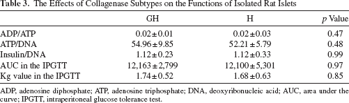
ADP, adenosine diphosphate; ATP, adenosine triphosphate; DNA, deoxyribonucleic acid; AUC, area under the curve; IPGTT, intraperitoneal glucose tolerance test.
The Effects of Sequential Injection of Collagenase Subtypes on the Yield of Isolated Rat Islets
To examine the role of the collagenase subtypes, ColG and ColH were sequentially injected into the pancreatic duct of rats. The islet yield was always highest when ColG and ColH were injected simultaneously. As shown in Figure 2A, an additional injection of ColG following an initial injection of ColH led to a slight increase in the islet yield. On the contrary, no beneficial effects were observed following an additional injection of ColH after ColG (Fig. 2B).

The effects of sequential injection of different collagenase subtypes on the yield of isolated rat islets. (A) The isolated islet yield in the H→G group (white bar: n = 6) was compared with the GH (black bar: n = 9) and H (gray bar: n = 9) groups. An increase of 20% in the islet yield was seen in the H→G group compared with the H group. (B) The isolated islet yield from the G→H group (white bar: n = 6) was compared with the GH (black bar: n = 9) and G (gray bar: n = 9) groups. No beneficial effects were observed after the additional injection of collagenase H (ColH) following ColG. All values are expressed as the means ± SD. ** p < 0.01.
Mass Analysis of Pancreatic Tissues Digested by ColH
Mass-analyzed peptide samples likely contain peptides digested by ColH, trypsin, and other pancreatic proteases, which were not perfectly denatured during the acetone precipitation process. The pancreatic tissue samples were labeled with TMPP after ColH digestion, because TMPP modification occurs at the N-terminal of the peptides (14) hydrolyzed by ColH. A database search was performed using the MASCOT software program (22) against nonspecific peptides. The mass analyses detected 59–69 proteins (Table 4). Focusing on fibril proteins and TMPP-modified peptides, collagen III (Col III) and Col I were included in the top 10 hits as search results (Fig. 3). Approximately 70% of the assigned peptides were TMPP modified in Col III, and 25% were TMPP modified in Col I. Although a few TMPP modifications were also observed in other proteins with low search scores, the search result of these proteins included only one to four unique peptides, and the TMPP modification might have been accidental (Fig. 3).
MASCOT Search Result for Mass Analyses

The score numbers (score1, 2, and 3) are trial numbers. Collagen peptides are in bold.

The plot score distributions in the mass spectroscopic analyses. (A) The search score was plotted for the modification ratio (2,4,6-trimethoxyphenyl) phosphonium bromide (TMPP)-modified peptide/detected peptide (p < 0.05). Higher search score indicates higher probability of protein identifications. The highest scored protein (pancreatic lipase) was omitted from the plot. Each analysis is indicated as a different point (●, ▲, and ■). In this plot, putative collagenase substrates appear in the diagonal area (from the origin to upper right). Collagen III and collagen I are indicated by red and green circles, respectively. (B) The score distribution of collagens. The plot is demonstrated only for the collagens used in plot (A).
Immunohistochemical Analysis
All rat pancreatic sections showed positive labeling for all collagen subtypes examined (collagens I, II, III, and VI). As shown by representative examples (Fig. 4), the lobular and acinar septa, blood vessels, and the pancreatic ducts were positively stained for Col I, III, and VI. In those areas, the staining for anti Col I and VI was moderate, whereas that for Col III was strong. Col II appeared to be diffusely located in the exocrine tissues. The peri-insular region displayed weak reactions for Col II and III.

The collagen staining of the pancreatic tissues. The lobular and acinar septa and the pancreatic ducts in the exocrine tissues were positively stained for Col I (A), III (C), and VI (D). In these areas, the reaction for anti-Col I and VI was moderate, whereas the reaction for anti-Col III was well developed. Col II (B) appeared to be diffusely located in the exocrine tissues. The peri-insular region displayed a weak reaction to Col II and III (arrow).
In Vitro Digestion of Collagens by Collagenase Subtypes
Collagen digestions were performed with ColG or ColH, and the results were analyzed by SDS-PAGE (Fig. 5). Based on bands that appeared after digestion, neither ColG nor ColH was able to digest Col VI, which is a nonfibril collagen. ColG digested Col I and III; however, less digestion was demonstrated for Col II. In contrast, ColH digested all three fibril collagens tested (Col I to III). Although Col I and III were digested by both types of collagenase, different digestion patterns were observed, which was consistent with previous reports (10,11).

In vitro collagen digestion by collagenases. Collagen digestion was performed for Col I, II, III, and VI using ColG (left panel) and ColH (right panel). Samples were run in three lanes for each collagen at different sampling times (left: 0 min, middle: 5 min, right: 10 min). The band of collagenase is indicated by a black arrow. The molecular weight was estimated using the marker (M).
Discussion
Numerous studies have been performed to elucidate the roles of ColG and ColH during pancreatic islet isolation (1,9,18,32), but the results have been contradictory. One possible explanation is the enzyme blends used in those studies. In the present study, we examined the roles of ColG and ColH using highly purified, TLA-free enzyme blends. The innovative recombinant technique introduced in this study provides more accurate and highly reproducible outcomes compared with the conventional anion exchange chromatography technique (9,32). We have clearly shown that ColH and TL can efficiently dissociate rat pancreatic tissues and release functional islets from the acinar cells. In agreement with the present study, it was previously reported that ColH plays a predominant role in rat pancreas dissociation, while ColG has a limited role (32). In contrast, Brandhorst et al. reported that neither ColG nor ColH alone was able to dissociate pancreatic tissues and release islets from the exocrine tissue (9). This discrepancy may be explained, at least in part, by the method that Brandhorst et al. used, which employed a marginal amount of neutral protease to detect the optimal ratio between ColG and ColH. Based on these and the present results, the function of ColG may be partially replaced by neutral protease. Of particular interest, in the present study, no pancreatic tissues were dissociated by the combination of ColG and TL, suggesting that the function of ColH cannot be replaced by ColG and/or neutral protease. Moreover, it seems likely that some ECM components of the pancreatic tissues can be digested only by ColH. However, these findings are still limited in the case of small animal models; further studies in other species may be needed to draw broader conclusions.
An additional injection of ColG following an initial injection of ColH led to a slight increase in the islet yield. However, no beneficial effects were observed following an additional injection of ColH. These results suggest that ColH may need to bind to the target matrix components before ColG binds to them.
Since ColH was proven to be crucial for rat islet isolation in the present study, we also investigated the molecular composition of the target ECM of ColH using an immunohistochemical analysis and mass spectrometry. In the mass spectrometry assay, a protease inhibitor cocktail was added to the pancreatic tissue before reacting them with ColH in order to prevent autolysis due to endogenous digestive enzymes, such as trypsin and lipase. Both TMPP labeling and the use of the protease inhibitor cocktail made it possible to narrow down the matrix components targeted by ColH. Mass spectrometry suggested that ColH specifically reacts with Col I and III, especially Col III.
In the immunohistochemical analysis, both Col I and III were located in the acinar septa and the pancreatic ducts. Col III was more intensely expressed in the exocrine area, while almost no expression of Col III was observed in peri-insular regions. This pattern was consistent with a previous report by Van Deijnen et al. (29). Furthermore, in support of the previous findings (20), the in vitro digestion of collagens also revealed that Col III was more effectively digested by ColH than by ColG. Taken together, these findings suggest that islet isolation is initiated by fragmentation of the pancreatic tissues by ColH mainly via the degradation of Col I and III expressed on the exocrine tissues. Thereafter, islets are released from the exocrine tissues, most likely by ColG and/or neutral protease. Detecting the target matrix components of ColG and TL are topics of interests for a future study.
In conclusion, the present study revealed that ColH is of great importance, while ColG plays only a supporting role, in rat islet isolation. In addition, the present data also suggest that one of the targets of ColH is Col III. Therefore, semiquantitation of Col III in pancreatic tissues may greatly contribute to a more reproducible and successful islet isolation by optimizing the level of ColH. Further investigations using a large animal model with highly variable matrix components (3,16,25,30) are required.
Footnotes
Acknowledgments
The authors thank Kozue Imura, Takahiro Ito, and Megumi Goto for their excellent technical assistance. The authors also acknowledge the support of the Biomedical Research Core of Tohoku University, Graduate School of Medicine and TAMRIC (Tohoku Advanced Medical Research and Incubation Center), and the Research Seeds Quest Program (JST) and the grant of “Coordination, Support and Training Program for Translational Research” from the Ministry of Education, Culture, Sports, Science and Technology. The founders played no role in the study design, data collection and analysis, decision to publish, or preparation of the manuscript. Kazutaka Murayama participated in the performance of the research and the writing of the paper. Youhei Yamagata participated in the performance of the research. Kimiko Watanabe participated in the writing of the paper. Takehiro Imura participated in the performance of the research. Akiko Inagaki participated in the performance of the research. Naomi Ohbayashi participated in the performance of the research. Hiroki Shima participated in the performance of the research. Satoshi Sekiguchi participated in the performance of the research. Keisei Fujimori participated in the performance of the research. Kazuhiko Igarashi contributed to the analysis. Noriaki Ohuchi participated in the writing of the paper. Susumu Satomi participated in the writing of the paper. Masafumi Goto participated in the research design, the performance of the research, and the writing of the paper. The authors declare no conflict of interest.