
Abstract
Following a stroke, the administration of stem cells that have been treated with granulocyte colony-stimulating factor (GCSF) can ameliorate functional deficits in both rats and humans. It is not known, however, whether the application of GCSF-mobilized peripheral blood stem cells (PBSCs) to human skin can function as an antiaging treatment. We used a Lanyu pig (Sus scrofa) model, since compared with rodents, the structure of a pig's skin is very similar to human skin, to provide preliminary data on whether these cells can exert antiaging effects over a short time frame. GCSF-mobilized PBSCs from a young male Lanyu pig (5 months) were injected intradermally into the cheek skin of aged female Lanyu pigs, and tissues before and after the cell injections were compared to determine whether this treatment caused skin rejuvenation. Increased levels of collagen, elastin, hyaluronic acid, and the hyaluronic acid receptor CD44 were observed in both dermal and subcutaneous layers following the injection of PBSCs. In addition, the treated skin tissue was tighter and more elastic than adjacent control regions of aged skin tissue. In the epidermal layer, PBSC injection altered the levels of both involucrin and integrin, indicating an increased rate of epidermal cell renewal as evidenced by reductions in both cornified cells and cells of the spinous layers and increases in the number of dividing cells within the basal layer. We found that the exogenous PBSCs, visualized using fluorescence in situ hybridization, were located primarily in hair follicles and adjacent tissues. In summary, PBSC injection restored young skin properties in the skin of aged (90 months) pigs. On the basis of our preliminary data, we conclude that intra dermal injection of GCSF-mobilized PBSCs from a young pig can rejuvenate the skin in aged pigs.
Keywords
Introduction
Skin is the largest organ of the human body and is composed of three layers: epidermis, dermis, and hypodermis. There are two distinct aging processes that affect the skin: photoaging and chronological aging. Chronological aging, which is also called intrinsic aging, represents the natural aging process. Intrinsic aging results in fine wrinkles, dryness, laxity, and elevated pigmentation of the skin. To determine the intrinsic age status of skin, levels of several key factors are typically evaluated. Fibrous collagen is important for skin strength and elasticity, and levels of this protein are generally decreased in aged skin (5,12). Elastin helps skin remain flexible but tight, providing a bounce-back reaction when the skin is poked or pinched (29). In humans, hyaluronic acid (HA) is abundant in skin and connective tissue and helps skin remain moisturized and smooth. Finally, CD44 is a receptor for HA. We use these intrinsic markers, therefore, to evaluate skin rejuvenation in our experimental system.
Therapeutic strategies that involve stem cells, including embryonic, induced pluripotent, and somatic, represent potential treatments for a wide variety of intractable diseases, such as skin degeneration. To clinically induce skin regeneration, for example, a wide variety of stem cells have been used (11,12,20). In 2011, Kim et al. reported that adipose-derived stem cells also may be useful for antiaging skin therapies. These cells primarily stimulate collagen synthesis in dermal fibroblasts and increase angiogenesis (12).
Hematopoietic stem cells (HSCs) are multipotent stem cells with self-renewal properties that can be isolated from the blood or bone marrow. HSCs can be divided into monocytes, granulocytes, lymphocytes, red blood cells, and platelets. Peripheral blood stem cells (PBSCs), a form of HSCs, are relatively easy to obtain, as the collection process inflicts minimal physical harm on the donor (11,20). Granulocyte colony-stimulating factor (GCSF) has been shown to induce migration of HSCs from the bone marrow into the peripheral blood (6,25,31). Among HSCs, GCSF-mobilized PBSCs are used preferentially for the majority of therapeutic applications (21,24,26). Furthermore, as GCSF-mobilized PBSCs are the most feasible clinical source of stem cells, they have also been used to regenerate nonhematopoietic tissues, such as skeletal muscle, heart (19), and neurons (25). There are very few reports, however, concerning the antiaging effects of PBSCs in skin. We hypothesized that GCSF-mobilized PBSCs have the capacity to rejuvenate aged skin, and we tested this idea in a preliminary study using the Lanyu pig (Sus scrofa) model system. The Lanyu pig is an indigenous breed from the Lanyu Islet southeast of Taiwan and is a well-characterized animal model for studies involving medical therapies and tissue engineering (3,10,13–16). The physiological skin structures of the pig and human are quite similar (2), making the pig an ideal model (often better than the mouse) for physiological studies.
Materials and Methods
Animals
One male (5 months) and three female (91.7 ± 0.2 months) Lanyu miniature pigs were purchased from the Taitung Animal Propagation Station (Taitung, Taiwan). This experiment was approved by the China Medical University Institutional Animal Care and Use Committee. Animals were allowed free access to water and quota diet and were monitored regularly.
PBSC Mobilization, Isolation, and Characterization
To mobilize HSCs, a male Lanyu pig was injected with human GCSF (hGCSF) (Filgrastim M300; Kyowa Hakko Kirin Co., Ltd, Takasaki-shi, Japan) via the central ear vein for 5 consecutive days (10 μg/kg/day). Peripheral blood was obtained from the male donor pig via jugular venipuncture 6 days after the final GCSF injection. We analyzed monocyte levels in peripheral blood using a hematology analyzer (Sysmex KX21N; Sysmex Corporation, Kobe, Japan). PBSCs were isolated via leukapheresis into BD Vacutainer CPT tubes (Becton Dickinson; Franklin Lakes, NJ, USA) that contained Ficoll Hypaque density gradient liquid.
Flow cytometry was used to determine the percentage of monocytes within the population of GCSF-mobilized PBSCs. To do this, GCSF-mobilized PBSCs were first washed with cold phosphate-buffered saline (PBS). Following centrifugation (400 × g for 5 min), approximately 5 × 10⁵ cells were incubated with 2 μl of anti-CD34 (Abcam, Cambridge, MA, USA) for 30 min at 4°C. Cells were collected via centrifugation (400 × g for 5 min) and washed with ice-cold PBS. Cell-bound CD34 was detected by incubating the cells with 1 μl of fluorescently labeled donkey anti-rabbit IgG mAb (Alexa Fluor 546; Invitrogen, Grand Island, NY, USA) for 30 min at 4°C. Following two washes, a BD LSR™ II flow cytometer (Becton Dickinson) was used to detect CD34+ monocytes. Data were read by BD FACSDiva software (Becton Dickinson).
PBSC Transplantation
Three female Lanyu pigs with a mean age of 91 ± 1 months were anesthetized with an intradermal injection of 50 mg/kg Zoletil (Zoletil 50; Virbac Animal Health, Carros, France). Each side of the face was divided into four quadrants, with one side treated with 1 × 10⁶ PBSCs in each quadrant and the other side administered saline. In order to track cells, PBSC nuclei were fluorescently labeled by incubating them with 1 μg/ml bis-benzimide (Hoechst 33258; Invitrogen) for 1 h at 37°C before injection. Skin biopsies were collected for analysis 1 and 2 weeks following the transplantation. All experiments were performed with three different Lanyu female pigs at four different locations, and the values were averaged.
Tissue Preparation
Tissue biopsies were fixed in a 10% neutral-buffered formalin solution for 24 h. Samples were then embedded in paraffin blocks using a paraffin embedding system (Tissue Block System 88; Medite, Burgdorf, Germany) and sectioned (3 or 5 μm in thickness) using a Shandon AS 325 microtome (Southeast Pathology, Folly Beach, SC, USA).
Histochemistry
Sections were deparaffinized in xylene and rehydrated through a graded ethanol series. To evaluate skin structure and thickness, sections were then subjected to hematoxylin and eosin staining (Medical Chemical Corporation, Los Angeles, CA, USA). For each section, skin thickness was measured at three representative locations (magnification 40x) with Microsoft Image Composite Editor Version 1.4.4.0 software (Microsoft, Redmond, WA, USA). Serial sections (3 μm thick) were also stained with Masson-trichrome stain (Sigma-Aldrich, St. Louis, MO, USA) or elastic stain (ScyTek Laboratories Inc., Logan, UT, USA) to detect collagen or elastin densities, respectively. Photographs were taken at 400x magnification.
Immunohistochemistry
Paraffin-embedded sections were obtained from the pig skin and were processed for immunohistochemical staining. Following deparaffinization and rehydration, serial sections (3 μm thick) were placed in 0.01 M citrate buffer (pH 6.0) at 121°C 10 min for antigen retrieval. After the sections were washed with rinse buffer (1 x PBS + 0.05% Triton X-100) and blocked with 5% fetal bovine serum (Gibco Life Technologies, Grand Island, NY, USA) for 30 min at room temperature, they were incubated with diluted primary antibodies for 16–18 h at 4°C. The following primary antibodies were used: anti-sheep HA polyclonal (1:200 dilution; Abcam), anti-rat CD44 monoclonal (1:100 dilution; Abcam), anti-mouse β1-integrin monoclonal (1:100 dilution; Abcam), anti-rabbit Ki67 monoclonal (1:100 dilution; Abcam), and anti-mouse involucrin monoclonal (1:200 dilution; Abcam). Following washes, sections were then incubated with biotinylated secondary antibodies (SuperSensitiv Link-Label IHC Detection System; BioGenex, Fremont, CA, USA) for 15 min at room temperature. Sections were washed twice in rinse buffer and then reacted with streptavidin-horseradish peroxidase. Staining was visualized with diaminobenzidine (DAB) (2-Component DAB Pack Kit; BioGenex). Finally, the sections were counterstained with hematoxylin solution.
Fluorescence In Situ Hybridization (FISH)
Skin sections (3 μm thick) were deparaffinized in xylene (twice for 5 min) and then incubated in PBS for 5 min at room temperature. Sections were digested with 0.02% pepsin (Riedel-de Haen, Seelze, Germany) for 30 min, dehydrated using a graded ethanol series, and incubated in PBS for 5 min at room temperature. Sections were then incubated with 10 μl of a pig Y chromosome DNA probe that was labeled with cyanine 3 (Cy3; Chromosome Science, Sapporo, Japan) for 10 min at 90°C using a hotplate. Samples were then hybridized overnight at 37°C in a moist chamber. After the slides were washed twice with saline-sodium citrate (SSC) buffer, they were stained with 1.5 μg/ml 4,6-diamidino-2-phenylindole (DAPI) (UltraCruz Mounting Medium; Santa Cruz Biotechnology) and mounted in Aqueous Mount (ScyTek Laboratories, Inc.). Serial sections were photographed with an inverted fluorescence microscope (IX71, OLYMPUS, Tokyo, Japan).
Statistical Analysis
Statistical analysis was performed with Microsoft Excel 2010 software (Microsoft). All experiments were performed with three different Lanyu female pigs. The data were expressed as the mean ± standard deviation (mean ± SD). The paired Student's t test was used to determine statistical differences between groups. Values of p < 0.05 were considered statistically significant.
Results
Structural Differences Between Young and Aged Lanyu Pig Skin
To analyze skin thickness and structure, samples from both young and aged Lanyu pigs were sectioned and subjected to hematoxylin and eosin staining (Fig. 1A). Measurements indicated that the corneum was thicker in aged skin (0.364 mm) compared to younger samples (0.156 mm). Skin from aged pigs also had more melanocytes and more cells within the spinous layer. In addition, Masson-trichrome and elastin staining revealed that the levels of dermal collagen and the density of elastin were also higher in young skin (Fig. 1C, D). Finally, to analyze the rate of keratinocyte proliferation, skin samples were immunohistochemically stained for the proliferation marker Ki67 (Fig. 1E). Ki67 expression was significantly higher in aged pig skin than in young pig skin.

Differences in skin structure between young and aged Lanyu pigs. (A–E) Representative skin samples from young and aged pigs (5 and 90 months, respectively) are shown. Hematoxylin and eosin staining was used to analyze (A) skin thickness (40x; scale bars: 1 mm) and (B) the structure of the skin (i.e., the quantity of melanocytes and spinous layer cells) (400x; scale bars: 0.2 mm). (C) Masson-trichrome staining was used to evaluate collagen density (400x; scale bars: 0.2 mm). (D) Elastin stain was used to evaluate elastin density (400x; scale bars: 0.2 mm). (E) Immunohistochemistry for Ki67 was used to measure keratinocyte proliferation (400x; scale bars: 0.2 mm).
hGCSF Injection Alters the Cellular Composition of Blood
After the donor pig (5 months) was injected with hGCSF for 5 consecutive days, complete blood samples were collected. We used a hematology analyzer and flow cytometry to analyze the cellular composition of these samples. Compared with baseline samples, the number of monocytes increased about sevenfold following hGCSF injections. In addition, the number of CD34+ cells was elevated after hGCSF stimulation (data not shown).
Distribution of PBSCs After Intradermal Transplantation
To visualize the distribution of injected PBSCs, the cells' nuclei were labeled with bis-benzimide prior to injection and we used FISH to label the Y chromosome of donor cells. Bis-benzimide visualization showed that during the PBSC injections, the needle was inserted to a depth of ~0.3 mm (i.e., the dermo-epidermal junction) (Fig. 2A). One week after the injection, the donor cells were still detectable by bis-benzimide imaging (Fig. 2B). FISH was also used to visualize the Y chromosome of the donor cells (which were derived from a male pig) within tissue of the female recipient. FISH results indicated that the donor cells were still present in the cornfield layer of the epidermis 2 weeks after the injection of PBSCs (Fig 2C). In addition, we found that the majority of injected PBSCs after 1 week were located in the dermis (Fig. 3A). Most injected cells were also found near a hair follicle (Fig. 3B). Moreover, transplanted PBSCs migrated from the hair follicle to the dermal area and synthesized high levels of support matrix (HA) in the hair follicle of the dermis (Fig. 3B). A similar distribution was seen after 2 weeks (data not shown).

Visualization of transplanted cells up to 2 weeks after transplant. In order to track the cells, male peripheral blood stem cell (PBSC) nuclei were fluorescently labeled by bis-benzimide before transplantation into female pigs. (A) Depth of needle insertion. (B) Distribution of PBSCs after 1 week. The arrows represent bis-benzimide-labeled (transplanted) cells (25x; scale bars: 100 μm). (C) Distribution of PBSCs after 2 weeks. The arrows represent cells positive for the Y chromosome by fluorescence in situ hybridization (FISH) (i.e., transplanted cells) (25x; scale bars: 0.2 mm).

Localization of transplanted PBSCs in recipient tissue via FISH. (A) Distribution of PBSCs in the dermis 1 week after the injection (25x; scale bars: 0.2 mm). The arrows represent cells positive for the Y chromosome by FISH. (B) Transplanted PBSCs exhibit high levels of hyaluronic acid (HA) in hair follicles. The arrows represent transplanted cells (400x; scale bars: 0.1 mm).
Subcutaneous Transplantation of GCSF-Mobilized PBSCs Into Aged Pig Skin Leads to Tissue Rejuvenation
After GCSF-mobilized PBSCs were injected into the skin of an aged Lanyu pig, a statistically significant decrease in dermal thickness was measured. This is consistent with skin rejuvenation, as the skin of young animals is typically thin and transparent (Fig. 4). At the same time, skin parameters that are associated with young or rejuvenated skin were increased following PBSC treatment. These parameters included collagen, elastin, HA, and CD44 (Fig. 5). We also examined the expression of involucrin and β1-integrin, which are markers of cornification and cell proliferation, respectively (25). Involucrin is a precursor of the cornified envelope and a useful marker for terminal differentiation. PBSC injection decreased the levels of involucrin. Integrins consist of one a and one β subunit, and function as receptors. Importantly, these proteins relay information from the extracellular matrix into the cell. Keratinocytes with high levels of β1-integrin exhibit stem cell properties, such as the enhanced ability to form colonies in vitro (24). The injection of PBSCs into aged skin increased the level of β1-integrin expression (Fig. 6). As such, when GCSF-mobilized PBSCs were isolated from a young pig and subcutaneously injected into an older pig, we observed skin regeneration and rejuvenation (Fig. 7). This led us to propose that β1-integrin might attract autologous PBSCs leading to skin regeneration. This theory will be studied in the future.

Injection of PBSCs affects dermal thickness. (A) Hematoxylin and eosin staining of skin from young, old, and PBSC-treated (2 weeks after the injection) pigs. (B) Quantification of dermal thickness in young, old, and PBSC-treated pigs.

PBSC treatment significantly elevated levels of collagen, elastin, HA, and CD44, compared to baseline. The arrows represent stained cells (+: <20%, ++: 20–40%, +++: 40–60%, ++++: 60–80%, +++++: >80%).

PBSC treatment reduced levels of involucrin and increased levels of β1-integrin, compared to baseline. The arrows represent stained cells (+: <20%, ++: 20–40%, +++: 40–60%, ++++: 60–80%, +++++: >80%).
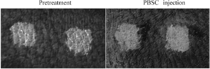
PBSC treatment reduced wrinkles. The clinical pictures demonstrate a reduction in wrinkles 2 weeks after the injection of PBSCs.
In this study, therefore, we have isolated GCSF-mobilized PBMCs from a young pig and injected them into the skin of an aged pig. As a result, recipient skin thickness was reduced, and skin parameters associated with rejuvenation and youth were elevated. In summary, PBSC injection reduced wrinkles and pigmentation, improved skin elasticity and tightness, and promoted epidermal cell renewal.
Discussion
In this study, we demonstrated for the first time that PBSCs derived from a young male pig rejuvenated the skin of female aged pigs. We documented the histological changes associated with the rejuvenation process (e.g., increases in collagen, elastin, polysaccharides, and HA) and traced the PBSCs after transplantation. Our data suggest that PBSC transplantation can be used to modify the microenvironment of aged skin. Finally, we used FISH to label the Y chromosome of donor cells and found that transplanted PBSCs migrated from the hair follicle to the dermal area and secreted HA. This represents a possible mechanism for the decrease in skin wrinkling that we observed.
PBSCs are being used more and more frequently as a source of HSCs for transplantation. Compared with bone marrow, adipose tissue, and umbilical cord blood, PBSC transplants are easier to administer and require less supportive care (28,30). The concentration of PBSCs in peripheral blood, however, is quite low (7). To address this problem, GCSF has been used to stimulate migration of HSCs from bone marrow into the peripheral blood (25). hGCSF was chosen because it stimulates the migration of hematopoietic progenitor cells from the bone marrow into the circulating bloodstream (24). When humans are treated with hGCSF (15 μg/kg for 5 consecutive days), leukocytes are increased 4–10 times, on average (1,4,8,17,23,24,27,32). In our study, treating a young donor pig (5 months old) with hGCSF (10 μg/kg for 5 consecutive days) increased monocytes 7-fold compared with baseline. These results suggest that hGCSF functions well in pigs.
Collagen and the HA receptor CD44 are expressed at very low levels in aged skin. Based on histological analyses, we found that levels of these proteins were significantly elevated in pigs that received PBSCs, compared to the elder pigs. To visualize the distribution of the injected male PBSCs, we labeled the cells' nuclei with bis-benzimide and used FISH to label the Y chromosome of the donor cells. This revealed that the transplanted PBSCs migrated from the subcutaneous injection site into hair follicles. In a variety of stem cell systems, the microenvironment of the stem cell niche is critical for stem cell maintenance (17,18,22). Among self-renewing compartments of the mammalian epidermis, the hair follicle provides a niche for both hair-follicle stem cells and melanocyte stem cells, two distinct stem cell populations with different origins (9). The concentration of the injected PBSCs around hair follicles suggests that PBSCs may be attracted by factors secreted by the follicle. Homing factors such as stromal cell-derived factor 1α (SDF-1α) promote neuroprotection and angiogenesis, as well as the mobilization and homing of bone marrow-derived cells in the ischemic brain (25). We propose that subcutaneously injected PBSCs home to the hair follicle where they secrete HA. This could potentially recruit endogenous stem cells, thus producing an environment that rejuvenates aged skin.
Numerous studies have suggested that adult stem cell populations decline with age (1,4,8,17,23,27,31). We have found that the injection of young-pig PBSCs into the skin of an aged pig led to a significant improvement in the health and life span of the skin. This provides experimental evidence that the loss of an adult stem cell population with aging may directly contribute to an age-related phenotype. Stem cells from younger animals may be able to repair the aging environment. For example, when muscle-derived stem/progenitor cells are isolated from young wild-type mice and provided (via intraperitoneal administration) to progeroid mice, a significant extension of both life span and health span are observed. During pregnancy, fetal CD34+ cells enter the maternal circulation and create a state of physiological microchimerism (fetal microchimerism). Fetal cells may respond to maternal injury by developing multi-lineage capacities within maternal organs. In this study, transplantation of young-pig PBSCs directly into an older pig's subcutaneous skin led to skin regeneration and rejuvenation. Our results also suggest a paracrine mechanism that may require secreted factors. Among these secreted factors are elastin, which helps the skin return to its original position when pinched, and collagen, which is primarily found in fibrous tissues such as skin and decreases with age. HA is also a major component of skin that is involved in tissue repair. CD44 is a receptor for HA that can also interact with other ligands, such as osteopontin, collagens, and matrix metalloproteinases. These factors are present at very low levels in aged skin. The transplantation of PBSCs, however, can upregulate these factors in aged skin. These observations suggest that transplanted PBSCs survive in the allograft and reinitiate metabolic functions within the skin via secreted factors. In the future, identification of these secreted factors and methods to rejuvenate adult stem cells ex vivo will be critical for regenerative medicine.
In conclusion, we show here that transplantation of young hGCSF mobilized PBSCs induced rejuvenation in the facial skin of aged pigs. Furthermore, our results suggest two mechanisms for this rejuvenation effect. First, transplanted PBSCs migrated into hair follicles where they restored skin metabolism. Second, transplanted PBSCs expressed collagen, elastin, polysaccharides, and HA, which seem to contribute to skin rejuvenation. Although the long-term effects of autologous PBSCs must be examined more closely, we expect that this approach will promote clinical trials in the future. It is worth noting the discrepancy in age and sex between the animals, the short time frame studied and the small number of animals, which was limited by availability and cost in this preliminary study. These influences need to be explored in future studies to verify our findings.
Footnotes
Acknowledgments
This work was supported by China Medical University (DMR-102-052); Taiwan Department of Health Clinical Trial and Research Center of Excellence (DOH101-TD-B-111-004); “Aim for the Top University Plan” of the National Chiao Tung University and Ministry of Education, Taiwan, ROC; and National Science Council (NSC100-2314-B-039-006-MY3). This work was also supported in part by a grant from Enhance Biomedical Ltd. We thank Yao-Horng Wang for his technical assistance. The authors declare no conflict of interest.