
Abstract
Cell constructs and culture methods are essential tools in tissue engineering. The cell construct should be equivalent to the native cartilage it is intended to replace. Thus, three-dimensional cell constructs are usually composed of a high density of cells and dense extracellular matrix. However, dense constructs suffer from a lack of passive nutrient supply, gas exchange, and removal of degraded debris. We have developed a novel hydrostatic pressure/perfusion culture system that improves the quality of neo-tissues, providing an automated and affordable system for clinical applications. We have also developed a semipermeable membrane pouch that contains a fragile amorphous cell carrier. Although amorphous material is difficult to handle, it is a useful medium in which to deliver cells to the desired site via injection. We evaluated phenotypes of bovine articular chondrocytes embedded in a collagen type I gel enclosed within membrane pouches permeable to molecules of various sizes. Constant or cyclic hydrostatic pressure was externally applied to the medium phase with a new culture system. Accumulation of cartilage specific matrix was promoted with a 500-kDa cutoff membrane pouch and cyclic hydrostatic pressure at 0.5 MPa, 0.5 Hz. This new method will be useful in the delivery of engineered cells to a desired tissue in regenerative medicine.
Keywords
Introduction
The first commercially available tissue engineering product for cartilage repair was an autologous chondrocyte suspension (1,25). Advanced products have been developed to improve methods of cell expansion and delivery, and surgical approach (12). Cell culture is a critical in vitro process for manufacturing cell/tissue constructs. Technological advancements have made it possible to implant cell constructs to replace damaged cartilage and promote subsequent regeneration (13,16). However, a three-dimensional (3D) construct creates its own problems if it has high cell density and causes large amounts of extracellular matrix (ECM) to accumulate; this blocks the supply of necessary nutrients and gas exchange. To solve such problems, commercially available or custom-made bioreactors have been used. Bioreactors are designed to optimize culture conditions by controlling oxygen concentration (19,26), shear stress (8,17), and hydrostatic pressure (HP). We and other researchers have studied the ability of HP to promote chondrocyte phenotypes (4,21,22) with a variety of pressure profiles: continuous versus intermittent (10,29) and constant versus cyclic (2,3,5,7,11,30). The results of these studies differed depending on the magnitude, duration, and profile of the HP.
Using our hydrostatic fluid pressure/perfusion culture system, we previously reported that HP promoted accumulation of cartilage ECM by bovine articular chondrocytes (bACs) in a porous collagen sponge (21). In addition, the manufactured chondrocyte construct has been studied for clinical applications (9). Besides the structural scaffold, injection of amorphous gel is another tool that can be used to augment tissue in general. However, amorphous gel is difficult to handle in culture and in surgery, problems that must be solved in order to explore new applications of such gels (27). Therefore, we developed a novel culture method using a semipermeable membrane pouch to handle amorphous gel and redesigned our culture system (23). We evaluated the accumulation of ECM by bACs seeded in the gel within semipermeable membrane pouches of varied molecular cutoff sizes and incubated with constant or cyclic HP. Combining the HP and pouches will be explored to develop useful applications (e.g., augmentation of tissue mass and minimally invasive cell/matrix delivery for tissue engineering).
Materials and Methods
Performance of Semipermeable Membrane Pouches
Pouches were constructed of semipermeable membrane tubing made of polyvinylidene difluoride (PVDF; 8-mm diameter, Spectra/Por Biotech®, Spectrum Laboratories, Rancho Dominguez, CA). We tested two tubings with different molecular weight cutoffs: 250 and 500 kDa. The tubing was cut into 15-mm pieces and folded 1–2 mm from one end (leaving the other end open), and the folded end was sealed with two stainless steel clips to construct a tube. The tubes were rinsed with culture-grade water (Sigma-Aldrich, St. Louis, MO), submerged in the water, and autoclaved at 121°C for 15 min.
For performance evaluation, the tubes were filled with sterile culture-grade water. The open end of the tube was folded at 90° from the opposite closed end and sealed with two stainless steel clips to construct a pouch like a tetra pack (Fig. 1A).

A pouch device and hydrostatic pressure (HP) culture system. Pouches were constructed of semipermeable membrane tubing. (A) Both ends of the tubing were folded and sealed with stainless steel clips to construct a pouch like a tetra pack. (B) The pouches were placed in a pressure-proof culture chamber unit. (C) The culture chamber unit and a medium bag were installed in the culture system. The culture unit has three components: a pump unit (1), a pressure-proof culture chamber unit (2), and a backpressure regulator unit (3). Medium is replenished with gas-permeable silicon tubing (4) and kept in a medium bag (5). The culture system is automatically controlled with a built-in computer control system. The backpressure regulator unit is fitted with a needle valve and attached to a spring-operated actuator of the control system. The set pressure is regulated by changing the distance between the valve and the actuator. The culture chamber unit has a flexible silicon film (500 μm thick) that separates the medium in the culture chamber from the adjacent water-filled compression chamber. The compression chamber has a piston connected to a spring-attached actuator. The piston compresses the water in the compression chamber and indirectly the medium through the silicon film. (D) The HP profile at 0.5 MPa, 0.5 Hz, 100 μl/min.
The pouches were placed in a pressure-proof culture chamber unit (Fig. 1B) connected to a medium bag (Transfer Pack Container, Baxter, Deerfield, IL) with gas-permeable silicon tubing. The bag was filled with 100 ml of 10 mg/ml sterile bovine serum albumin (BSA, Sigma-Aldrich). The culture chamber unit and a medium bag were installed in the culture system (Fig. 1C, TEP-02, Takagi Industrial, Shizuoka, Japan). In a series of experiments, the pouches were incubated at 1) static conditions (no pressure and no perfusion) in the BSA; 2) perfusion alone at 100 μl/min with continuous replenishment of the BSA; 3) constant HP at 0.5 MPa with 100 μl/min continuous replenishment of the BSA; or 4) cyclic HP at 0.5 MPa, 0.5 Hz, with 100 μl/min continuous replenishment of the BSA using the culture system (Fig. 1D). All incubation was conducted under the same conditions as regular cell culture, at 37°C and 5% CO2. Four pouches each were harvested after 3, 6, 12, and 24 h of incubation. The outside of each harvested pouch was quickly flushed with water, and excess water on the outside of the pouch was absorbed with Kimwipes. One end of the pouch was cut, and the sample was immediately aspirated. The protein content of each sample was measured with a protein assay kit (BCA protein assay, Bio-Rad, Hercules, CA).
Chondrocyte Isolation
A bovine forelimb (from a calf 2–3 weeks old) was purchased from a local abattoir. Pieces of articular cartilage (5 × 5 × 2–5 mm) were harvested from the weight-bearing area of the humeral chondyle (18). Slices of the middle zone (MZ) layer were isolated by removing a layer of surface zone (–100 μm thick from the top surface) and a layer of deep zone (–300 μm thick from subchondral bone) using a scalpel (#15, BD, Franklin Lakes, NJ) under a stereomicroscope (Nikon Instruments, Melville, NY). The MZ slices were digested with 0.15% collagenase (CLS-1, Worthington, Lakewood, NJ) dissolved in Ham's F-12 medium with 100 U/ml penicillin and 100 μg/ml streptomycin (Invitrogen, Carlsbad, CA) at 37°C with gentle shaking for 12 h. The bACs of the MZ were collected through a cell strainer (70-μm mesh, BD), followed by rinsing with D-PBS.
Constructing Cell/Collagen Gel in a Semipermeable Membrane Pouch
One tenth volume of 10× Dulbecco's modified Eagle medium (DMEM, Invitrogen) and 1/10 volume of 0.1 N NaOH were added to 8/10 volume of 0.3% pepsin-digested acid-soluble collagen solution from bovine skin (Pure-Col™, Cohesion, Palo Alto, CA). This collagen solution was neutralized (7 < pH < 8) with additional 0.1 N NaOH, confirming the pH using pH paper (EM Science, Gibbstown, NJ). Cells were suspended in the neutralized collagen solution (750,000/50 μl), and 50 μl of the cell suspension was injected into the semipermeable membrane tube. The tube was incubated at 37°C for 1 h to let the collagen form an amorphous cell/gel. Finally, the tube was filled with serum-free DMEM/F12 to eliminate air bubbles, and the open end of the tube was folded at 90° from the opposite end, constructing a tetra pack, and sealed with two stainless steel clips (Fig. 1A).
Optimization of Culture Conditions with HP
We assessed culture conditions with two profiles of HP treatment: 1) constant HP and 2) cyclic HP (Fig. 1D). For a static-controlled culture, the amorphous cell/gel was ejected from each pouch and incubated in DMEM/Ham's F-12 medium (F-12, Invitrogen) with 10% fetal bovine serum (FBS, Invitrogen), 100 U/ml penicillin, and 100 μg/ml streptomycin (Invitrogen) at 37°C and 5% CO2 for 7 days. Pouches were incubated with continuous medium replenishment at 100 μl/min (designated “perfusion”), with constant HP at 0.5 MPa with continuous medium replenishment at 100 μl/min, and with cyclic HP at 0.5 MPa, 0.5 Hz with continuous medium replenishment at 100 μl/min. The magnitude and cycle of HP were programmed and automatically controlled with our new culture system. Ten milliliters of medium was used for each pouch. After combinations of multiple culture conditions were evaluated, static control, constant HP, and cyclic HP culture conditions were evaluated simultaneously without normalization.
Biochemical Evaluation
After 7 days of culture, the pouches were harvested for biochemical and histological evaluation. Both ends of each pouch were cut, and the cell/gel was ejected and digested in 450 μ l of 125 μg/ml papain (Sigma-Aldrich) at 60°C for 8 h (14). Four cell/gels from each group were measured for DNA and sulfated glycosaminoglycan (S-GAG) content. For DNA content, the samples were incubated with 10 ng/ml bisbenzimide H 33258 (Hoechst 33258, Molecular Probes) and diluted in TNE buffer (Tris-EDTA sodium chloride, pH 7.4). Fluorescence intensity of the Hoechst 33258 was measured at ex 365 nm and the em blue region with a fluorometer (TBS-380, Turner, Sunnyvale, CA). Salmon sperm DNA (Sigma-Aldrich) was used as a standard. S-GAG was measured with 1,9-dimethyl-methylene blue (DMB, Sigma-Aldrich) (6). DMB (200 μl) was added to 2 μl of sample, and the optical density was immediately measured at 540 and 570 nm (Bio-Rad 550). Chondroitin sulfate C from a shark (Sigma-Aldrich) was used as a standard.
Histological Evaluation
Two samples of cell/gel from each test condition were fixed with 2% paraformaldehyde in 0.1 M cacodylic acid, pH 7.4 (Polysciences, Warrington, PA), and embedded in paraffin. Rehydrated sections (5 μm) were stained with safranin-O (Saf-O) to identify negatively charged ECM. Accumulation of collagen type II (Col 2) and keratan sulfate (KS) was evaluated immunohistochemically. The immunohistochemistry sections were stained with an anti-Col 2 antibody (1:20, Chemicon, Temecula, CA) and then with a biotinylated second antibody kit following the manufacturer's instructions (Vectastain ABC Elite kit, Vector Laboratory, Burlingame, CA). For KS staining, the sections were digested with 0.1 U/ml chondroitinase ABC (Seikagaku America) for 1 h at 37°C before staining. The sections were stained with an anti-KS antibody (Seikagaku America, Falmouth, MA) and then a biotinylated second antibody kit following the manufacturer's instructions (Vectastain ABC Elite kit). For color development, the sections were incubated with DAB (DAB substrate kit, Vector Laboratory). Counterstaining was performed with Harris's hematoxylin (Sigma-Aldrich).
Semiquantitative Assessment of Amount of Saf-O-Positive Matrix and Cell Number in Histological Section
We randomly selected five square areas (200 × 200 μm) at the exterior, periphery, and interior of each section stained with Saf-O under 33× optical magnification. Two sections were also randomly selected from both samples. We measured optical density of the area at fixed video mode (100 ns) with a longpass filter (590 nm, Omega), a CCD camera (ORCA-AG, Hamamatsu, Bridgewater, NJ), and the OpenLab™ image acquisition system (Improvision, Waltham, MA). The filter emphasized the red color of Saf-O and minimized the interference of other colors (shorter wavelength). The optical density of each area was automatically converted to pixel number. The pixel number/area of the periphery and interior of a section was subtracted from the mean number of the exterior (background) of each section. The number of nuclei in the same area was counted and converted to the number per mm2. Because these measurements were taken from only two samples, statistical analysis was not performed. However, the data were used to support our understanding of the heterogenic spatial distribution of Saf-O-positive matrix and cells.
Data Analysis
The performance of the semipermeable membrane pouch was assessed by a mean value and SD representing four samples. S-GAG and DNA data were analyzed using one-way analysis of variance (ANOVA) followed by Dunnett's test for comparing all cutoff size and HP profiles versus control (static) with p < 0.05 considered statistically significant (GraphPad InStat ver. 3.00, San Diego, CA). The optical density of Saf-O-positive matrix and the cell number in the randomly selected five areas in each sample were measured and counted, respectively.
Results
Performance of the Semipermeable Membrane Pouch
Infiltration of BSA (MW 70 kDa) was used to measure the performance of semipermeable membrane pouches in two MW cutoff sizes and culture under static conditions (no pressure), with cyclic HP/perfusion, with constant HP/perfusion, and perfusion alone. Under static conditions, less than 5% of BSA infiltrated the pouches (regardless of cutoff size) in 24 h (Fig. 2). With perfusion, 18% of BSA infiltrated the 500-kDa pouch in 24 h, but less than 10% of BSA infiltrated the 250-kDa pouch. With constant HP for 24 h, 40% and 20% of BSA infiltrated the 500- and 250-kDa pouches, respectively. With cyclic HP for 24 h, 99.5% and 15% of BSA infiltrated the 500- and 250-kDa pouches, respectively.

Performance of semipermeable membrane pouches with cutoff of (A) 250 kDa and (B) 500 kDa. The pouches were incubated in 0.1% BSA at static, perfusion at 100 μl/min, constant HP at 0.5 MPa, or cyclic HP at 0.5 MPa, 0.5 Hz, each with replenishment at 100 μl/min. BSA concentration within each pouch was converted to a percentage in a culture chamber. Data represent the mean and SD of four samples.
Biochemical Evaluation
The amount of S-GAG produced by bACs under static conditions without a pouch (used as a control) was 85.4 ± 5.4 μg/gel (Fig. 3A). With cyclic HP, bACs in the 500-kDa pouch accumulated significantly more S-GAG (101.6 ± 7.7 μg/gel, p < 0.01) than the static control. S-GAG in the 250-kDa pouch was slightly less than in the control. With constant HP, the amounts of S-GAG in pouches of the 250 and 500 kDa cutoff sizes were similar to the static control. With perfusion, S-GAG in pouches of the 250 and 500 kDa cutoff sizes was significantly less than the control (p < 0.01).
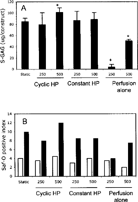
(A) S-GAG accumulation by bACs cultured in gel within pouches for 7 days. Bars represent the mean ± SD of four samples. ∗Significant difference relative to static control, p < 0.01. (B) Optical density of Saf-O-positive matrix by bACs cultured in gel within pouches for 7 days. The optical density in randomly selected 200 × 200 μm square at periphery (blank bar) and interior (closed bar) of each section was measured with a CCD camera. The density was converted to pixel number, the background subtracted, and expressed as an index.
The optical density of the Saf-O-positive matrix was converted to pixel number and expressed as an index (Fig. 3B). In all cutoff sizes, regardless of HP profiles and static control, the density at the interior section was greater than at the periphery. With perfusion, the periphery and interior of the 250-kDa pouch had similar density. However, there were differences between the interior and periphery of the 500-kDa pouch.
DNA content of bAC/gel after 7 days of culture under static conditions was 4.0 ± 0.5 μg/gel (Fig. 4A). With cyclic HP, constant HP, and perfusion alone, DNA was 4.1–4.7, 3.8–4.2, and 3.7–4.7 μg/gel, respectively. DNA contents were similar across cutoff sizes and HP profiles.

(A) DNA content in gel within pouches at day 7. The pouches are exposed to cyclic HP 0.5 MPa, 0.5 Hz; 0.1 ml/min, constant HP 0.5 MPa, 0.5 Hz, 0.1 ml/min, and perfusion alone 0.1 ml/min. Bars represent the mean ± SD of four samples. (B) Cell density in gel within pouches at day 7. The cell number in randomly selected 200 × 200 μm square at periphery (open bars) and interior (filled bars) of each section was counted under a microscope. The density was converted to cell number per area (mm2).
The cell number within randomly selected areas at the periphery and interior of each section were counted and normalized per mm2 (Fig. 4B). The cell density at the periphery and interior was similar under all HP profiles. However, the cell number in the 250-kDa pouch with perfusion alone was greater than in the 500-kDa pouch.
Histological Evaluation
Characteristics of accumulated ECM differed notably between constant and cyclic profiles of HP. After 7 days of culture, bAC/gel without a pouch under the static condition showed empty spaces and dense Saf-O-positive ECM (Fig. 5A). With a pouch under the static condition, Saf-O-positive ECM had less color intensity than that without a pouch (data not shown). In addition, nuclei of the bACs in the pouch were pyknotic. Therefore, the bAC/gel under static conditions was always incubated without a pouch in further studies, and that was defined as the static control. With perfusion, bAC/gel accumulated Saf-O-positive ECM at much lower levels in the 250-kDa pouch (Fig. 5B) than in the 500-kDa pouch (Fig. 5C). In addition, the volume of the gels with perfusion was smaller than under static (without pouch) or HP conditions. In order to compare a complete set of all cutoff sizes, perfusion culture was eliminated from further studies because necessary metabolic activity was not shown with the 250-kDa cutoff pouch. With both constant and cyclic HP, bACs accumulated dense Saf-O-positive ECM in gels within pouches of both 250-and 500-kDa cutoff sizes. Less accumulation was seen with constant (Fig. 5D, E) than with cyclic HP (Fig. 5F, G). In particular, notably large amounts of Saf-O were found within the 500-kDa pouch with treatment of cyclic HP at 0.5 MPa, 0.5Hz (Fig. 5G). The 500-kDa pouch showed both intense and fibrous accumulation of Saf-O-positive ECM (Fig. 5G).
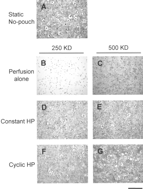
Photomicrographs of bACs in collagen gel after 7 days of culture. (A) A static culture control without a pouch, (B, C) perfusion at 0.1 ml/min, (D, E) constant HP at 0.5 MPa, 0.1 ml/min, and (F, G) cyclic HP at 0.5 MPa, 0.5 Hz, 0.1 ml/min. Intensity of red indicates S-GAG accumulation (a bar indicates 100 μm, Safranin-O, 5-μm-thick section).
bACs accumulated Col 2 in the gel with treatment of constant or cyclic HP (Fig. 6A–D). Both constant and cyclic HP showed dense Col 2 accumulation within the 500-kDa pouch, particularly pericellularly.

Photomicrographs of immunohistochemistry of (A–D) collagen type II (Col 2) and (E–H) keratan sulfate (KS) in gel after 7 days of culture. Intense brown indicates accumulation of each matrix component. Nuclei were counterstained with hematoxylin (a bar indicates 100 μm, 5-μm-thick section).
The bACs accumulated KS, one of the components of aggrecan, with treatment of constant or cyclic HP (Fig. 6E–H). Constant HP elicited dense and fibrous accumulation of KS in the 500-kDa pouch. Cyclic HP produced dense and fibrous accumulation of KS in the 500-kDa pouch.
Discussion
Semipermeable membrane pouches have varied permeability to BSA depending on their molecular cutoff sizes and the profiles of fluid HP to which they are subjected. The best performance was found in the 500-kDa cutoff pouch with cyclic HP at 0.5 MPa, 0.5 Hz, allowing infiltration of 99.5% of BSA in 24 h. Although performance was evaluated for only 24 h and was limited to a typical serum molecule, we speculate that essential nutrients infiltrated the pouch under the aforementioned culture conditions.
Most bioreactors were designed to promote nutrient supply to a 3D cell construct (28,31). However, it is vital that the neo-tissue build the ECM within the cell construct, and the high rate of medium flow creates the risk of washing away the ECM. A semipermeable membrane pouch was exposed to fluid-flow shear stress through replenishing the medium. However, the stress was thought to be negligible, because the replenishment rate was 0.1 ml/min, and the culture chamber has an obstacle platen to prevent jet flow through the injection of medium. Thus, using a semipermeable membrane pouch is a useful method to reduce such risk.
Histological evaluation indicated that nutrient supply to the pouches was probably restricted by the semipermeable membrane in both static and perfusion culture. On the other hand, constant and cyclic HP apparently allowed for the maintenance of cell viability and ECM production by bACs in a pouch. The ECM in constant and cyclic HP showed slightly different morphology, indicating that interstitial fluid motion due to each HP profile affected both the assembly and the amount of ECM (31). Medium volume shrinks by 0.022% with HP at 0.5 MPa. Even though this change in volume is quite small, the cyclic HP seems to increase permeability of soluble molecules through the semipermeable membrane. HP application involves medium perfusion at the same replenish rate as perfusion alone. Thus, HP should be recognized as a major stimulator to promote ECM accumulation by bACs. Although determining the mechanism of the effects of HP was not our aim in this study, extended studies using pouches and HP indicate that HP stimulates both anabolic and catabolic mRNA expression (20). The mechanism of HP stimulation is under investigation. Besides direct effects of the HP, we speculate that eventually nutrients and gases could be supplied, allowing large-molecule products to be retained within the pouch and small-molecule debris to be exuded (Fig. 7). This speculation is partially supported by Klein and Sah's model by diffusion or convection using a semipermeable membrane (15). The amount of S-GAG in the 500-kDa pouch with cyclic HP was 18% (p < 0.01) greater than in the static control without a pouch. However, the small difference in the amounts was hard to correlate with histological changes (Fig. 5F, G).

Schematic drawing of a semipermeable membrane pouch device.
The cell density of the cell/gel construct with perfusion was higher than with other culture conditions (Fig. 4B). The properties and shape of the gel were not considered in this study, because it was amorphous and formed within a flexible plastic tube. It was difficult to determine whether the gel was degraded or shrank. One possibility was that the gel shrank and made the cell density appear higher. Thus, we have to carefully evaluate cell density and determine the optimal culture conditions for secure clinical applications.
Recently, we developed a structural 3D construct to replace damaged and/or defective cartilage, because the goal of engineering tissue was to build a cell construct similar to native cartilage in vitro. By extending the concept from replacement to repair of tissues, the clinical applications can be broadened. One proposal was to inject cells/gel into fibrocartilage to repair insufficient regeneration of hyaline cartilage or to augment defective cartilaginous tissue (e.g., ears and nose). The injected chondrocytes/gel are expected to release cytokines and recruit appropriate cells from adjacent tissue. Other ideas may include injecting cell/gel into the vertebral space for intervertebral disc repair. By combining new methods, even uncontrollable and fragile amorphous cell carriers can be used to develop further applications. In this study, we focused on a profile of HP and the permeability of semipermeable membrane pouches for tissue engineering applications. Recently, we demonstrated that S-GAG accumulation by chondrocytes or chondrogenic adipose-derived stem cells increased with cyclic HP at 0.5 MPa, 0.5 Hz followed by static culture for 3 weeks (15,24). Beyond our current studies, applications using a semipermeable membrane pouch and HP will be explored for other applications. In addition, deformation of the amorphous construct within a semipermeable membrane can be made with a strain module. Cartilage physiology in response to weight bearing and joint loading will be reproduced with those apparatus.
In conclusion, we developed an HP/P culture system that applies HP by compressing the medium fluid (23). We also developed a semipermeable membrane pouch to incubate chondrocytes in amorphous gel. We found that constant or cyclic HP maintained viability and increased dense ECM by bACs in gel. Combining the HP and pouches will be explored to develop useful applications for tissue engineering.
Footnotes
Acknowledgments
This research was partially supported by CIMIT (Boston, MA) and Takagi Industrial, Co., LTD. (Shizuoka, Japan). Special thanks to Dr. Toshimi Murata for technical assistance.