
Abstract
The potential use of stem/progenitor cells as alternative cell sources to mature hepatocytes remains basically dependent on their ability to exhibit some, if not all, the metabolic liver functions. In the current study, four major liver functions were investigated in adult derived human liver stem/progenitor cell (ADHLSCs) populations submitted to in vitro hepatogenic differentiation: gluconeogenesis, ammonia detoxification, and activity of phase I and phase II drug-metabolizing enzymes. These acquired hepatic activities were compared to those of primary adult human hepatocytes, the standard reference. Amino acid content was also investigated after hepatogenic differentiation. Differentiated ADHLSCs display higher de novo synthesis of glucose correlated to an increased activity of glucose-6 phosphatase and mRNA expression of key related enzymes. Differentiated ADHLSCs are also able to metabolize ammonium chloride and to produce urea. This was correlated to an increase in the mRNA expression of relevant key enzymes such arginase. With respect to drug metabolism, differentiated ADHLSCs express mRNAs of all the major cytochromes investigated, among which the CYP3A4 isoform (the most important drug-metabolizing enzyme). Such increased expression is correlated to an enhanced phase I activity as independently demonstrated using fluorescence-based assays. Phase II enzyme activity and amino acid levels also show a significant enhancement in differentiated ADHLSCs. The current study, according to data independently obtained in different labs, demonstrates that in vitro differentiated ADHLSCs are able to display advanced liver metabolic functions supporting the possibility to develop them as potential alternatives to primary hepatocytes for in vitro settings.
Keywords
Introduction
Liver cell transplantation can transfer a missing metabolic function in a deficient native liver (7,11, 27,38,40). However, the technique faces various problems, such as lack of supply due to organ shortage, poor proliferation capacity of mature hepatocytes, poor resistance to cryostorage, and limited durability of the metabolic effect (29). An alternative cell source that could overcome these limitations is needed. Stem/progenitor cells are increasingly considered due to their ability to proliferate and to differentiate and the hope to provide unlimited supply for both in vitro needs and clinical human use. The well-described intrahepatic stem/progenitor cells, as for instance oval cells (31), are difficult to isolate and to maintain longer in vitro (9,16,18). Extrahepatic stem/progenitor cells, such as mesenchymal phenotype, show good potential for proliferation and are easy to manipulate in vitro, whereas their functional differentiation capability remains quite variable and incomplete (5,8,19, 22,24,25).
In contrast to fetal and embryonic stem/progenitor cells (13,26), increased interest for somatic cells has been documented for several reasons, including safety concerns as well as the level of their functional differentiation and immunogenic profile (33). We recently isolated mesenchymal stem/progenitor cell populations from healthy adult human livers (28). The cells we isolated from enzymatically digested healthy liver are able to proliferate in vitro and show a predetermination for hepatogenic differentiation but not for osteogenic or adipogenic differentiation. At the basal level, the liver stem/progenitor cells express, besides mesenchymal proteins (vimentin and α-smooth muscle actin), hepatic-specific markers such as albumin and CYP3A4 in accordance with their liver origin. They do not express biliary markers such as cytokeratin 19 (CK19). In vivo, when intrasplenically transplanted into uPA+/+-SCID and SCID mice, the undifferentiated cells are able to migrate, to engraft into the recipient liver parenchyma, and to function up to 10 weeks posttransplantation (28).
Liver cell transplantation has mainly been developed for the treatment of inborn errors of liver metabolism (37). This condition remains ideal for proof of concept demonstration as only one single enzyme is missing and cell supply efficacy can be easily followed. The use of stem/progenitor cells as alternative cells for the clinical development of liver cell-based therapies remains closely dependent on their metabolic activity potential. Accordingly, evaluation of basal and/or acquired liver metabolic functions, after in vitro hepatogenic differentiation, is mandatory. These properties, if acquired and demonstrated, are also important to propose differentiated stem/progenitor cells as an alternative in vitro cell model for the evaluation of acute and chronic drug metabolism as well as xenobiotic screening. The development of these cell models will provide the possibility to better understand cell responses to drugs. It also may allow and facilitate the classification of several compounds according to their specific induced signaling pathways and tissue activities.
In this work, we evaluate the main important hepatic functions that are acquired in ADHLSCs differentiated in vitro. We investigate their ability to synthesize glucose, and to metabolize ammonia. We furthermore analyze the activity of phase I and II drug-metabolizing enzymes as well as the content of amino acids. These analyses were independently performed in different labs and used human mature hepatocytes as controls. The data of the current study demonstrate that all the investigated metabolic activities were increased after in vitro ADHLSCs hepatogenic differentiation, which opens new perspectives for the development of these cells as alternative candidates to mature hepatocytes for in vitro metabolic screening assays.
Materials and Methods
Chemicals
The following chemicals were used: Glucose oxidase type II from Aspergillus (Sigma); peroxidase type II from horseradish (Sigma); ethylenediaminetetraacetic acid disodium salt dehydrate (EDTA) (Sigma); 2-dianisidine (Ucb); sodium L-lactate (Fluka); sodium pyruvate (Sigma); potassium dihydrogen phosphate (KH2PO4) (Merck); potassium chloride (KCl) (Fluka AG); sodium chloride (NaCl) (Acros); sodium hydrogen carbonate (NaHCO3) (Merck); magnesium sulfate heptahydrate (MgSO4•7H2O) (Merck); ethanol (Merck); Tris[hydroxymethyl]aminomethane (Tris-acetate) (Sigma); D-glucose 6-phosphate sodium salt (Sigma); lead nitrate (Acros); ammoniac sulfide solution (Sigma); phenobarbital (Sigma); 4′,5,7-trihydroxyflavanone 7-rhamnoglucoside (Naringin) (Sigma); rat tail collagen type I (BD Biosciences); 0.05% Trypsin-EDTA 1× (Invitrogen); L-ornithine hydrochloride (Janssen chimica); ammonium chloride (Sigma); P450-Glo™ assays (Promega); paraformaldehyde 4% (Merck); Triton X-100 (Sigma); urea colorimetric assay (Gentaur); Dulbecco's modified Eagle medium (Invitrogen); Iscove's modified Dulbecco's media (IMDM) with and without phenol red (Invitrogen); and Williams' medium E (Invitrogen). Chemicals used for phase I and II enzyme activity measurement were obtained from Sigma (München, Germany).
Hepatogenic Differentiation
All experiments of the current study were approved by the ethical committees of the St-Luc Hospital and faculty of Medicine of Université Catholique de Louvain.
ADHLSCs isolated from different healthy livers of cadaveric donors (n > 5) were seeded at a density of 104 cells/cm2 in six-well plates coated with rat tail collagen type I and in vitro differentiated into hepatocyte-like cells after sequential incubation with growth factors and cytokines, as previously described (28). Medium was changed every 3 days and the cells were microscopically observed every 24–36 h. At the end of the differentiation process, cells with hepatocyte-like morphology were harvested for metabolic activity analyses. Human hepatoblastoma HepG2 cells and primary cultures of freshly isolated human hepatocytes were used as controls.
Gluconeogenesis Analysis
Cells (n = 6, isolated from five different liver donors) were washed two times with 2 ml of PBS preheated at 37°C and then incubated with Krebs solution (0.15 M NaCl, 5.8 mM KCl, 1.5 mM KH2PO4, 2.6 mM MgSO4• H2O, and NaHCO3 0.2%, pH 7.4) containing 10 mM of sodium L-lactate and 1 mM of sodium pyruvate for 24 h in a fully humidified atmosphere containing 5% CO2. The culture medium was harvested for glucose assay and adherent cells were collected for protein quantification. Samples were analyzed in duplicate and data were presented as nmol glucose/mg protein∗24 h. Five hundred microliters of each sample was incubated with 3 ml Tris-HCl 1 M, pH 8, containing 25 units glucose oxidase, 39 purpurogallin units peroxidase, 2 nM EDTA, and 0.03 ml 2-dianisidine saturated in ethanol (100 mg 2-dianisidine dissolved in 10 ml ethanol) for 1 h at 37°C. Glucose oxidase converted present glucose to gluconolactone and produced hydrogen peroxide. In the presence of peroxidase, 2-dianisidine reacted with hydrogen peroxide and formed brown dianisidine oxides, which could be measured using spectrometry at 420 nm (20). Glucose concentration was indirectly determined using standard curve with dilutions ranging from 0 to 240 mM glucose. Adherent cells were washed two times with 2 ml PBS and incubated with 1 ml Trypsin-EDTA for 5 min at 37°C. Cell suspension was recovered and centrifuged at 3000 rpm for 3 min. The pellet was washed two times with 2 ml PBS and then centrifuged. The cells were lysed in 1 ml NaOH 0.1 M for 24 h at 4°C; total protein was measured using Bio-Rad Protein Assay according to the manufacturer's instruction.
Glucose-6-Phosphatase Activity Assay
Cells (n = 6, isolated from five different liver donors) were washed two times with PBS and incubated in 2 ml of Tris-acetate buffer 0.1 M, pH 6.5, containing 2.08 mM glucose-6-phosphate and 2.4 mM nitric lead at 37°C in a fully humidified atmosphere containing 5% CO2 for 4 h. Glucose-6-phosphatase transformed glucose-6-phosphate into glucose and produced intracellular lead nitrate precipitates. The supernatant was eliminated and cells were rinsed for 10 s in 1 ml of ammonium sulfide 5% to convert lead nitrate into lead sulfate in brown (39). Cells were examined using an HP50 inverted microscope coupled to a DFC camera (Leica, Switzerland) and digital images were acquired using Leica IM50 Image Manager Software.
Phase I and II Enzyme Activity Assays
For fluorescent-based assays, intact undifferentiated and differentiated ADHLSCs (n = 3) were washed with D-PBS to remove residual medium, detached using Trypsin-EDTA and transferred to 96-well plates at 20,000 cells/well. After 4-h attachment in serum-free culture conditions, the medium was removed and immediately 100 μl of reaction solution [1 mM Na2HPO4, 137 mM NaCl, 5 mM KCl, 0.5 mM MgCl2, 2 mM CaCl2, 10 mM O-(+)-glucose, 10 mM HEPES, pH 7.4] containing the appropriate amount of fluorogenic substrate (see Table 1) was added per well. In phase I enzyme activity assays, metabolism of phase II was prevented using appropriate inhibitors (Table 1). Fluorescence intensities were measured over a period of 2 h at 37°C using a Fluoroskan Ascent fluorescence microplate reader (ThermoLabsystems, Egelsbach, Germany). Results are given as picomoles of metabolite formed per minute normalized to the cell number. Methanol fixed cells were used as negative control.
Fluorogenic Substrates Used for Phase I and Phase II Metabolic Studies

For luminescence-based assays, cells (n = 7, isolated from seven different donors) were lifted with 1 ml Trypsin and seeded on 48-well plates coated with rat tail collagen type I at density of 105 cells/well. CYP3A4 activity was analyzed using P450-Glo™ assays according to the manufacturer's instruction as previously described (4). Freshly isolated human hepatocytes served as positive controls. For the evaluation of CYP3A4 activity regulation, cells were treated with 1 mM phenobarbital (inducer) or two concentrations (50 or 100 μM) of naringin (inhibitor) (10) for 48 h at 37°C in a fully humidified atmosphere containing 5% CO2.
Immunocytochemistry
Cells plated on collagen I-coated glass coverslips were fixed using paraformaldehyde 4%. After washing with PBS, fixed cells were permeabilized for 15 min using 1% Triton X-100 (Sigma) in PBS buffer at room temperature. Endogenous peroxidase was eliminated using methanol containing hydrogen peroxide 0.5% for 1 min. Nonspecific immunostaining was prevented by 1-h incubation in PBS buffer containing 1% bovine serum albumin at 37°C. The cells were incubated with rabbit polyclonal antiserum to human CYP3A4 (Biomol) for 1 h at room temperature. Detection was performed after incubation with peroxidase-labeled polymer and substrate chromogen (Envision-DAB system, Dako). After washes, counterstaining was performed using Mayer's hematoxylin for 10 min. Preparations were then washed and mounted for microscopic analysis (DMIL, Leica, Belgium).
Urea Synthesis
Forty thousand cells (n = 5, isolated from four different liver donors) were cultured per well on 48-well plates coated with rat tail collagen type I. Cells were cultured in IMDM without phenol red supplemented or not with 1 mM ornithine and 5 mM ammonium chloride for 24 h at 37°C in a fully humidified atmosphere containing 5% CO2. Supernatants were centrifuged at 1200 rpm for 5 min and harvested for urea colorimetric assay (Gentaur, Brussels, Belgium) according to the manufacturer's instructions.
Reverse Transcription-Polymerase Chain Reaction (RT-PCR)
Total RNA was prepared using Tripure isolation Reagent (Roche) and cDNA was generated using the Thermoscript™ RT kit (Invitrogen). PCR amplification was performed using specific primers (Table 2) and Elongase® Enzyme Mix (Invitrogen). PCR products were subjected to 1.5% agarose gel electrophoresis and visualized after ethidium bromide staining.
Primers Used for Conventional RT-PCR Analysis

Semiquantitative Real-Time PCR
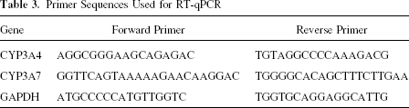
First-strand cDNA was synthesized using a superscript II cDNA synthesis kit according to the manufacturer's instructions (Invitrogen), and subsequently diluted with nuclease free water (Invitrogen) to 10 ng/μl cDNA. RT-PCR amplification mixtures (25 μl) contained 25 ng template cDNA, 2× SYBR Green I Master Mix buffer (12, 5 μl) (Applied Biosystems), and 300 nM forward and reverse primer PCR reactions were run in duplicate and performed on a StepOnePlus Real-Time PCR (Applied Biosystems). The cycling conditions comprised 10-min polymerase activation at 95°C and 40 cycles at 95°C for 15 s and 60°C for 20 s and 72°C for 60 s. Each PCR was followed by a melting curve analysis. The primer sequences used are reported in Table 3. Relative quantification was normalized against the house keeping gene (GAPDH). After normalization, the samples were plotted relative to the first sample in the data set.
Primer Sequences Used for RT-qPCR
Statistical Analysis
Results are expressed as mean ± SEM. Statistical differences were determined by Student's t-test for two groups' comparison or by one-way ANOVA followed by the Newman-Keuls post hoc test for multiple comparisons between more than two groups. Differences were considered significant when values were p < 0.05, p < 0.01, or p < 0.001.
Results
ADHLSCs Increase Their Ability to Synthesize Glucose After In Vitro Hepatogenic Differentiation
ADHLSCs from at least five different donors were subjected to an in vitro hepatogenic differentiation using specific growth factors and cytokines (4,28). Cells were then recovered for metabolic activity analyses. Differentiated ADHLSCs were both compared to undifferentiated cells and primary mature hepatocytes. To evaluate their ability to synthesize glucose, undifferentiated and differentiated ADHLSCs were incubated 24 h with lactate and pyruvate. Culture medium was recovered for the evaluation of glucose concentration. These levels were normalized to corresponding protein levels. As shown in Figure 1A, in vitro differentiated ADHLSCs produced glucose ~8 times more efficiently than undifferentiated cells (81.6 ± 6.1 vs. 11.4 ± 1.6 nmol glucose/mg protein∗24 h, respectively). Such increased glucose production was observed in cells at passages 3–4 of culture (n = 5) and can be maintained up to passage 8 (n = 1). However, this de novo synthesis of glucose remains lower than the one measured in primary culture of freshly isolated adult human hepatocytes and plated for 24 h (527.4 ± 139.1 nmol glucose/mg protein/24 h) (Fig. 1A). Nevertheless, ADHLSC synthesized glucose levels were comparable to those measured on 4-day plated primary hepatocytes (data not shown). In parallel, we demonstrated that in vitro de novo glucose synthesis is accompanied with an upregulation of the mRNA expression of key enzymes as, for instance, pyruvate carboxylase (PC), phosphoenolpyruvate carboxy kinase (PEPCK), and glucose-6-phosphatase (G6-Pase) (Fig. 1B).

Upregulation of gluconeogenesis pathway in differentiated ADHLSCs. (A) The novo glucose synthesis was evaluated after 24-h incubation with L-lactate and pyruvate. Analysis of incubation medium revealed that in vitro differentiated ADHLSCs produced glucose ~8 times more than undifferentiated cells (p = 0.0002, unpaired t-test). These levels remained lower than detected using freshly isolated human hepatocytes (hLCs). Data shown represent the mean ± SEM of six different experiments. (B) Upregulation of the mRNA expression of key enzymes of gluconeogenesis pathway, PC (pyruvate carboxylase), PEPCK (phosphoenolpyruvate carboxykinase), and G6Pase (glucose-6-phosphatase) in differentiated ADHLSCs. U: undifferentiated ADHLSCs; D: differentiated ADHLSCs; hLCs: freshly isolated human hepatocytes. (C) Activity of G6Pase was assessed using cytochemistry and based on the conversion of glucose-6-phosphate to glucose by G6Pase and the consequent production of intracellular lead nitrate precipitates. If G6Pase is active, cells will be stained in brown. Compared to undifferentiated which are not stained (C1), in vitro differentiated ADHLSCs all dyed deep brown (C2), suggesting high activity of G6Pase compared to primary mature hepatocytes (C3). ADHLSCs isolated from glycogenosis type 1a and submitted to the same hepatogenic differentiation protocol did not show active G6Pase both in undifferentiated (C4) and differentiated groups (C5). Original magnification 200×.
To confirm the ability of differentiated ADHLSCs to synthesize glucose, we analyzed the activity of G6-Pase, the final key enzyme of the gluconeogenesis pathway. This was performed using cytochemistry after incubation with the specific substrate glucose-6-phosphate (38). Analysis performed on live cells demonstrated that specific positive staining (intracellular brownish precipitates of ammonium phosphate) was detected in a higher level within the in vitro differentiated ADHLSCs (Fig. 1C). The intensity of staining was comparable to that observed in primary human hepatocytes. These data were obtained on cells previously cultured at passages 3–7 (n = 5) and can be observed at passage 9 (n = 1).
To demonstrate the specificity of our data, we evaluated such a parameter on ADHLSCs isolated from glycogenosis type 1a human liver (characterized by an inactive G6Pase) (36) and differentiated in vitro according to the same differentiation protocol. From such a liver, no specific staining was detected both in undifferentiated and differentiated ADHLSCs (Fig. 1C). This shows that in vitro differentiated ADHLSCs acquire the ability to synthesize glucose, a function correlated to an increase in both expression and activity of specific key gluconeogenesis enzymes.
ADHLSCs Display Ability to Metabolize Ammonium Into Urea
Urea metabolism and secretion capacities were also investigated in ADHLSCs differentiated in vitro. In serum-free culture conditions, culture medium of undifferentiated and differentiated ADHLSCs was analyzed for the detection of urea amounts produced after 24 h and normalized per cell. Such parameter was investigated in basal conditions as well as after 24-h treatment with ornithine and ammonium chloride. As shown in Figure 2A, in basal conditions both undifferentiated and differentiated cells weakly produced urea (11.3 ± 4.6 vs. 10.8 ± 7.8 pg/cell/24 h, respectively, p = 0.13). Incubation of the undifferentiated cells with specific substrates did not change the levels of detected urea (7.9 ± 3.8 pg urea/cell/24 h). However, a significant approximately threefold increase in urea levels was measured in differentiated ADHLSCs after ornithine and ammonium chloride treatment (28.2 ± 8.6 pg urea/cell/24 h, p = 0.02) compared to corresponding controls. The effect of ornithine and ammonium chloride on urea production by differentiated ADHLSCS has been obtained on cells that were previously cultured at passages 3–6 (n = 4). Such effect has also been observed with cells cultured at passage 9 (n = 1). With respect to primary mature hepatocytes, this capacity remains lower both in basal conditions (342.4 ± 52.8 pg urea/cell/24 h) and after ammonium chloride and ornithine treatment (553.3 ± 113.0 pg urea/cell/24 h) (Fig. 2A). Such data also importantly demonstrate that ornithine and ammonium chloride treatment equivalently induces a comparable fold increase both in primary hepatocytes and in vitro differentiated ADHLSCs. The acquisition of this key hepatic function is confirmed by an upregulation in the expression of major urea cycle involved enzymes. As shown in Figure 2B, the analysis of their mRNA expression in differentiated ADHLSCs revealed an upregulation of all studied key urea cycles enzymes, among which were NAGS, ornithine transcarbamylase, arginase, and argininosuccinate lyase.

In vitro differentiated ADHLSCs acquire the ability to metabolize ammonia. After in vitro hepatogenic differentiation process, cells were incubated for 24 h with or without 5 mM NH4Cl and 1 mM ornithine before measuring urea concentration in the recovered culture supernatants. No changes were detected in basal conditions whereas ornithine and ammonium chloride supply induced an increase in secreted urea levels of differentiated ADHLSCs (D) compared to undifferentiated cells (U) (p < 0.05, one-way ANOVA test) and nontreated cells (p = 0.006). Data were compared to primary human hepatocytes (hLCs) (n = 3). (B) Effect of hepatogenic differentiation of the mRNA expression of key enzymes of the urea cycle. Using conventional RT-PCR, an increased expression was demonstrated for the five analyzed enzymes: N-acetylglutamate synthase (NAGS); carbamoyl-phosphate synthetase 1 (CPS1); ornithine transcarbamylase (OTC); argininosuccinate lyase (ASL); and arginase. Freshly isolated human hepatocytes (hLCs) were used as positive controls. U: undifferentiated ADHLSCs; D: differentiated ADHLSCs.
Expression and Activity of Phase I and Phase II Drug-Metabolizing Enzymes in ADHLSCs Differentiated In Vitro
We also analyzed the ability of differentiated ADHLSCs to express active phase I and phase II drug-metabolizing enzymes. Expression of P450 cytochromes was firstly evaluated using conventional RT-PCR. The three first families of P450 cytochromes are considered to be the major enzymes in the liver. Such proteins are responsible for the metabolism and the clearance of most endogenous or exogenous substances, notably drug and chemical compounds. As shown in Figure 3A, we demonstrated that the expression of three genes of the CYP1 family (CYP1A1, CYP1A2, and CYP1B1), involved in metabolism of polycyclic aromatic hydrocarbons, is weakly detected in undifferentiated ADHLSCs. In vitro hepatogenic differentiation induced a pronounced increase for CYP1A1 and CYP1B1 and a weak one for CYP1A2. Five genes of the CYP2 family were also analyzed. CYP2A6 (4% of total CYP content and plays a major role in the metabolism of nicotine) and CYP2B6 (1–2% of total hepatic CYP and participates in the metabolism of cyclophosphamide) mRNA expression was also upregulated after in vitro hepatogenic differentiation. Analysis of CYP2C subfamily cytochromes (20% of the human total liver CYP content) revealed that expression of both CYP2C8 and CYP2C9 also increased after in vitro hepatogenic differentiation. The expression of CYP2E1, involved in metabolism of ethanol and some tobacco-specific nitrosamines, was also enhanced in differentiated ADHLSCs. The analysis of mRNA expression of two members of the CYP3 family (CYP3A4 and CYP3A7) revealed their presence in undifferentiated cells and an increase of their levels after in vitro hepatogenic differentiation (Fig. 3A).

In vitro differentiated ADHLSCs express active phase I drug-metabolizing enzymes. (A) The expression of 10 different cytochrome enzymes that are principally involved in xenobiotic metabolism was investigated using RT-PCR. After in vitro hepatogenic differentiation, mRNA levels of all analyzed cytochromes significantly increased. Isolated human hepatocytes and HepG2 cells were used as controls. U: undifferentiated ADHLSCs; D: differentiated ADHLSCs; hLCs: freshly isolated human hepatocytes; HepG2: human hepatoblastoma cells. (B) Phase I P450 cytochrome enzyme activities were measured in differentiated and undifferentiated ADHLSCs (donors N = 8; independent measures n = 3). Twenty-four-hour plated primary human hepatocytes (hLCs) (N = 6; n = 4) were used as positive controls. All cells were serum starved for 4 h prior to measurement. Methanol-fixed cells were used as negative controls. Enzyme activities are given as pmol product formed (conjugated) per minute from million cells.
Importantly, the upregulated mRNA expression of these P450 cytochromes is correlated to an increase in their activity as independently demonstrated using fluorescence-based assays (Fig. 3B). ADHLSCs used for this part of the study were independently differentiated in A. Nüssler laboratory according to our developed protocol conditions. Except for CYP2C8/9, in which no statistical difference has been obtained, all the other investigated P450 cytochromes of the three families presented enhanced activity in differentiated ADHLSCs (Fig. 3B). Primary hepatocytes were used as positive controls whereas their analysis was not performed the same time as ADHLSCs due to irregular organ availability and hepatocyte isolation.
We therefore focused on the analysis of CYP3A4 enzyme, the major isoform responsible for the metabolism of about 50% of drugs. This isoform is also considered to be original and specific marker for ADHLSC in expansion culture conditions (28). In our study, we confirmed that the increased mRNA expression and activity of CYP3A4 were correlated to an enhancement of its protein levels as demonstrated using immunocytochemistry (Fig. 4A). Because CYP3A4 was known to be highly inducible by xenobiotics, undifferentiated and differentiated ADHLSCs were treated 48 h using 1 mM phenobarbital. The analysis of both activity and mRNA expression of CYP3A4 was assessed. As shown in Figure 4B, besides confirming the increased CYP3A4 mRNA levels in differentiated cells (~ 10-fold), we demonstrated that phenobarbital induces a two- and threefold increase of these levels in differentiated and undifferentiated ADHLSCs, respectively. In parallel, differentiated ADHLSCs showed 20-fold increase in CYP3A7 mRNA levels without treatment whereas phenobarbital induced threefold increase of these levels (Fig. 4B).

In vitro differentiated ADHLSCs expressed an active CYP3A4, the major P450 enzyme. (A) CYP3A4 protein expression was in parallel increased in differentiated ADHLSCs as revealed using immunocytochemistry. Original magnification 200×. (B) CYP3A4 and CYP3A7 mRNA expression was increased in differentiated cells. Forty-eight-hour phenobarbital treatment stimulates mRNA levels expression of both CYP3A4 and CYP3A7 isoforms (one-way ANOVA followed by Newman-Keuls post test ∗∗p < 0.01, ∗p < 0.05). (C) CYP3A4 catalytic activity was analyzed after in vitro hepatogenic differentiation process. Cells were incubated for 4 h with luciferin-PFBE substrate and luciferase activity thereafter assessed. For treatment experiments using inducers and inhibitors, cells were treated for 48 h with 1 mM phenobarbital or naringin (50 and 100 μM) before the assay. Results are expressed as fold increase activity of the relative luminescence unit (RLU) detected in the analyzed groups. Data shown are the mean ± SEM of at least five independent experiments (one-way ANOVA followed by Newman-Keuls post test ***p < 0.001, ∗∗p < 0.01, ∗p < 0.05). (D) The induced CYP3A4 activity measured after in vitro hepatogenic differentiation was not altered if ADHLSCs were previously cryopreserved/thawed.
To investigate the CYP3A4 activity, an exogenous substrate luciferin-PFBE was added in the culture medium of undifferentiated and differentiated ADHLSCs. The metabolism of this substrate allows the measurement of luminescence intensity, which proportionally reflected the activity of this protein. As shown in Figure 4C, the activity of CYP3A4 in differentiated ADHLSCs was significantly increased (~8-fold) compared to undifferentiated cells. When studied ADHLSCs were incubated with the inducer agent phenobarbital, a ~13-fold CYP3A4 activity increase was observed compared to undifferentiated ADHLSCs. Forty-eight-hour treatment of undifferentiated and differentiated ADHLSCs with naringin (an inhibitor of CYP3A4) revealed no difference of CYP3A4 activity in undifferentiated ADHLSCs (Fig. 4C). However, CYP3A4 activity of differentiated ADHLSCs was dose-dependently inhibited with this inhibitor (Fig. 4C).
When looking at procedures to preserve as long as possible the viability of cryopreserved cells, the maintenance of cellular metabolic activities is a critical issue. Interestingly, the acquisition of this capacity to be induced was maintained when evaluated on in vitro differentiated ADHLSCs previously cryopreserved and thawed (Fig. 4D). Phase II activity has also been investigated in ADHLSCs and data revealed that UGT and GST activities have been upregulated in differentiated ADHLSCs compared to undifferentiated cells (Fig. 5A, B).

In vitro differentiated ADHLSCs express active phase II drug-metabolizing enzymes. Phase II GST and UGT enzyme activities were measured in differentiated and undifferentiated ADHLSCs. Methanol-fixed cells were used as negative controls. Primary human hepatocytes plated 24 h were used as positive controls. Enzyme activities are given as pmol product formed (conjugated) per minute from million cells. Average SEM and statistical analysis were calculated with Excel and the GraphPad Prism Software. (A) GST activity. (B) UGT activity.
In Vitro Differentiated ADHLSCs Display Significant Amino Acid Levels
Finally, we investigated the content of amino acids in undifferentiated and differentiated ADHLSCs using liquid chromatography in combination with tandem mass spectrometry (LC/MS/MS, Agilent Technologies; Applied Biosystems) and the AA45/32 kit (Applied Biosystems). Results show that differentiated ADHLSCs present higher amounts of most amino acids analyzed than undifferentiated cells (Fig. 6). These data also confirm our results regarding urea cycle activity (see above) as differentiated ADHLSCs show significant increased levels of ornithine and arginine, metabolic products of arginase and argininosuccinate lyase enzymes, respectively. The levels of branched chain amino acids leucine, isoleucine, and valine widely involved in protein synthesis are also significantly increased. The levels of glycine, which is a part of the powerful glutathione and involved in liver detoxification, are also significantly enhanced. Serine involved in direct synthesis of glycine is also increased. However, we do not detect differences in the level of other amino acids such as aspartate, glutamate, and taurine (Fig. 6).

In vitro hepatogenic differentiation of ADHLSCs is correlated with a significant increase of amino acid levels. Most essential amino acids such as methionine (Met), phenylalanine (Phe), threonine (Thr), tryptophan (Trp), valine (Val), leucine (Leu), and isoleucine (Ile) or conditionally essential amino acids such as arginine (Arg), glycine (Gly), glutamine (Gln), proline (Pro), serine (Ser), and tyrosine (Tyr) were significantly higher in differentiated ADHLSCs compared to undifferentiated cells. Amounts of some non essential amino acids such as aspartate (Asp), cysteine (Cys), taurine (Tau), hyproxyproline (Hyp), citrulline (Cit), and homocysteine (Hcy) 2 are not statistically different. ∗p < 0.05; ∗∗p < 0.01.
Discussion
We demonstrate in our current study that differentiated ADHLSCs display hepatic metabolic activities. Advanced functional hepatogenic differentiation of ADHLSCs suggests their potential to be developed for toxicopharmacological screening use. Furthermore, such ability to acquire liver metabolic functions combined to self-renew potential and resistance to cryopreservation may allow investigating these hepatic stem/progenitor cells as potential alternative cells in liver cell transplantation studies at least for metabolic diseases.
Current detailed demonstration of their functional differentiation level confirms the previous report in which ADHLSCs have been shown to acquire hepatic features at the morphological and genetic levels both in vitro and in vivo after transplantation (28). Differentiated ADHLSCs were studied to evaluate their ability to synthesize glucose, to metabolize xenobiotic as well as ammonia. All these functions were compared to those of primary human mature hepatocytes, the reference standard. Levels of amino acids were also evaluated in undifferentiated and differentiated ADHLSCs.
Hepatic gluconeogenesis, the major source of fasting hyperglycemia, contributes to 50–60% of the endogenous glucose production (17). This function is regulated by the rate-limiting enzymes PEPCK and G6Pase (30). We firstly analyzed the potential of in vitro differentiated ADHLSCs to synthesize glucose in the culture medium. Undifferentiated and differentiated ADHLSCs were incubated with pyruvate and lactate in glucose-free medium and glucose concentrations were analyzed 24 h later. As shown in Figure 1, in vitro differentiated ADHLSCs were able to significantly synthesize higher levels of glucose than undifferentiated cells. However, this de novo production remained lower than values obtained with primary mature hepatocytes plated for 24 h but is comparable to those obtained with 96-h plated hepatocytes (data not shown). In comparison with other mesenchymal stem cells isolated in our laboratory such as those obtained from bone marrow (23), these levels remained significantly higher, suggesting that the hepatic origin of ADHLSCs could influence the quality of their functional differentiation. In parallel, we demonstrated that in vitro glucose production is correlated to an increase of G6Pase, PEPCK, and PC mRNA expression levels.
In order to investigate if this de novo production was the result of an active gluconeogenesis pathway, we demonstrate using cytochemistry that G6Pase activity was highly increased after in vitro differentiation of ADHLSCs (Fig. 1C). The specificity of these data was demonstrated after using ADHLSCs isolated from glycogenosis type 1a adult liver (characterized by the absence of glucose-6-phosphatase activity) (36). From that liver, we demonstrated that both primary hepatocytes (data not shown) and in vitro differentiated corresponding ADHLSCs are not able to synthesize glucose. This de novo produced glucose measured in our experiments is mainly not originated from glycogenolysis pathway as incubation of ADHLSCs without lactate/pyruvate only shows few amounts of glucose (data not shown). Together these data demonstrate that in vitro differentiated ADHLSCs display an increased expression of gluconeogenesis key enzymes correlated to an augmented de novo glucose production.
We also demonstrated that in vitro differentiated ADHLSCs displayed ammonia detoxification capacity and urea secretion. Importantly, we demonstrated that besides this acquired function, ADHLSCs express the mRNA of key enzymes involved in the urea cycle and upregulate their expression after in vitro hepatogenic differentiation. The expression of these genes is in accordance with the hepatic origin of ADHLSCs as well as their advanced hepatogenic differentiation potential compared to the other mesenchymal stem cells we isolated and studied in our laboratory (bone marrow and umbilical cord).
Metabolism of intracellular and extracellular compounds and chemicals is one of the major and critical functions of the liver. Such activity is mainly managed by enzymes belonging to the family of cytochrome P450 in the mammalian liver (12,14,21,32). In the current study, we analyzed both their expression and activity after in vitro hepatogenic differentiation of ADHLSCs. Using conventional RT-PCR, we observed that mRNA levels of the analyzed cytochrome isoforms (three families) were increased in differentiated ADHLSCs. Such data reveal that some CYP isoforms (e.g., CYP1A1, CYP1B1, CYP2C8, and CYP2E1) were slightly expressed in undifferentiated ADHLSCs in accordance with their hepatic origin.
The expression analysis was performed in our laboratory in Brussels while the investigation of P450 cytochromes activity using fluorescence-based assays was independently performed in Germany on ADHLSCs differentiated by the corresponding team according to our developed protocol. These data demonstrated that our hepatogenic differentiation protocol is reproducible, something that strengthens the quality and the value of our cell-based model. Phase II enzyme activities were also studied and data demonstrated the ability of ADHLSCs to acquire these functions after in vitro hepatogenic differentiation.
Using luminescence-based assay, we focused our analysis on CYP3A4 isoform, the predominant hepatic form involved in the metabolism of the largest variety of substrates. This marker is specifically expressed in ADHLSCs in expansion culture conditions, whereas its mRNA expression is positively regulated by CYP3A4 inducers like phenobarbital (Fig. 4B). Our current data reveal that the upregulated CYP3A4 mRNA expression observed after in vitro hepatogenic differentiation is correlated to an increase in its protein expression as demonstrated using conventional RT-qPCR and immunocytochemistry, respectively. Besides increased expression, we also demonstrated that differentiated ADHLSCs displayed an increased CYP3A4 activity by using luminescence-based assay. CYP3A4 activity is found to be ~8 times higher in differentiated ADHLSCs compared to undifferentiated cells. Such increase is dose-dependently inhibited using naringin and augmented using phenobarbital. We also demonstrated that CYP3A7 mRNA levels, the major CYP isoform of the fetal liver but also present at significant levels in adult, are enhanced in differentiated ADHLSCs. CYP3A7 mRNA expression is also inducible after phenobarbital treatment. Such data suggest that the activity we detected using luminescence-based assay (luciferin-PFBE) may also reflect the involvement of CYP3A7. Nevertheless, the use of 7-benzyloxy-4-trifluoromethylcoumarin, a substrate primarily metabolized by CYP1A2 and CYP3A4, in our fluorescence-based assays (Fig. 3B) indicates that differentiated ADHLSC express active CYP3A4.
Because the liver is a target organ in systemic toxicity, liver cells are sensitively used as in vitro alternative models for pharmacotoxicological and metabolic studies (3,6). If primary hepatocyte cultures are preferentially used for in vitro studies (2,34,35), the instability of their differentiation properties in vitro, their poor resistance to in vitro culture, and cryopreservation conditions as well as the increasing liver shortage significantly hamper their routinely based use. Briefly, we observed in differentiated ADHLSCs an increased enzyme expression, phase I and phase II enzymes activities, and ability of CYP3A4 isoform activity to be induced and inhibited. The metabolic activities acquired in differentiated ADHLSCs have been mainly demonstrated with cells at passages 3–6 and can be maintained up to passages 8–9 (unfortunately with limited number of samples). Accordingly, additional studies are mandatory to evaluate the stability of these functions and their correlation to protein expression levels in differentiated cells before and after cryopreservation/thawing.
This battery of specifications demonstrated in differentiated ADHLSCs leads to propose them as a promising system to develop for in vitro screening and metabolism studies.
Finally, amino acid content analysis showed significant increase in differentiated ADHLSCs, suggesting an augmentation in the metabolic activity of differentiated ADHLSCs. Such enhancement could be the result of an increase in amino acid synthesis, and/or transport from extracellular medium. Amino acids have been shown to stimulate protein synthesis and to inhibit protein degradation. Their transport has also been demonstrated to increase in primary cultured hepatocytes during liver development (1). In our study, the increase of their content remains positively correlated to the other acquired investigated metabolic activities as well as total protein levels (data not shown). For instance, the levels of glutamine and alanine, the major amino acid precursors of glucose production (15), are significantly increased after in vitro hepatogenic differentiation.
In conclusion, we demonstrated in the current study that in vitro differentiated ADHLSCs displayed advanced hepatocytic metabolic functions. The induced expression of key corresponding genes and the revealed modulated activity reflect the level of in vitro hepatogenic differentiation acquired in ADHLSCs. Advanced quality of differentiated ADHLSCs as well as expression of most needed genes involved in xenobiotic metabolism obviously support their potential development as alternatives to mature hepatocytes. Furthermore, such step remained strongly necessary before exploiting them for the conception and the development of human liver cell-based future therapies.
Footnotes
Acknowledgments
The current study was financially supported by Région Wallonne-DGTRE (Grant Waleo-HEPATERA). D.N.K. and I.S. are Télévie recipients. M.N. is a scientific collaborator for FNRS.