
Abstract
The porcine circovirus type 1 (PCV1) has been identified within lymphoid tissues of experimental infected pigs and suggested to induce an immunosuppressive stage in pigs. The virus does not induce a cytophatic effect in the pig-derived cell line PK-15. Because PCV1 is prevalent in many pig cells and tissues, the risk of inducing a viral xenozoonosis by PCV1 was raised for the xenoimplantation of pig cells into human hosts. The present work evaluated if PCV1 is able to replicate in mice tissues after xenoimplantation of PCV1-infected pig cells. Active growing PK-15 cells harboring PCV1 with or without microencapsulation in sodium alginate were implanted into the peritoneal cavity of mice. After 1 month postimplantation in mice, peritoneal macrophages, spleen, and lymph nodes were harvested and analyzed with the polymerase chain reaction technique (PCR). No evidence of circovirus type 1 DNA was detected within the mice tissues.
Introduction
Porcine circovirus (PCV) was first encountered in the cell culture of porcine kidney line PK-15 and was described as a suspected picornavirus-like cell contaminant with no cytopathic effects (21). The characterization of the picornavirus-like virus showed a new small icosahedral nonenveloped virus containing a single stranded circular DNA genome and proposed the name porcine circovirus (19). Because of its morphologic and physico-chemical properties, it was classified as a new animal virus belonging to the Circoviridae family and consequently named porcine circovirus type 1 (PCV1) (16).
Serum antibodies to PCV1 have been demonstrated in pigs in Germany (19), Canada (6), New Zealand (10), Great Britain (8), Northern Ireland (2), and Spain (5,18), suggesting that PCV1 is widely distributed on the pig population around the world. On the other hand, the experimental infections of six antibody-negative minipigs seroconverted to PCV1, and virus was recovered from nasal swabs and from feces but no clinical disease was observed (20). A similar observation was obtained from the experimental infection with PCV1 to 2-day-old colostrums-deprived piglets. The authors showed virus antigen on cryostat sections stained by immunofluorescens in tissues from the lymphoid system, intestine, and lung (3), confirming results of virus isolation from nasal swabs and feces previously described (20).
PCV1 has been considered as a nonpathogenic virus of pigs but an immunosuppressive role has been suggested (1,3). Moreover, PCV1 DNA from a lymph node from a pig in France with wasting disease was isolated (15). A serum and lymphoid node survey from 135 swine samples in Spain showed three cases of PCV1 identification, suggesting that PCV1 has a low prevalence (5).
In this study we aimed to investigate the infectious potential of PCV1 in animal species other than pigs, describing a xenotransplantation with or without microencapsulation in sodium alginate of PK-15 cells harboring PCV1. The cells were implanted into the abdominal cavity of adult healthy mice for further evaluation to determine the presence of PCV1 in peritoneal macrophages and lymphoid tissue using molecular means (PCR).
Materials and Methods
PK-15 Cells
The pig kidney cell line PK-15 on its culture passage number 221 was obtained from the In Vitro Company (In Vitro, Mexico D.F.) and used for this study. The cells were grown in plastic bottles with minimal essential medium (MEM) with 10% fetal bovine serum and Pen-Strep (Sigma Aldrich, USA). Persistent cell infection with PCV1 was monitored before implantation in mice by PCR.
NIH Mice
Ten NIH mice were selected for implantation of PK-15 cells into the abdominal cavity. Mice were slightly sedated with xylazine HCl and bled from the tail blood vessels.
DNA Extraction
After subcultures of PK-15 cells, DNA was extracted from 25 mg of cells using the Wizard Genomic DNA Purification kit and following the manufacturer' instructions (Promega Corporation. Madison, WI. USA). Extracted DNA was stored in Eppendorf tubes at −20°C until used. Extracted DNA concentration was estimated after analysis in 0.7% agarose gel electrophoresis and adjusted to 0.1 mg/ml per sample.
PCV Detection and PCR Conditions
For the PCR amplification method of PCV1 we used two sets of nested primers already tested specific to PCV1 and without cross-reaction with PCV2 or any other known porcine virus. We followed the PCR conditions reported in Kim et al. (11,12). Briefly, 1 μl of extracted DNA was used as PCR templates in a first reaction and 10 μl of the product was used for the second reaction. The amplification was performed in a 50-μl reaction mixture containing 1.25 mM MgCl2, 1x PCR buffer, 0.2 mM of each dNTP, 1 μM of each primer, and 2.5 U of Taq DNA polymerase. Both reactions were run in a thermocycler (Px2 Thermal cycler, Thermo Electron Co.) under similar conditions: 35 cycles of 45 s at 95°C, 60 s at 63°C, and 60 s 72°C.
Microencapsulation of PK-15 Cells
Encapsulation was carried out according to previous described technique (7) with modifications. After culture, cells on monolayer at 90% confluence were trypsinized and the enzymatic effect was blocked with 5% fetal bovine serum, washed three times with MEM and ~ 10,000 suspended cells were mixed with 2% (w/v) sodium alginate (61%, high M alginate; PRONOVA Norwegian). The microcapsules were prepared using an electrostatic generator (Nisco Electrostatic Encapsulation Unit type VI, Nisco Engineering, Zürich, Switzerland). The polymerization of alginate occurred when alginate dropped into a barium solution. After orange-acridine staining, samples of 10 microcapsules were examined by fluorescent microscopy to test cell viability.
Intraperitoneal Implantation in Mice
Ten male mice of the strain NIH, 2 months old and weighing 30 g, were used for cell implantation. Each mouse received a combination of 2.5 mg/kg of xylazine HCl and 4 mg/kg of ketamine HCl and no presurgical fasting was used. All mice were placed in dorsal decumbency over sterile surgical boards, shaved aseptically, and draped for surgery. After reaching proper anesthetic status 1 cm medial incision was performed up to the peritoneum. Five mice received unencapsulated 50,000 PK-15 cells into the peritoneal cavity, and five mice received a similar quantity of cells previously microencapsulated in sodium alginate. The body wall was closed using and absorbable suture material, while a simple interrupted pattern was applied for wound skin closure using 4–0 polypropylene monofilament USP.
Postsurgical Care and Housing
After surgery was completed mice were kept warm using a newborn incubator (Air Shields Mod. 201 M) for immediate postsurgical care and maintained at 28°C until they became ambulatory and initial recovery was assessed. Once fully recovered they were housed in a conventional animal facility in single polypropylene cages and maintained at 21°C with 55% relative humidity. Animal health was evaluated daily and all animals were fed a constant pelleted formula (Formulab Chow 5008, Nutrimix, S.A. de C.V. Mexico D.F.) and fresh water ad libitum.
Tissue Collection
After ether anesthesia each mouse was killed by cervical dislocation and the peritoneal cavity washed with 5 ml of saline solution with a gentle massage in the abdomen. With the aid of a 5-ml syringe coupled with plastic tubing the saline solution was recovered and transferred into Eppendorf tubes. Pieces of the spleen and submaxilar lymphoid node were also obtained and placed into Eppendorf tubes. All tissues were kept frozen at −20°C for further analysis.
Results
Preimplantation Cell Viability
After trypsin treatment the PK-15 cells were able to maintain more than 90% viability, either with or without microencapsulation treatment. Cells having a predominantly green fluorescence after acridine orange staining were interpreted as live cells (Fig. 1).
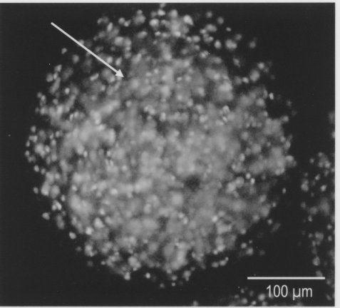
Photomicrography of a sodium alginate microcapsule with numerous PK-15 cells. Acridine orange stain shows most observed cells are viable (live cells in green fluorescence, arrow; dead cells in orange) 10x.
PCV Detection
The first round of amplification by PCR in PK-15 DNA template was unable to amplify a detectable PCV1 349-bp product. However, the second round of amplification using as template the product from the first round, a nested PCR amplification, resulted in a detectable 317-bp product. Also, serial dilutions of PK-15 DNA up to 10−3 dilution used as template in nested PCR also resulted in a positive 317-bp product (Table 1, Fig. 2). Peripheral blood cells obtained from experimental mice previous to PK-15 cells implantation tested negative to PCV1. Mice from the same colony as in this experiment have also been tested previously by nested PCR for the presence of PCV1 in blood, peritoneal macrophages, spleen, and lymph node tissues, with no PCV1 DNA detected (data not shown).

Nested PCR for PCV1 in PK-15 cells at different dilutions. M: 100 bp molecular weight marker. Templates for PCR are as follows: C1, positive control PCV1; lane 1, PK-15 at 10−1 dilution; lane 2, PK-15 at 10−2 dilution; lane 3, PK-15 at 10−3 dilution.
PCV1 Detection in PK-15 Cells

One month after intraperitoneal implantation of PK-15 cells, mice from both groups were tested by nested PCR for the presence of PCV-1. None of the analyzed mice harbored detectable PCV-1 in peritoneal macrophages or in spleen or lymph node tissues.
Discussion
Culture and encapsulation on agaroseagarose macrobeads of adult porcine islets implanted into diabetic BB rats for extended times has proved their functional assessment up to 24 weeks (9). The study conclude that following the transplantation of porcine islet macrobeads, rats fed a 20% carbohydrate diet had significantly lower daily blood glucose values compared with similar experimental animals fed a 64% carbohydrate diet. Moreover, the ability to culture porcine islets up to 67 weeks and further intraperitoneal implantation in agar-agar macrobeads into diabetic BB rats resulted in response to intraperitoneal glucose challenge and maintained the rats free of insulin therapy for 6 months. No microbiological tests were performed in either study but the authors suggest that the long-term viability of the porcine islets into their host may be suitable for such studies (22). On the other hand, microencapsulated neonatal porcine islets cultured for 50 days as xenografts in immune competent streptozotocin-induced diabetic BALB/c mice showed a reverse of the hyperglycemic stage after 2 days of implantation but mice eventually rejected their grafts after 15 days posttransplantation (13).
Attempts to detect PCV1 DNA in lymphoid cells after PK-15 cell implantation, with or without microencapsulation in alginate beads, was negative for the implanted mice of this study. It is estimated that implantation of pig cells into different animal recipients, including mice, mimics the events developed after xenoimplantation of pig cells in humans or any other mammal. The current PCR test methodology employed for detection of PCV1 in our study is accepted by other authors as an appropriate and highly reliable diagnosis technique (11,12,14). According to Kim et al. (11,12), first round of PCV1 DNA PCR amplifies a 349-bp product, whereas the second round amplifies a 317-bp product. The first round of PCR was able to detect 1.8 × 10⁻³ TCID50/ml of PCV1, while the second detected 2.5 × 10⁻⁴ TCID50/ml of PCV1. Our analysis detected PCV1 DNA in 0.5 pg of template DNA from infected PK-15 cells, considered as a high detection value. Our results are in disagreement with those reported by Quintana and coworkers for experimentally inoculated immunosuppressed mice, with PCV1, at 101.75 TCID50 into the abdominal cavity. This group found a PCV1 204-bp nucleotide fragment amplified from the serum of various mice between days 3 and 20 postinoculation. Only one mouse from the experimentally PCV1-inoculated group developed a weak seroconversion (17). Our different results can be explained in the sense that mice used in the present experiment were immunocompetent, different biological material was analyzed, and the nature of circovirus challenge was different (PK-15-infected cells vs. purified PCV1 virus).
A cell coculture of PK-15 cells harboring PCV1 with human polymorphonuclear cells and lymphoid T cells showed infection in T-CD4+, CD8+, CD14+, CD19+, and CD56+ cells. A word of caution was given from the authors concerning the possibility of creating a xenozoonosis if PCV-contaminated cells are xenoimplanted into humans (4). Our results suggested that pig-to-mouse xenoinfection is probably not easily accomplished, because a series of enabling conditions must be met to facilitate infection, like the use of immunosuppressed host animals or a high viral titer inoculation. It may be controversial that due to the interspecies-specific recognition, the PCV1 may or may not represent a risk of infection for inducing a xenozoonosis. Nevertheless, accurate identification of PCV1 and PCV2 must be inco-porated in a viral detection protocol in the herd of pig tissue donators.