
Abstract
BACKGROUND:
Many different squatting techniques have been recommended, but few studies tried to identify how different muscle groups contributed to this movement in each technique.
OBJECTIVE:
To compare the electromyographic activation (EMG) of the thigh, hip and trunk muscles during maximal voluntary isometric contractions in the back and front squat performed in different degrees of knee flexion, while also comparing the levels of force produced during different ranges of motion.
METHODS:
Ten healthy men (30.7
RESULTS:
At 60
CONCLUSIONS:
A greater squat depth did promote a decrease in EMG activity When executed in isometric contraction, parallel squatting offers better ratio of force X recruitment of primary motor muscles. Therefore, this amplitude can be used in training or rehabilitation strategies, both in frontal and posterior variations (with higher level of strength), observed the most convenient option for the practitioner.
Introduction
Despite its complex execution, the squat is regarded as one of the best exercises in strength training, since it allows the recruitment of various muscle segments in one single motion [1] For this reason, it is one of the most used multi-joint exercises in strength training protocols, both by recreational practitioners and athletes [2, 3, 4] The complexity of the squat, together with its many variables related to performance, demand the understanding of its biomechanics [5] in order to allow ideal muscle development while minimizing the risk of injuries due to poor form [6]. Among the many different squatting techniques, both the back squat (BS, performed with a barbell resting on the shoulders, over the trapezius, slightly above the posterior deltoid), and the front squat (FS, performed with a barbell resting on the anterior deltoids and clavicles, sustained with the help of both arms), have been recommended [4, 7].
Both variations provide important activation of the torso and lower limb muscles. However, the differences in technique also create differences in muscle recruitment, which still need to be investigated [8, 9]. Gullett et al. [8] and Contreras et al. [10] found no significant differences between FS and BS exercises regarding hip and thigh muscle activation. The first study analyzed two sets of three repetitions at 70% of 1 repetition maximum (1RM), while Contreras et al. [10] found no statistical differences in muscle activity between the two variants (both parallel and full range of motion) with a 10RM load in healthy women. On the other hand, Yavuz et al. [9] reported higher muscle activity in the vastus medialis in the FS when compared to the back squat, as well as greater muscle activity in the erector spinae when trained males performed BS using a 1RM load. Korak et al. [11] reported higher muscle activity during the FS (M
To better understand the biomechanical aspects of these exercises, a number of studies have aimed to analyze the effects of different squat depths, in order to identify how different muscle groups contribute to perform this motion [12]. For instance, some have reported that the gluteus maximus muscle shows higher activation when squatting with higher degrees of knee flexion [13, 14]. Regarding other muscles, Jaberzadeh et al. [15] showed that the ratio of the EMG activity between the vastus medialis oblique and the vastus lateralis was higher when performing deeper squats. However, since these studies only addressed the BS, we cannot infer that the same would be observed when performing the FS.
The relationship between muscle activation level and the force generated in different types of squats seems to vary as a function of the angular relationships of the joints involved – specially the hip, knee and ankle – as well as the position of the torso [16]. In this sense, some studies reported that higher loads can be lifted during the BS when compared to the FS [8, 9]. This may be due to higher tibiofemoral compressive forces and a bigger moment arm of the extensor muscles [8]. These differences may also be related to variables interfering in the relationship between electromyographic activation and strength, such as angular and contraction velocity [17].
In general, most of the studies that compared EMG activity and the amount of force generated between different types of squats did so during dynamic actions. However, a more concrete association between these measures can be obtained during maximal isometric actions, since there is an increase in the firing rate of motor units in this type of muscle action, even when their recruitment reaches a saturation, causing the muscle to keep producing energy for the EMG signal leading, in turn, to an increase in amplitude [17].
Moreover, when considering the mutual interference between the range of motion of the different types of squat, muscle activation and the force produced in maximal isometric actions, there is a lack of sufficient evidence for clarifying the relationship between these variables. Additionally, different angles of exercise influence the inclination angle of the trunk and, consequently, muscle recruitment during squatting. Such information may play a large role in the prescription of these exercises in strength training programs and in understanding lower body muscle activation patterns between squat variations. These are important factors in strength and conditioning for isolating or creating greater activation of selected lower body muscles for training, performance increases, injury prevention, or rehabilitation techniques.
Therefore, the aim of our study was to compare the EMG activation of the muscles of the thigh, hip and torso during maximal isometric contractions in both BS and FS, performed at different degrees of knee flexion. Our hypothesis was that in both kinds of squats the lower the degree of knee flexion, the higher the level of muscle activation, mainly in the posterior hip and thigh muscles. We also believe that the strength of the triple ankle, knee and hip extension would be inversely proportional to the yours flexion angle and will be higher during the higher degrees of knee flexion, particularly in the BS. Furthermore, we hypothesized, due to previous findings, that the FS would produce greater muscle activity of the GM.
Methods
Subjects
The participants were 10 healthy men (30.7
Design
In this cross-over trial, subjects performed three experimental sessions with a washout period of 48 hours. In the first session, subjects performed a maximum voluntary isometric contraction (MVIC) test, from which we verified the signals corresponding to the root mean square (RMS) peaks regarding each evaluated muscle using isolated exercises, with the goal being to establish parameters for posterior normalization. Still in the first session, subjects went through a familiarization with the tools and equipment that would be posteriorly utilized in the FS and BS sessions, both which were assessed through the maximal isometric contraction.
In the second and third sessions, subjects performed the FS and BS, in random order. Subjects rested for five minutes before all maximal isometric actions, with the order also being random for the 60
Positioning of the barbell and adjustment of the angles of knee flexion during the front and back squats. A – Front squat at 60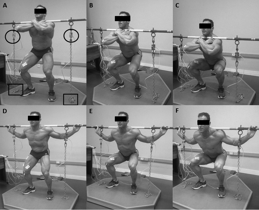
To measure the MVIC, we fixed surface electrodes on the right side of the body above the muscle belly of the following muscles: rectus femoris (RF), vastus lateralis (VL), vastus medialis (VM), biceps femoris (BF), gluteus maximus (GM) and erector spinae (ES). The electrodes were placed according to Broer and Houtz [18].
To verify the relative RMS value for each of the assessed muscles, subjects were oriented to perform MVIC during knee extension limited to 60
Acquisition of the EMG signal and maximum isometric force
The EMG signal and maximum isometric force recordings started at the same time, following a verbal command issued by the researcher. Participants were requested to perform a 10-second MVIC for every angle of knee flexion. We selected the 5 seconds corresponding to the period where the highest force was employed over the load cells (
To assess the maximum isometric force generated by subjects in both squat variations, we utilized two free load cells (Miotec
EMG data
Six pairs of Ag/AgCl surface electrodes, model 2223BRQ, 3 M brand, were fixed by the same researcher (avoiding possible inter-rater variability) over the bellies of all muscles, parallel to the muscle striations of the respective muscle fibers, after previous cleansing and trichotomy of skin surface. The procedure of electrode fixation and positioning followed the same criteria adopted in the first session, where subjects performed the MVIC tests. A distance of 2 cm between the centers of electrodes was kept. We assessed muscle activation utilizing an 8-channel electromyograph Miotool (Miotec
Statistical analysis
Data were typed originally in the database of the SPSS
Isometric force production between the squat variations at different degrees of knee flexion. KGF – Kilograms force. Values are shown in median and interquartile range. *Significant difference between squat variations; †Significant difference at 90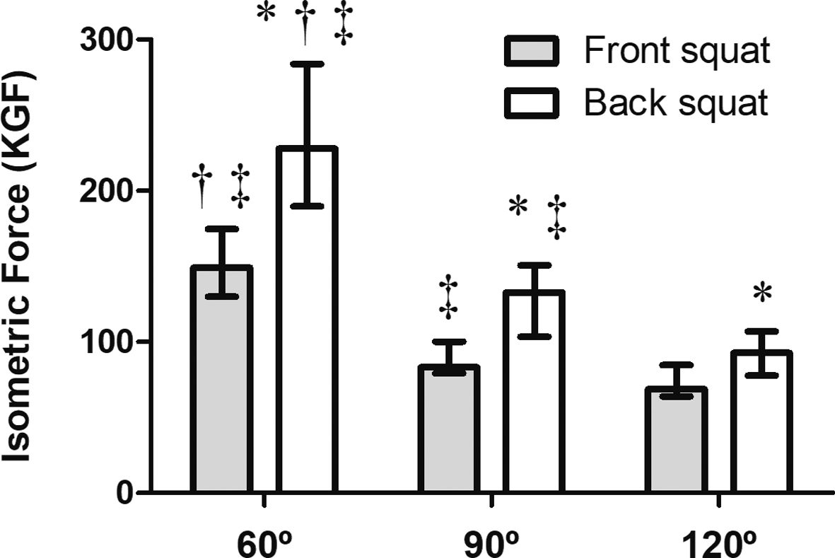
A larger magnitude of force was produced in the BS, in all assessed angles (Fig. 2). We verified that the maximum voluntary isometric force decreased progressively as the degree of knee flexion increased.
Figure 2 shows results related to the isometric force producted.
Mean values of muscle activity of different muscles during back and front squats, represented by the percentage of maximal voluntary isometric contraction (%MCIV). A – 120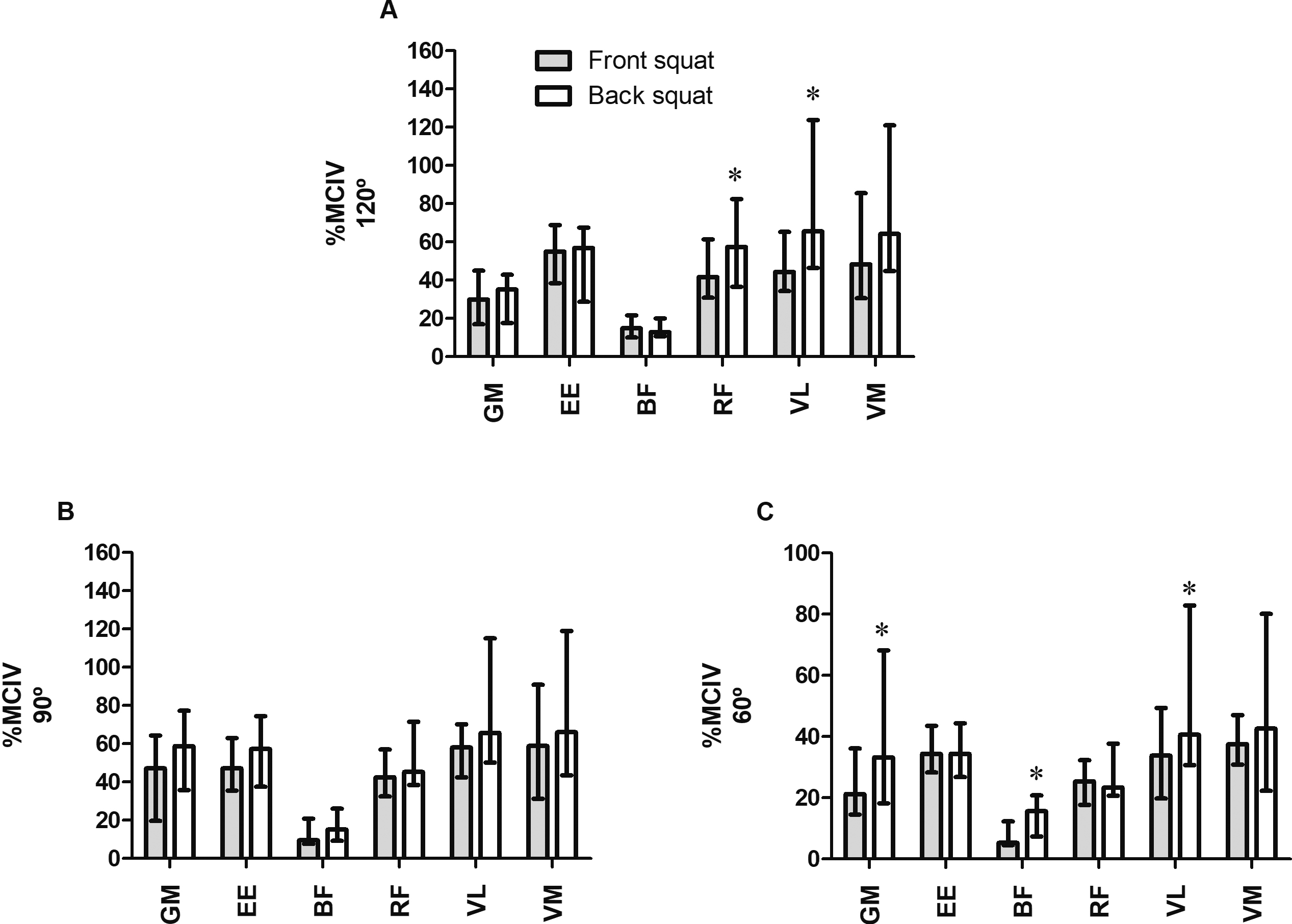
Electromyographic activity referring to the percent of maximum voluntary isometric contraction (%MVIC) for the front and back squats in different angles of knee flexion
GM
The results of the comparison of muscle activity between the FS and the BS are shown in Fig. 3. At 60
Table 1 outlines the normalized values of the EMG signals between the different angles. The gluteus maximus had a higher activation when knee flexion was at 90
Our results indicate that, in both the FS and BS, the lowest electrical activity of the assessed muscles was observed when the knee was flexed at 60
Mechanical conditions, as opposed to neural ones, have a much bigger influence on the reduction electrical activity of the assessed muscles observed during knee flexion at 60
Despite the lack of a kinematic analysis in our study, it is possible to affirm that squatting with knees flexed at 60
The similarity in activation of the assessed muscles during MVIC at 90
Our findings do not match those obtained in a previous study [13] which had indicated a direct relationship between gluteus maximus activation and the depth of the dynamic squat. We suggest that this finding was due to the utilization of submaximal loads, a fact that may have altered the recruitment pattern of high threshold motor units, especially in more experienced individuals [9]. In lower load conditions, stronger muscles may compensate the activation of weaker ones, promoting variations in their respective EMG activities, which can compromise the results found. Furthermore, in this study the loads were not balanced for comparison of deep and partial squats, which might have compromised the results.
The rectus femoris has manifested lower EMG activation in all variations and ranges of motion when compared to the other muscles of the quadriceps – the vastus lateralis and vastus medialis. This may be due to a possible stabilization of its length, since its mutual function as a knee extensor and hip flexor promotes a simultaneous shortening of one of its ends while stretching the other [5, 7, 9]. Regarding the low activation of the biceps femoris in all assessed ranges and variations, we suggest that the squat may not be the best exercise option for the work of the hamstring muscles, as proposed by Contreras et al. [10] and Gullett et al. [8]. In this context, Ebben et al. [20] indicated that knee flexion exercises and the stiff-legged deadlift (performed with both knees fully extended) might be better options.
One of the more significant clinical implications of the current results lies in the relationship between lifted load and muscle activation. Considering that the use of lower loads when performing the front squat did not generate a decrease in the activation of the targeted muscles this variation may be safer and better suited for practitioners who want to prioritize the preservation of joints that have a potential to be vulnerable to external load.
The main limitation of our study is due to the manner through which we performed the analysis. All squat exercises were performed as maximal isometric contractions, which reduces the similarity of the tests we analyzed with the practices observed in gyms and training centers, where squats are commonly performed as dynamic exercise. Additionally, the normalization of the EMG signals from MVIC obtained in various angles and variations may have interfered with our results, despite the existing precedents in literature [4]. The normalization obtained through own maximal isometric contractions, performed during the execution of the assessed squats, might have provided more reliable parameters for the assessment of the proposed variables. Lastly, anthropometric and kinematic differences between sexes exist, so future research is needed to fill in this gap [21].
Conclusion
Our results indicate that in both the front and back squats, muscle activation was lower at 60
Footnotes
Conflict of interest
The authors of this study declare no conflicts of interest.