
Abstract
BACKGROUND:
Squats are considered one of the main exercises for the lower limbs and are used in resistance training under different contexts, including rehabilitation and sports performance.
OBJECTIVE:
To compare the EMG activity of different muscles in back squat and lunge exercises in trained women.
METHODS:
Ten healthy women experienced in resistance training performed back squat and lunge exercises on a Smith machine (total work: 70% of 1RM, 1 set, 10 repetitions and 2-s/2-s of execution speed) with an interval of 20-min between exercises. Both exercises were standardized in relation to the trunk inclination and were performed with an erect trunk parallel to the cursor of the guided bar.
RESULTS:
The EMG activity of the vastus medialis (VM), vastus lateralis (VL), biceps femoris (BF), and gluteus maximus (GM) were analyzed. There were no significant differences in the EMG activity of the VM, VL, and BF muscles between the back squat and lunge exercises (
CONCLUSIONS:
Lunges were more effective in recruiting the GM when compared to back squats. However, both exercises can be recommended when the goal is knee extensor and flexor muscle activity.
Introduction
Squats are considered one of the main exercises for the lower limbs and are used in resistance training [1]. The importance of squats is justified by application in different contexts, including rehabilitation and sports performance, among other aspects [1, 2]. There are a series of variations in performing squats in the literature, among them: front or back squat [3], with feet positioned at different widths [4] or in different degrees of medial and lateral rotation [5] in relation to the trunk, with free weights or guided bars [6], and even with a different range of motion at the knees and/or hips [7, 8]. These variations seek to stimulate the muscle groups which compose the lower limbs in a more selective or effective manner in order to promote better results for resistance training practitioners [1]. Electromyographic (EMG) activity additionally appears as one of the most explored variables in these types of studies and is minimally affected by the squat variations mentioned above, especially with regard to the action of the primary knee agonists [1, 4, 5].
Although primary knee agonists are most often not significantly affected by squat variations, primary hip agonists such as the biceps femoris and gluteus maximus seem to be more sensitive to such variations, especially in positioning the feet in relation to the torso [1]. Accordingly, a variation of the squat which has been the subject of research is the performance of lunges [9, 10, 11, 12]. Although lunges are widely used by resistance training practitioners [10], few studies have compared the EMG activity of primary knee and hip agonist muscles between variations of lunge squats (with one foot in front) and back squat (with feet parallel). Stuart et al. [2] compared squats in each of the aforementioned conditions and noted that the lunge exercise provided higher EMG activity values for the knee extensors and flexors were compared to the back squat and front squat, but there was poor control and standardization of the conditions tested. For instance, only the bar was used as an external load, thereby compromising the study results since the intensity was not standardized for each of the tested conditions. Additionally, the position of the trunk was different, since there is an anterior inclination of the trunk when executing the back squat with free bar, while the trunk remains perpendicular to the ground during lunges, and this modifies the EMG activity [12, 13, 14]. In this sense, it is not clear whether there is a difference in EMG activity when the exercises are performed with the same trunk alignment.
Thus, the objective of the present study was to compare the EMG activity of the knee extensor and flexor muscles and hip extensors between the lunge and back squat exercises in women when both exercises are standardized by the intensity, amplitude, and inclination of the trunk. It was hypothesized that lunges would activate the muscles more because it is an exercise in which the lower limbs are positioned in a way which requires greater balance in relation to back squats, and therefore (according to Dwyer et al.) [15] the disposition of one leg in front of the other provides a pre-stretching condition of the posterior thigh and gluteal muscles from the kinesiological point of view, with greater myoelectric activation therefore being expected under these conditions.
Methods
Subjects
A total of 10 women (mean
The sample size was calculated using the G*Power 3.1.9 software program (Franz Faul, Germany) and based on the literature [1, 15, 16, 17]. As it is a multiarticular exercise which involves large muscle groups, an effect size (
Procedures
Two visits to the laboratory were carried out with an interval of 48 to 96-h in between. Anthropometric measurements, the maximum repetition (1RM) test and familiarization with exercises and repetition speed using the metronome were performed on the first visit. The participants did the experimental session on the second visit, which consisted of performing the back squats first and then the lunges, with an interval of 20-min between exercises [19].
Prior to the first visit, participants were instructed to abstain from exercise for a minimum of 24-h and to eat a light meal 2-h before the visit. Anthropometric measurements including height (m), body mass (kg), and skinfold thickness (mm) were determined. Body mass index was calculated by dividing weight by height squared (kg/m
The load percentage used in the experimental session was determined by performing the 1RM test according to the recommendations of Kraemer et al. [21]. The order of the test in the exercises was the same as that used in the experimental session. Values of 52.2
The subjects performed a set of 10 repetitions at 70% of 1RM for each exercise during the experimental session. The repetition speed was 2-s for each concentric and eccentric phase controlled by the metronome in both exercises. Subjects performed a warm up on a cycle ergometer for 5-min before starting the exercise protocol.
Back squats and lunges were performed on the Smith machine based on the procedures by Stuart et al. [2] and Caulfield and Berninger [22] for free bar exercises. During the back squats the barbell was placed in the high bar position across the shoulders on the trapezius, slightly above the posterior aspect of the deltoids, with the subjects in the vertical posture and their feet fixed and positioned parallel to the shoulder width with their toes pointed forward or slightly outward. The subjects performed the descending phase by flexing their hips and knees until their thighs were parallel to the floor (Fig. 1). Lunges were performed with the bar positioned in the same way as the back squats, the subjects took a large step forward keeping the torso upright and during the descending phase the leg was positioned down until the knee approached the floor, and the knee of the anterior leg was flexed until the thigh was parallel to the ground (Fig. 2). Both exercises were standardized and performed with the trunk erect parallel to the cursor bar guided on the Smith machine, and both in the eccentric and concentric phase in order to eliminate the influence of the trunk inclination on the EMG activity [12, 13, 14]; thus, measuring tapes were placed on the floor both in the sagittal and in the frontal plane to standardize the positions of the feet between the exercises (Figs 1 and 2). The only difference was the posterior leg in the lunge exercise. It is important to highlight that these same procedures were performed on the first visit, and the feet adjustment at that moment to enable the trunk to remain erect during the exercises was determined and recorded.
Back squat execution.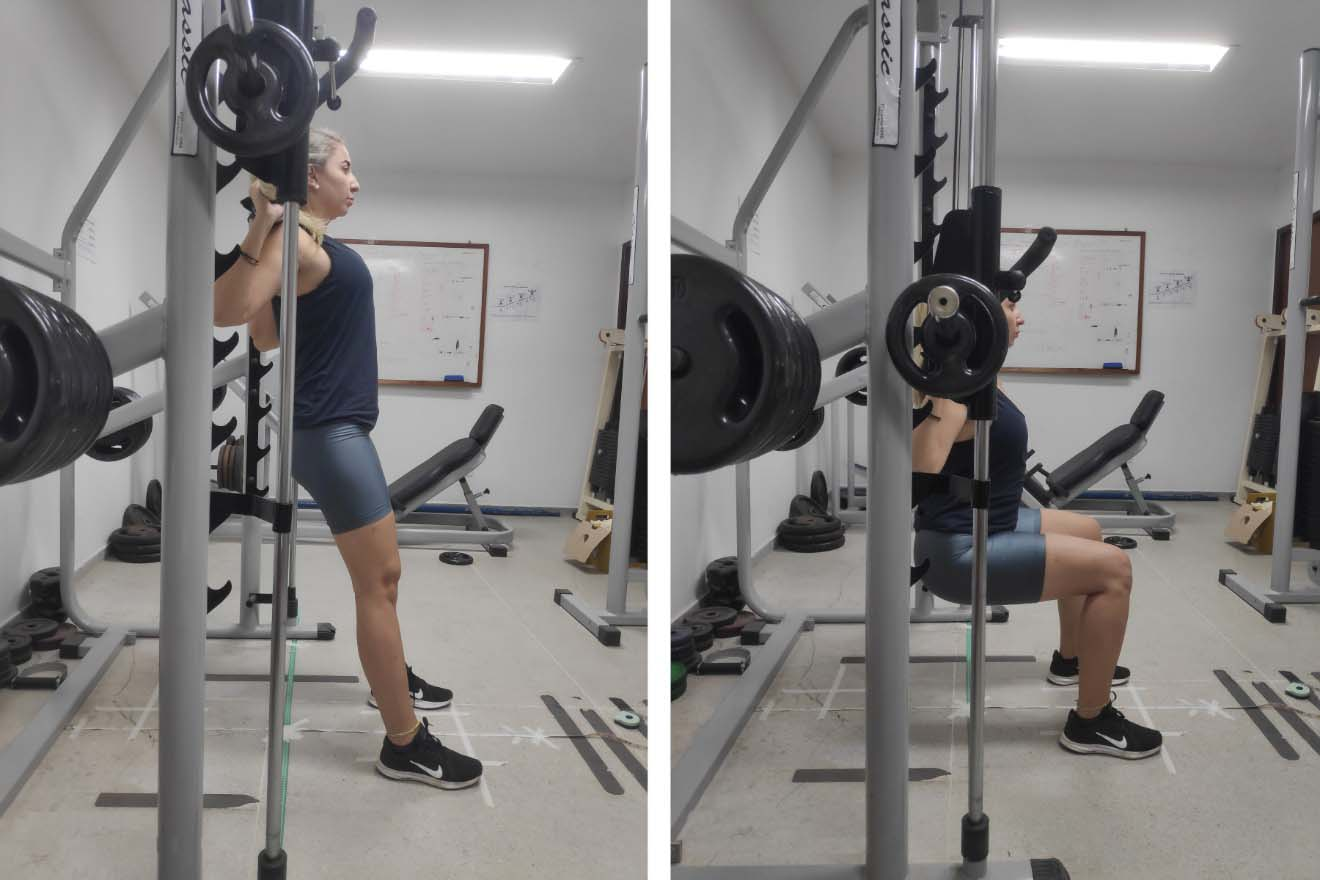
Lunges execution.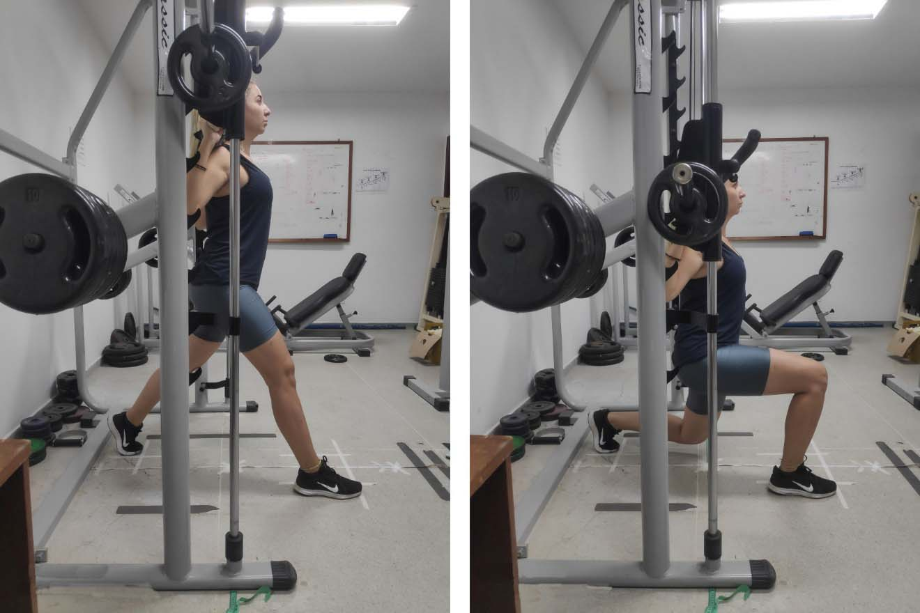
Comparison of the mean peak electromyographic activity of different muscles between back squats and lunges performed on the Smith machine (
Note. VM
Next, Ag/AgCl surface electrodes (Maxicor Produtos Médicos, Pinhais, PR, Brazil) with 1 cm in diameter arranged in a bipolar configuration with 2 cm of inter-electrode distance were used to collect the EMG activity. Before placing the electrodes on the skin, excess hair was removed with a razor, and skin was cleaned and abraded using cotton and 70% alcohol. The electrodes were attached to the subjects’ dominant side [6] on the belly of the vastus medialis (VM), vastus lateralis (VL), biceps femoris (BF), and gluteus maximus (GM) muscles according to the recommendations of Hermens et al. [23] After the electrodes were secured, a quality check was performed to ensure EMG signal validity [24].
The electrodes were connected to an electromyograph (Miotec Miotool 400, Porto Alegre, RS, Brazil) with its respective software (Miotec Miograph 2.0, Porto Alegre, RS, Brazil), which had been previously calibrated according to the manufacturer’s recommendations. The EMG signals were amplified 1000 times with a common mode rejection rate of 110 dB. The sampling frequency was 2000 Hz and a 20 Hz high pass filter and a 500 Hz low pass filter were applied. After data collection, the magnitude of the EMG signal was calculated in root mean square (RMS) values in millivolts (mV) using the peak contraction of each repetition to obtain the average peak of the 10 repetitions.
The normality and homogeneity of the data were confirmed by the Shapiro-Wilk and Levene test, respectively. The paired Student’s
Results
The data on Table 1 demonstrate there was no significant difference between the back squats and lunges in relation to the EMG activity of the VM, VL, and BF muscles (
Discussion
The results of the present study showed that the GM muscle showed higher levels of EMG activity in lunges when compared to back squats, with no significant differences between the exercises for the other analyzed muscles. These findings partially confirmed the hypothesis of the present study, considering only the GM was more activated in lunges than in back squats; a fact which did not occur with the BF.
According to findings in the literature [4, 27, 28, 29], the external loads applied in squats have a direct impact on muscle recruitment. Thus, there is an increase in EMG activity as loads are increased on the equipment bar. As we are aware of this information, the loads in the present study were normalized individually. Each subject performed the 1RM test for the back squat and the lunge and subsequently underwent 10 repetitions at 70% of 1RM. Accordingly, this procedure enabled comparing the exercises in an equal manner.
Another important point refers to the trunk positioning. Performing back squats and lunges on the Smith machine in the present study enabled the distances of the lower limbs to be standardized according to the length of the limbs for the sagittal plane and based on the hip width for the frontal plane. Thus, the only difference between the exercises was positioning of the base line of the feet in relation to the trunk. This standardization enabled the subjects to perform both exercises with the same trunk alignment. The trunk posture in the upright position is important because the muscle contraction of the hamstrings [12, 13] increases as the anterior flexion angle of the trunk increases. Accordingly, the fact that there were no differences in the trunk position may explain the similarities between the EMG activity of VM, VL, and BF.
The findings of the present study do not corroborate the results reported by Stuart et al. [2]. Although the methodological differences between Stuart et al. [2] and the present study limit some comparisons, such as the EMG analysis of the GM, it is important to emphasize studies with a closer methodological approach are scarce in the literature. Stuart et al. [2] compared the EMG activity of knee extensors (VM, VL, rectus femoris, vastus intermedius) and flexors (semitendinosus, semimembranosus, biceps femoris long head) during three closed kinetic chain exercises with the distal segment fixed: front squats, back squats, and lunges. The authors concluded lunges showed higher EMG activity levels for both knee extensor and flexor muscles compared to back squats and front squats. However, the authors used an equal load for the three exercises and did not standardize the trunk position; these limitations compromised these results and their extrapolation.
In the present study it was found that lunges activate the GM musculature more than in back squats; this result perhaps occurred because lunges are an exercise of greater complexity and instability, considering that the primary action of the GM is the hip extension and as a secondary action to assist in the trunk stability, especially during coordinated actions of the knee and hip joints [30]. Thus, feet positioning spread in the anteroposterior direction favors GM recruitment, similar to running and/or walking movements, in order to guarantee the trunk alignment as upright as possible. In addition, the lower limbs in front are overloaded in lunges, to the detriment of the limb positioned behind. Conversely, the load in the back squat is equally distributed between the two lower limbs, thus decreasing GM activation [31, 32].
This was the first study to compare back squats and lunges on the Smith machine in trained women. The findings demonstrate both back squats and lunges were similar for recruiting the VM, VL and BF muscles; however, performing lunges is the most suitable exercise if the practitioner’s goal is to obtain higher GM recruitment levels. Finally, the importance of standardizing the tested conditions is emphasized, given that this procedure ensures the analyzes are not influenced by other factors which may have an impact on the results, such as inadequate execution in the movement, underestimation or overestimation of the external training load in different exercises, as well as the volume (series, repetitions, break time, etc.) of the training session.
Author contributions
CONCEPTION: Rodrigo Ramalho Aniceto and Hélen Cristina Ferreira da Silva.
PERFORMANCE OF WORK: Rodrigo Ramalho Aniceto and Hélen Cristina Ferreira da Silva.
INTERPRETATION OR ANALYSIS OF DATA: Rodrigo Ramalho Aniceto, Hélen Cristina Ferreira da Silva and Diego Mesquita Silva.
PREPARATION OF THE MANUSCRIPT: Rodrigo Ramalho Aniceto, André Luiz Torres Pirauá, Leonardo da Silva Leandro, Diego Mesquita Silva and Leandro Cândido de Araújo.
REVISION FOR IMPORTANT INTELLECTUAL CONTENT: Pablo Brando Costa and Heleodório Honorato dos Santos.
SUPERVISION: Pablo Brando Costa and Heleodório Honorato dos Santos.
Ethical considerations
This study was approved by the Research Ethics Committee of the Integrated Colleges of Patos (protocol: 190/2012; August 15, 2013). All participants signed a written informed consent.
Funding
The authors report no funding.
Footnotes
Acknowledgments
The authors would like to thank Prof. Elvis Costa Crispiniano of Integrated Colleges of Patos for his assistance in collecting electromyography data and all the participants for their effort and commitment during the study period.
Conflict of interest
The authors have no conflicts of interest to report.