
Abstract
Tuberculosis (TB) is an airborne infection affected by Mycobacterium TB. It is vital to identify cases of TB quickly if left untreated; there exists a 70% possibility of a patient dying in 10 years. An essential for extra device has been enhanced in mid to low-income countries because of the growth of automation in the field of medical care. The already restricted resources are being greatly assigned to control other dangerous infections. Modern digital radiography (DR) machines, utilized to screen chest X-rays (CXR) of possible TB victims. Combined with computer-aided detection (CAD) with the support of artificial intelligence (AI), radiologists employed in this domain actual support possible cases. This study presents a Hybrid Deep Learning Assisted Chest X-Ray Image Segmentation and Classification for Tuberculosis (HDL-ISCTB) diagnosis. The HDL-ISCTB model performs Otsu’s thresholding, which segments the lung regions from the input images. It effectually discriminates the lung areas from the background, decreasing computational complexity and potential noise. Besides, the segmented lung regions are then fed into the CNN-LSTM architecture for classification. The CNN-LSTM model leverages the powerful feature extraction capabilities of CNNs and the temporal dependencies captured by LSTM to obtain robust representations from sequential CXR image data. A wide experiments are conducted to calculate the performance of the presented approach in comparison to recent methods.
Introduction
Tuberculosis (TB) is a pulmonary infectious disease caused by bacterium Mycobacterium TB. In 2018, a report given by the World Health Organization (WHO) stated that 1.5 million people died of TB [1]. TB ranks as the second leading factor of infectious disease. Accurate and rapid diagnoses and prompt treatments are highly significant to control and prevent TB. One of the typically used techniques for identifying TB is Chest X-ray (CXR) and it allows screening for TB at an initial phase [2]. Chest radiographs (CRs) are broadly available and relatively cheap and have served a significant part in diagnosing active TB [3]. In spite of its effectual solution, detection of TB on CR is a labour and time-intensive task that necessities an interpretation of an expert [4], which is a limited commodity in underdeveloped nations but medical sources and skilled radiologists are scarce [5]. So, automatic recognition of active pulmonary TB on CRs is of great clinical efficacy.
In this regard, chest radiographs (CRs) had a main role in screening active TB [6], and are comparatively broadly accessible and inexpensive CNNs to detect disease [7], but their application to TB identification is limited [8]. Research in the application of deep learning (DL) to radiology was a fast-growing domain because of its promising performance in disease detection [9], like cardiomegaly detection and pleural effusion on chest radiographs, and lung nodule detection and mediastinal lymph node on computed tomography (CT). The author has realized that AI-based CXR was an auspicious tool to diagnose TB, particularly in resource-limited rural areas [10].
This study presents a Hybrid Deep Learning Assisted Chest X-Ray Image Segmentation and Classification for Tuberculosis (HDL-ISCTB) diagnosis. The HDL-ISCTB model performs Otsu’s thresholding, which segments the lung regions from the input images. It effectually discriminates the lung areas from the background, decreasing computational complexity and potential noise. Besides, the segmented lung regions are then fed into the CNN-LSTM architecture for classification. The CNN-LSTM model leverages the powerful feature extraction capabilities of CNNs and the temporal dependencies captured by LSTM to obtain robust representations from sequential CXR image data. Extensive experiments are conducted to estimate the performance of the presented approach in comparison to recent methods.
Related works
Capellán-Martín et al. [11] projected a multi-view DL-related solution accompanied by presented template, targets to automatically extract and regionalize mediastinal and lung regions of interest from pediatric CXR imageries but TB findings are presented. Ahmad and Shin [12] devise a PE technique that is appropriate for both grayscale and color images. The author has concerned with a smart clinic that provides health-care cloud services for outsourcing their storage needs and DL computations as an application of presented technique. For automatic diagnosis of TB in CXR images, the EfficientNetV2-based methodology was applied. As well, to solve data deficiency in medical image analysis, the author has presented noise-related data augmentation techniques.
Ammar et al. [13] presented a new technique using MRI images called hybrid optimal DL-related method for TB disease recognition. For extracting, the related features from MRI images, quite a few DL methods were integrated. Specifically, aiming to get the best out of classification accuracy, the author established Efficient-Net models and vision transformers (ViTs). Dey et al. [14] devised a technique using CXR images to screen TB and with the help of the type-1 Sugeno fuzzy integral related ensemble method, the choices from 3 base learners will be merged. The author had been utilized meta-heuristic optimizer techniques to optimally set the fuzzy measures in the model training, to solve such manual tuning.
In [15], an innovative method is devised using CXR image classification for TB detection, using a mixture of 2 common pre-trained vgg19 and vgg16 applying the block attention module, and ImageNet data for acquiring spatial dataset. In [16], a systematic review has taken place on DL-based CAD systems that can be utilized for examining CXRs to identify pulmonary TB. DL is currently added to the list of best performing methods, mainly in the analysis of medical images. In DL-CNNs were broadly applied for detecting TB. A CNN method is formed from pooling layers or sub-sampling, fully connected (FC) layers, and convolutional layers.
The proposed model
This study has introduced a novel HDL-ISCTB system for Image Segmentation and Classification for TB diagnosis. The HDL-ISCTB model performs Otsu’s thresholding, which segments the lung regions from the input images. Besides, the segmented lung regions are then fed into the CNN-LSTM architecture for classification. Figure 1 illustrates the overall flow of HDL-ISCTB algorithm.
Image segmentation
Overall flow of HDL-ISCTB algorithm.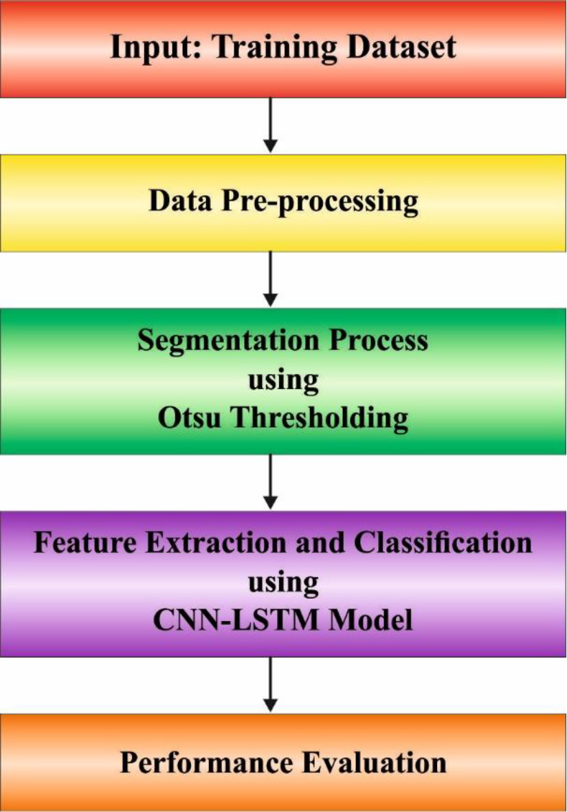
Here, the Otsu model is applied, which effectually discriminates the lung areas from the background, decreasing computational complexity and potential noise. The segmentation steps use Otsu thresholding following preprocessing [17]. Data were segmented dependent upon region through the automated threshold selection technique called Otsu. The non-parametric Otsu threshold method is unsupervised and exploits grey levels. Otsu threshold condition applies the grey level histogram of the picture, and the threshold method produces a normal value within
In Eq. (1),
Then, the class mean which is represented as
Equations (4) and (5) are substituted in Eqs (6) and (7) correspondingly. The preceding calculation was used to construct the class variance equation:
The performance rate can be utilized for assessing how well the segmentation performed after using the Otsu method on synthetic images shown in Eq. (9).
Usually, images were considered to be effectively segmented once each pixel makes up the item is isolated in the backdrop without addition or subtraction of pixels. During image segmenting process, the results of lesser than
At this stage, the CNN-LSTM model is used for classification purposes. DNN is known as CNN. It tries to find the intrinsic and fundamental traits through the guided analysis of 2D or 3D images [18]. These properties are beneficial for spotting abnormal features and categorizing anatomical structures. Convolutional, output and several pooling layers are associated with an input layer in a classical CNN model.
Convolutional layer
The convolution layer conducts a convolution function through the convolution kernel and raw input dataset to generate new feature values. In comparison to input matrix, the convolutional kernel is considered a small window that arranges coefficient into matrix. A characteristic variable named a convolved structure has been constructed by the filter’s allocated dimension element and coefficient value. The convoluted features are created by using multiple convolution kernels on the input data that are often more useful than the essential characteristics of the original data. The convolution layer serves as a basis of CNN because they are where most of the computations are done. By using the filters, the local features can be extracted.
In Eq. (10), the operator
Mostly, the pooling layer is put after the convolution layer. The pooling layer generates a compressed mapping feature utilizing the information from mapping feature from the convolution layer. Maximum and average pooling are the more commonly used techniques. A filter of size
Where
In the dense layer, the LSTM method was utilized. RNNs especially, LSTM-NN, have the ability to learn over time through feedback connection. This technique generates short-term memory and gathers information from it through cyclic linkages on hidden layer (HL). Also, it collects information via time sequences and series. The LSTM component includes memory cell input, forget, and output gates. By using Eq. (13), the functioning of LSTM unit can be predicted.
In Eq. (13),
The neuron of resultant layer, also called as FC layer, are completely reliant on the area of the prior layer of the brain. Here, information was turned into 1D matrix. The overall amount of FC layers in every model might differ.
In Eq. (14),
In this section, the TB classification results of the HDL-ISCTB algorithm can be tested on the CXR database comprising 4037 instances as defined in Table 1. Figure 2 depicts the sample images.
Description of database
Description of database
Figure 3 signifies the classifier outcomes of the HDL-ISCTB methodology on test database. Figures 3a–3b describes the confusion matrix attained by the HDL-ISCTB algorithm on 70:30 of TR set/TS set. The outcome inferred that the HDL-ISCTB methodology has recognized and classified 2 classes accurately. Next, Figs 3c–3d implies the TB detection outcomes of the HDL-ISCTB algorithm on 70:30 of TR set/TS set. The outcomes identified that the HDL-ISCTB methodology properly identifies the normal and pneumonia classes.
In Table 2 and Fig. 4, the TB detection outcome of the HDL-ISCTB methodology are reported. The result identified that the HDL-ISCTB system properly identifies the normal and pneumonia classes. With 70% of TR set, the HDL-ISCTB system offers average
TB detection outcome of HDL-ISCTB algorithm on 70:30 of TR set/TS set
Sample images.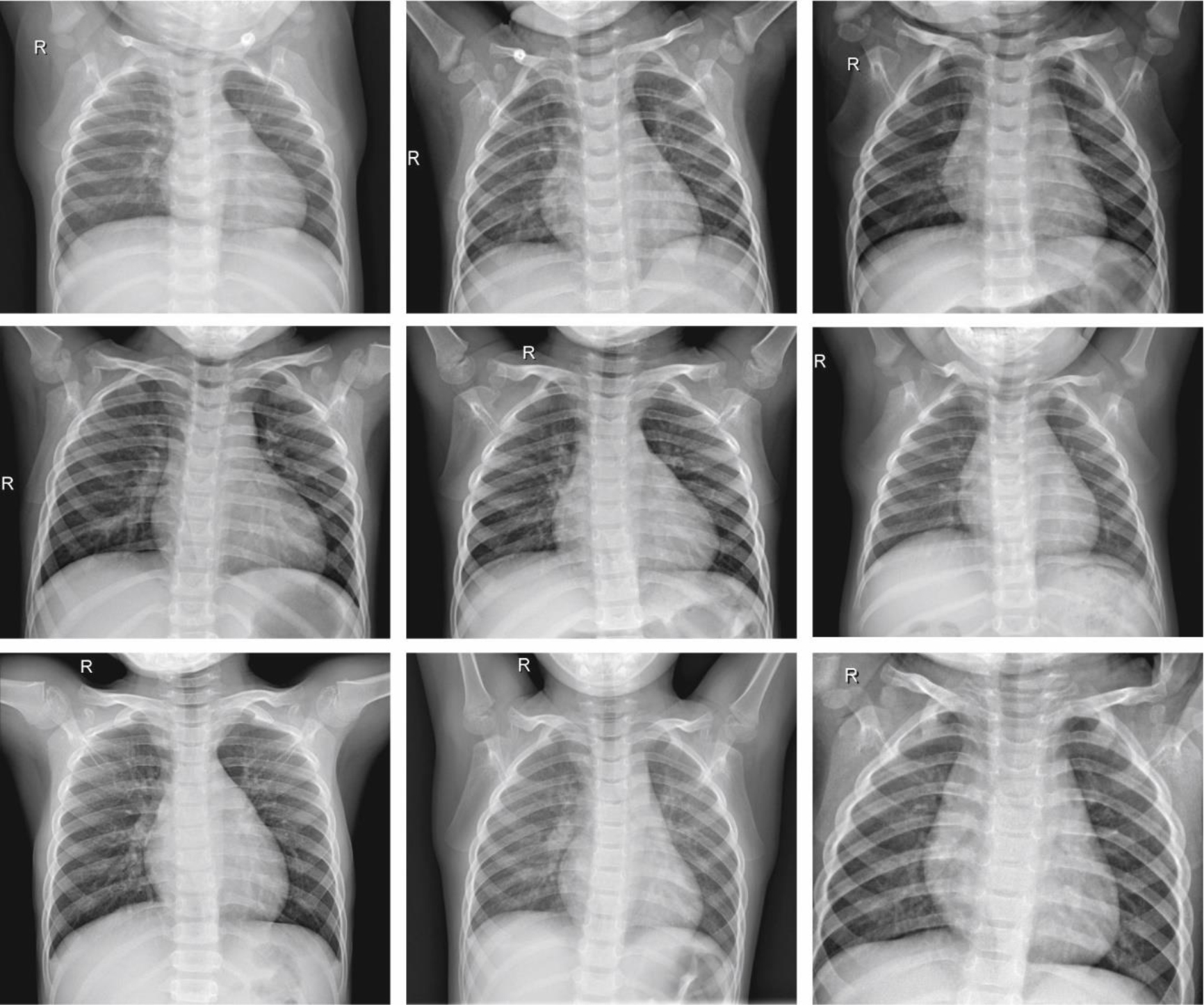
The performance of the HDL-ISCTB method with existing approaches is compared in Table 3 and Fig. 5. The table values inferred that the HDL-ISCTB system reaches improved outcome on TB classification performance. Based on
Comparative outcome of HDL-ISCTB algorithm with recent approaches
Performances of (a–b) Confusion matrices and (c–d) 70:30 of TR set/TS set.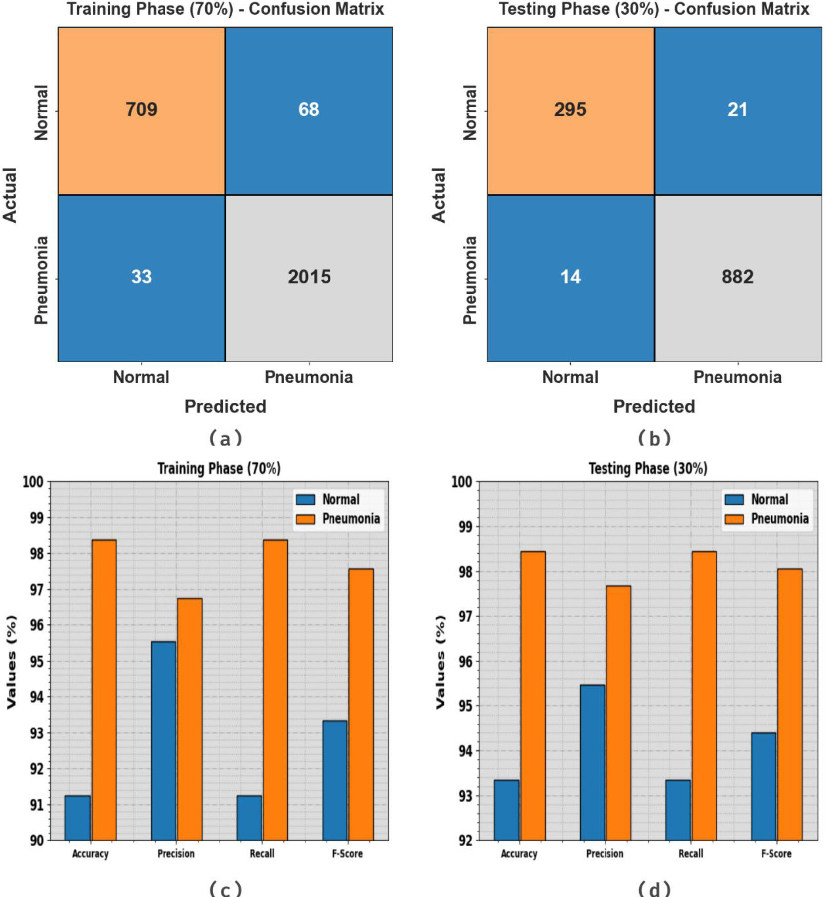
Average outcome of HDL-ISCTB algorithm on 70:30 of TR set/TS set.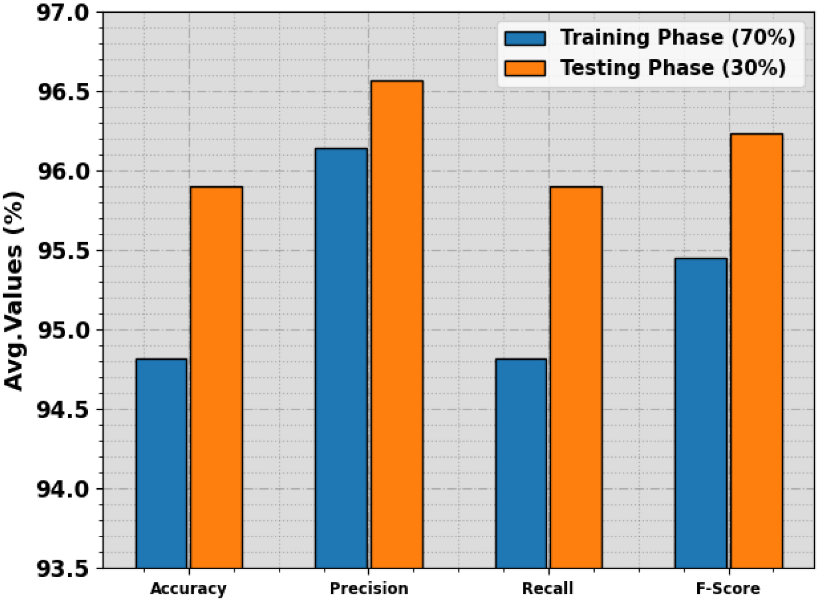
Comparative outcome of HDL-ISCTB algorithm with recent approaches.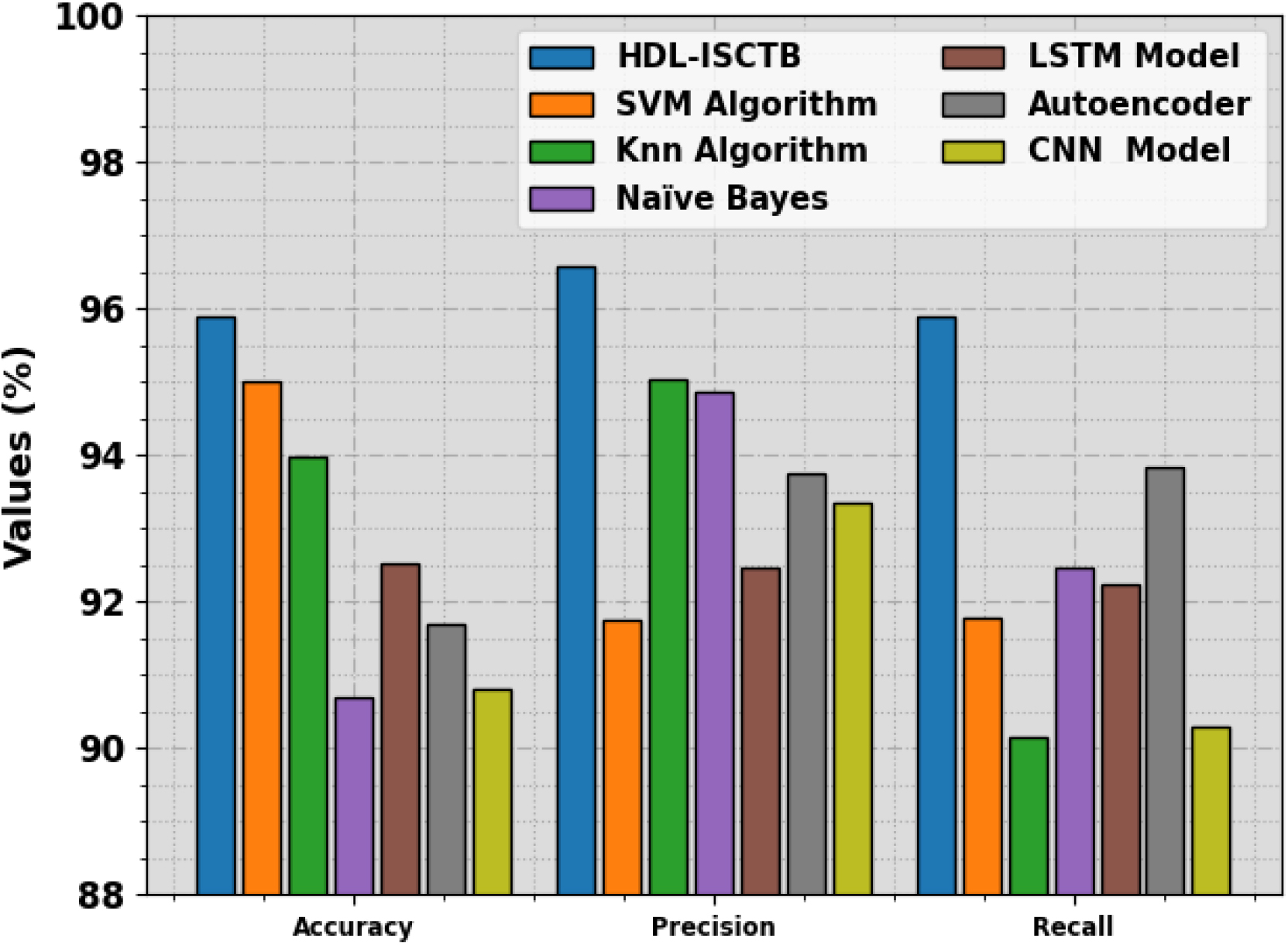
This study has introduced a novel HDL-ISCTB approach for Image Segmentation and Classification for TB diagnosis. The HDL-ISCTB model performs Otsu’s thresholding, which segments the lung regions from the input images. It effectually discriminates the lung areas from the background, decreasing computational complexity and potential noise. Besides, the segmented lung regions are then fed into the CNN-LSTM architecture for classification. The CNN-LSTM model leverages the powerful feature extraction capabilities of CNNs and the temporal dependencies captured by LSTM to obtain robust representations from sequential CXR image data. A wide experiment can be conducted to estimate the performance of the presented algorithm in comparison to recent methods.