
Abstract
OBJECTIVE:
To investigate the impact and potential mechanisms of serum extracellular nano-vesicles (sEVs) miR-412-3p released from sub-centimeter lung nodules with a diameter of
METHODS:
A total of 87 participants were included and divided into a mnLC group (
RESULTS:
The expression level of sEVs-miR-412-3p in the mnLC group was significantly higher than that in the BLN and healthy groups (
CONCLUSION:
sEVs-miR-412-3p could promote the biological process of EMT, and lead to the occurrence of malignant biological behavior in sub-centimeter lung nodules. This provides evidence for the miR-412-3p/TEAD1 signaling axis as a potential therapeutic target for mnLC.
Keywords
Introduction
Lung cancer is one of the most common and poorly prognostic cancers worldwide, and also one of the most burdensome malignant tumors worldwide, seriously endangering human health [1, 2]. Therefore, the early diagnosis and treatment of lung cancer are of great significance for improving the quality of life of patients and reducing mortality [3, 4]. With the widespread application of low-dose spiral computed tomography (LDCT) in clinical practice, the detection rate of sub-centimeter lung nodules with a diameter of
Serum extracellular nano-vesicles (sEVs) are extracellular vesicles secreted by cells with a diameter ranging from 30 to 150 nm, widely present in various bodily fluids [7]. After release, sEVs are absorbed by neighboring or distant cells, and their miRNAs are involved in regulating tumor immunity and microenvironment, which may further promote tumor growth, invasion, metastasis, angiogenesis, and drug resistance [8, 9, 10]. miRNAs have become important biomarkers in non-invasive liquid biopsy of various tumors [11, 12]. Previous studies reported that sEVs-miR-519a-3p secreted by gastric cancer cells could induce M2-like macrophage-mediated angiogenesis in the liver and promote liver metastasis [13]. In lung cancer, sEVs-miR-942 derived from M2 macrophages may promote lung cancer cell invasion and migration, as well as angiogenesis, making it a new therapeutic target for metastatic lung cancer [14]. Related fibroblasts in lung cancer tissue might promote tumor progression by releasing sEVs-miR-142-5p [15]. The above studies indicate that the enhanced proliferation, migration, and invasion ability of tumor cells involve changes in the microenvironment, and sEVs are important mediators in regulating the tumor microenvironment. In particular, sEVs-mediated functional miRNAs play an important role in mediating tumor occurrence and development. However, the biological function of sEVs miRNAs released from sub-cellular lung nodules in mnLC is currently unclear. In the preliminary research of this study, sEVs were isolated from the peripheral serum of patients with pulmonary nodules, and the expression profile of miRNAs contained in them was analyzed by sequencing. Our previous studies discovered that some sEVs miRNAs could be used as new biological detection markers to distinguish between benign and malignant pulmonary nodules [16, 17], among which miR-412-3p is highly expressed in the peripheral serum of early lung cancer patients. Therefore, we will further investigate the role and mechanism of miR-412-3p in the malignant biological behavior of sub-centimeter pulmonary nodules.
Materials and methods
Clinical sample collection
This study collected 87 participants from the Department of Respiratory, Thoracic Surgery, and Oncology at the Affiliated Zhongda Hospital of Southeast University from January 2022 to June 2023. They were divided into an mnLC group (
Purification and identification of sEVs
Referring to our previous research [16, 17], 4–5 mL of peripheral blood was extracted from all participants, and sEVs were purified. Observe the morphological characteristics of sEVs using a transmission electronic microscope (TEM) (Tecnai G2 Spirit, 120 KV, Dawson Creek Drive Hillsboro, OR, USA). Evaluate the particle size distribution of sEVs through nanoparticle tracking analysis (NTA). The data was processed using a particle tracking program (Zetasizer Ver. 7.03, Malvern Instruments, Ltd, UK).
Cell culture
Normal lung epithelial cell line (BEAS-2B) and lung adenocarcinoma cell line (A549, NCI-1299, PC-9, and H1975) (ATCC, Virginia, USA), human renal epithelial cell line 293T (Wuhan Punosai, CL-0005). Lung adenocarcinoma cell lines were cultured in RPMI-1640 (A549, NCI-1299, PC-9) and DMEM complete medium (BEAS-2B, 293T, and H1975) containing 10% fetal bovine serum (FBS; Sigma Aldrich, St Louis, MO), 100 U/mL penicillin, and Cultivate streptomycin with a concentration of 100
RT-qPCR
Total sEVs and BEAS-2B cells, as well as lung cancer A549, NCI-1299, PC-9, and H1975 cells, were extracted using Trizol LS (Invitrogen Life Technologies, MA, USA) reagent. After extraction, the total RNA concentration was measured using Nanodrop2000, and gene expression levels were quantitatively analyzed using Applied Biosystems 7500 Fast. Relative quantitative determination of each gene using the double standard curve method, with a relative expression level of 2 for each gene-
Cell transfection
Synthesize mimics and inhibitors of miR-412-3p (synthesized by Shanghai Shenggong). When the cell fusion reaches 90%, it is passaged. Inoculate cells into a 6-well plate and culture for 24 h. When the cells fuse to 70%, intervene according to the reagent instructions. After transfection, continue to culture for 48 h and collect cells for subsequent experiments.
Marking of sEVs
Follow the product manual of sEVs green fluorescent labeling dye (PKH67) for subsequent operations (Shanghai Noning Biotechnology Co., Ltd.). Firstly, quantify the extracted sEVs, then add dye working solution and cover the centrifuge tube tightly. Mix well with a vortex oscillator for 1 min, then let it stand and incubate for 10 min; Add 10 mL of PBS to the incubated sEVs dye complex and mix well. Extract sEVs again using the sEVs extraction method to remove excess dye; Take 200
CCK-8 cell viability experiment
Inoculate logarithmic growth phase cells in a 96 well plate, and transfect them when the cell growth fusion rate reaches 50%. Count 2000 cells per well, and set up 3 wells in each group. After transfection, continue to culture for 24, 48, 72, and 96 h. Add CCK-8 reagent 10
Clone formation assay
Take each group of cells with a logarithmic growth phase, digest them with 0.25% trypsin, and blow them into individual cells. Inoculate them on a 6-well plate with 700 cells per well, and culture in a 37∘C incubator with a CO2 content of 5%. Change the culture medium every three days until 21 days after cultivation, wash with PBS three times, then add 4% formaldehyde, fix for 10 min, and wash again three times. Add 1 mL of 1% crystal violet for staining for 15 min, wash three times with PBS solution, observe, take photos, and calculate the number of clones formed.
Transwell test
Take each group of cells in the logarithmic growth phase, digest them with 0.25% trypsin, blow them into a single cell suspension, adjust the cell count to 5
Stem cell sphere-forming assay
Digest and treat A549 and PC-9 lung cancer cells with 0.25% trypsin, centrifuge cell suspension at 300
Double luciferase reporter gene detection
Using software (
Western blot
Collect transfected A549 and PC-9 cells and shake them on ice with lysis buffer for lysis. Quantify with BCA protein, separate by SDA-PVDF electrophoresis, and transfer to PVDF membrane. Seal the PVDF membrane at room temperature using 8% skim milk for 2 h. The ratio of primary antibodies: CD81 (1:1000, ab155760, Abcam), TSG101 (1:1000, ab225877, Abcam), Calnexin (1:1000, ab92573, Abcam), E-cadherin (1:10000, ab40772, Abcam), N-cadherin (1:5000, ab76011, Abcam), TEAD1 (1:1000, D3F7L, CST),
Statistical analysis
Statistical analysis was conducted using the SPSS 20.0 (SPSS Inc., Chicago, USA) software system, and the experimental data was expressed as mean
Results
The expression level of miR-412-3p in sEVs of patients with sub-centimeter pulmonary nodules
Morphological characteristics and particle size distribution of sEVs in patients with sub-centimeter pulmonary nodules, as well as the expression level of miR-412-3p in sEVs. (A) TEM observation of the morphological characteristics of sEVs; (B) Evaluation of sEVs particle size distribution using nanoparticle size analysis method; (C) Surface positive markers (TSG101 and CD81) and negative markers (calnexin) protein bands of sEVs; (D) RT-qPCR was used to detect the transcription level of miR-412-3p in sEVs. ** P< 0.01, compared to the BLN and Healthy groups. All the results are representative of three independent experiments.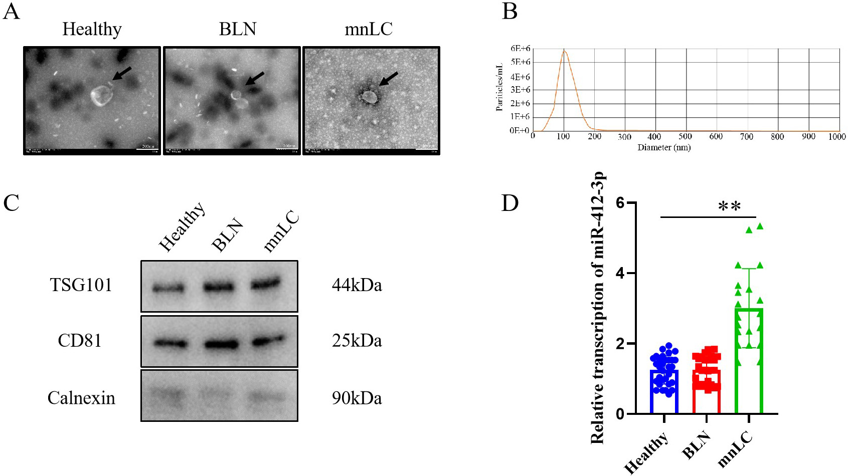
TEM intuitively confirmed the presence of sEVs of all three groups of participants, with a diameter of 40
miR-412-3p expression in lung cancer cells and its effect on cell viability. (A) RT-qPCR detection of miR-412-3p transcription levels in lung cancer cells; (B, C) RT-qPCR was used to detect the transcriptional levels of miR-412-3p in A549 and PC-9 cells transfected with miR-412-3p mimics and miR-412-3p inhibitor, respectively; (D, E) CCK-8 was used to detect the effect of transfection of miR-412-3p mimics and miR-412-3 inhibitor on cell viability. * P< 0.05, ** P< 0.01, compared to the control group. All the results are representative of three independent experiments.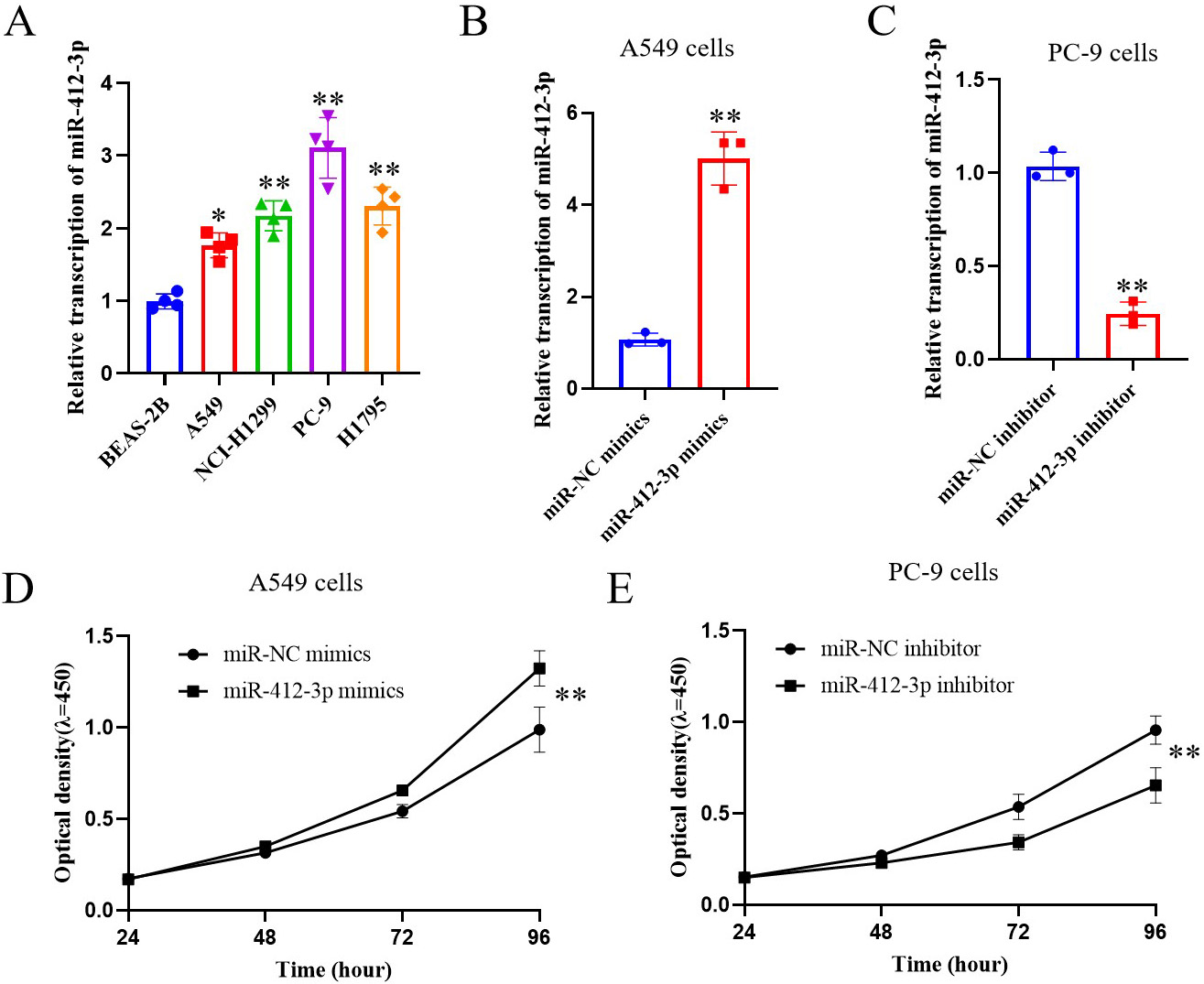
Research has reported that over 85% of malignant lung nodules are non-small cell lung cancer (NSCLC), with the majority being lung adenocarcinoma [18]. Therefore, lung adenocarcinoma cell lines (A549, NCI-1299, PC-9, and H1975) were selected as experimental cell lines in this experiment. The results showed that miR-412-3p was highly expressed in sEVs in the mnLC group, further exploring the impact of miR-412-3p on the malignant biological function of lung cancer cells. The RT-qPCR detection results showed that compared with normal lung cancer epithelial cells (BEAS-2B), the expression level of miR-412-3p in lung cancer cells (A549, NCI-1299, PC-9, and H1975) was significantly increased, with the highest expression level in PC-9 cells and the lowest expression level in A549 cells (
The synthesized miR-412-3p mimics and miR-412-3p inhibitor were transfected into A549 and PC-9 cells, respectively. RT-qPCR detection results showed that compared with the miR-NC mimics group, the transfected miR-412-3p mimics group significantly promoted the expression level of miR-412-3p in lung cancer A549 cells (
The effect of miR-412-3p on clone formation and tumor stemness ability of lung cancer cells. (A, B) Plate clone formation assay to detect the effect of transfection of miR-412-3p mimics and miR-412-3p inhibitor on cell clone formation ability; (C, D) Stem cell sphere-forming assay was used to detect the effects of transfection of miR-412-3p mimics and miR-412-3p inhibitor on cell stemness ability. *P< 0.05 and ** P< 0.01, compared to the control group. All the results are representative of three independent experiments.
Further cloning experiments showed that compared with the miR-NC mimics group, the miR-412-3p mimics group significantly promoted the cloning ability of lung cancer in A549 cells (
The results of the stem cell sphere-forming assay showed that compared with the miR-NC mimics group, the miR-412-3p mimics group significantly promoted the stemness ability of lung cancer A549 cells (
The effect of miR-412-3p on the migration and invasion ability and related proteins in lung cancer cells. (A, B) Transwell assay was used to detect the effect of overexpression of miR-412-3p on the migration and invasion ability of lung cancer A549 cells (A) and PC-9 cells (B); (C, D) The effect of miR-412-3p on the expression of migration and invasion related proteins in lung cancer A549 cells (C) and PC-9 cells (D). *P< 0.05 and ** P< 0.01, compared to the control group. All the results are representative of three independent experiments.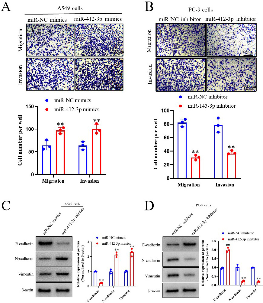
Transwell test results showed that compared with the miR-NC mimics group, the miR-412-3p mimics group significantly promoted the migration and invasion of lung cancer A549 cells (
Further WB analysis showed that in A549 cells, transfection of miR-412-3p mimics significantly inhibited the expression of E-cadherin protein in lung cancer cells compared to miR-NC mimics, while promoting the expression of N-cadherin and Vimentin proteins (
The effect of miR-412-3p on downstream target gene transcriptional enhancer associated domain proteins in lung cancer cells. (A) Design luciferase reporter gene vectors for wild-type (TEAD1 Wt) and mutant (TEAD1 Mut), and analyze the binding sites of miR-412-3p to TEAD1; (B, C) Detection of cell luciferase activity of miR-412-3p binding to TEAD1 using luciferase reporter gene in A549 and 293T cells; (D) Western blot was used to detect the effects of miR-412-3p mimics and miR-412-3p inhibitor transfection on the expression of TEAD1 in lung cancer cells; (E) Statistical analysis of the expression level of TEAD1 protein in Figure D. *P< 0.05 and **P< 0.01, compared to the control group. All the results are representative of three independent experiments.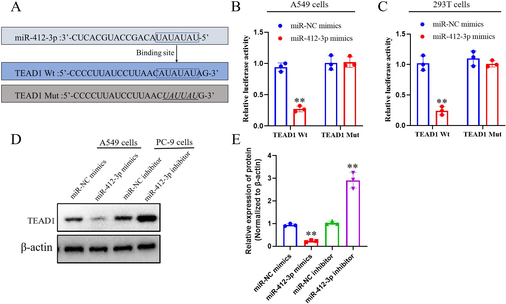
Using software (
Further validation was conducted in 293T human renal epithelial cells, and similar results were obtained. In the wild-type (TEAD1 Wt) vector, compared with the miR-NC mimics group, the transfected miR-412-3p mimics group significantly inhibited cell luciferase activity (
The effect of miR-412-3p on the proliferation and migration ability of lung cancer cells by regulating TEAD1. In the experiment, miR-412-3p mimics group and miR-NC mimics were established; Based on the miR-412-3p mimics group, a transfer with pcDNA-NC and pcDNA-TEAD1 were established. (A) The clone formation experiment detected that miR-412-3p affects the proliferation ability of lung cancer cells by regulating TEAD1; (B) Statistical analysis of clone formation experimental bar charts; (C) Transwell migration assay detected that miR-412-3p affects the migration ability of lung cancer cells by regulating TEAD1; (D) Statistical analysis of Transwell test cell migration bar chart. *P< 0.05, **P< 0.01, compared to the control group. All the results are representative of three independent experiments.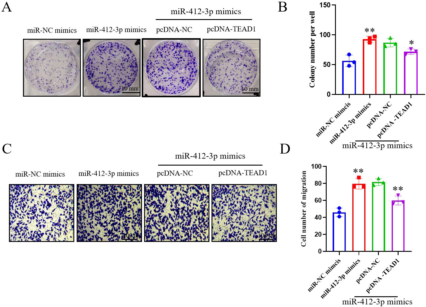
Next, we will further investigate whether miR-412-3p affects the proliferation and migration ability of lung cancer cells by regulating TEAD1. Based on the above research results, it can be inferred that miR-412-3p negatively regulates the expression of TEAD1 in lung cancer cells. To further validate this inference, the constructed blank vector (pcDNA-NC) and overexpressing TEAD1 vector (pcDNA-TEAD1) were transfected into lung cancer cell A549 cells, respectively, and miR-412-3p mimics were co-transfected. The results of clone formation and migration experiments showed that compared with miR-NC mimics, transfection of miR-412-3p mimics significantly promoted the clone formation (
sEVs enhance the proliferation, stemness, migration ability, invasion and TEAD1 related protiens of lung cancer A549 cells. (A) Immunofluorescence observation of the localization of green fluorescent labeled sEVs (PKH67) after incubation with lung cancer cells; (B) The effect of adding sEVs on cell clone formation ability in clone formation detection; (C) The effect of adding sEVs on cell stemness ability was detected through Stem cell sphere-forming assay; (D) Transwell assay was used to detect the effect of adding sEVs on cell migration ability. (E) Representative lung cancer cell A549 invasion related protein band diagram; (F) Histogram of expression of invasion related proteins in A549 lung cancer cells; (G) Intracellular TEAD1 protein band diagram of representative lung cancer cell A549; (H) Histogram of TEAD1 protein expression in A549 lung cancer cells. ** P< 0.01, compared to the BLN and Healthy groups. All the results are representative of three independent experiments.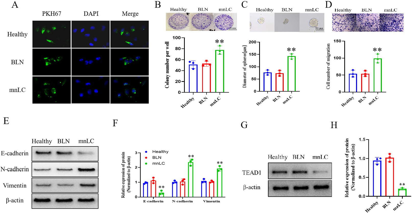
Select A549 cells with the lowest expression level of miR-412-3p from lung cancer cells (A549, NCI-1299, PC-9, and H1975) as the experimental cell model for detecting the effect of sEVs on lung cancer cells. Separate sEVs from the mnLC group, BLN group, and healthy group, and incubate them with A549 cells respectively. The changes in sEVs uptake by A549 cells can be clearly observed using immunofluorescence, and the results show that green fluorescent labeled sEVs (PKH67) can enter A549 cells (Fig. 7A). Further cellular biology functional experiments showed that compared with the sEVs of the BLN and healthy people groups, sEVs in the mnLC group significantly promoted the ability of lung cancer cells to form clones, dry and migrate (
Further WB analysis showed that compared with the sEVs of the BLN and health groups, the sEVs of the mnLC group significantly inhibited the expression of E-cadherin and TEAD1 proteins in lung cancer cells, while promoting the expression of N-cadherin and Vimentin proteins (
Schematic diagram of sEVs miR-412-3p promoting early lung cancer development through targeted regulation of TEAD1 signaling axis in micro-nodular lung cancer.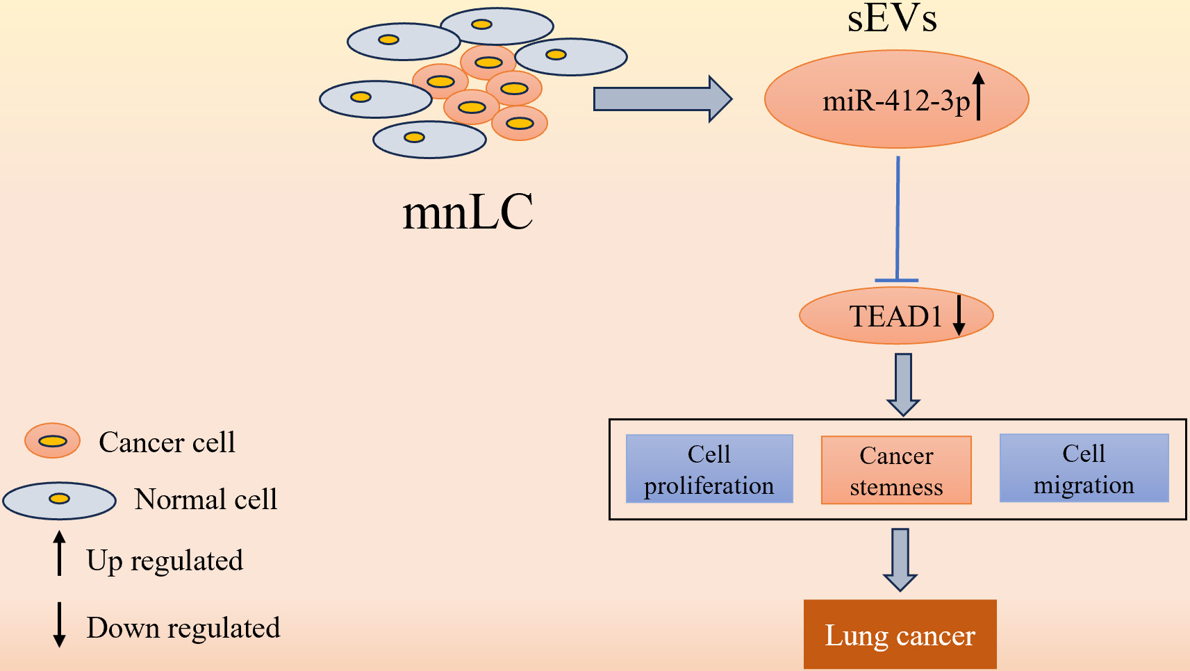
In summary, the results of this study found that sEVs-miR-412-3p was highly expressed in the serum of mnLC patients, suggesting that miR-412-3p may play an important role in the occurrence of early lung cancer. The results of in vitro cell experiments confirmed our hypothesis: miR-412-3p targeted regulation of the TEAD1 signaling pathway plays an important role in early lung cancer (Fig. 8). The high expression of miR-412-3p in lung cancer cells promotes the expression of N-cadherin and Vimentin proteins, inhibits the expression of E-cadherin proteins, induces the epithelial-mesenchymal transition (EMT) process in lung cancer cells, and enhances their proliferation, stemness, and migration abilities by inhibiting TEAD1.
Lung cancer is one of the most widely distributed malignant tumors worldwide, with an age-standardized 5-year survival rate of only 10% to 20% [19]. The EMT process is related to tumor occurrence and development, and tumor cells enhance their migration and invasion abilities through the EMT process [20]. During the process, EMT not only manifests as changes in cellular morphology, but is also accompanied by changes in related molecular markers such as E-cadherin, N-cadherin, and Vimentin [21]. Early lung cancer screening for high-risk populations has become an important link in reducing lung cancer mortality and reducing the burden of lung cancer in various countries. With the widespread application of LDCT in early lung cancer screening, the detection rate of sub-centimeter lung nodules with a diameter of
The sEVs are important mediators of intercellular communication, carrying abundant miRNAs and proteins that play important roles in information transmission between tumor cells and their microenvironment. Meanwhile, sEVs participate in regulating tumor cell proliferation, stemness, migration, and invasion ability, and can serve as a potential biomarker for early diagnosis of tumors [22, 23, 24]. Previous research reported fibrinogen
miR-412-3p is a newly identified class of non-coding miRNAs that are involved in regulating the occurrence and development of colorectal and renal cancer [29, 30]. In colorectal cancer, miR-412-3p acts as a pro-oncogene and promotes tumor progression by negatively regulating the expression of MYL9 which may mediate canceration through the TGF
The results of this study’s cell function experiment showed that transfection of miR-412-3p mimics significantly promoted the proliferation, stemness, migration, and invasion ability of lung cancer cells, while transfection of miR-412-3p inhibitor significantly inhibited the biological functions of lung cancer cells mentioned above, suggesting that miR-412-3p, as an oncogenic gene, significantly promote the progression of lung cancer. Therefore, we speculate that sEVs-miR-412-3p released by mnLC may significantly promote the proliferation of lung cancer cells, enhance the stem cell characteristics of lung cancer cells themselves, and enhance their ability to invade and migrate surrounding tissues. Biological information software prediction and luciferase reporter gene results show that one of the target genes of miR-412-3p is TEAD1, and miR-412-3p binds and negatively regulates the expression of TEAD1. Previous studies have reported that TEAD1 could regulate cell proliferation and stem cell function [33, 34], and reducing TEAD1 expression should significantly affect Hippo, Wnt, and TGF-
In summary, this study constraints that miR-412-3p targets negative regulation of TEAD1 and plays an important role in the malignant proliferation, stemness, invasion, and migration ability, as well as EMT process of lung cancer cells, exhibiting a progression in promoting the malignant biological behavior of sub-centimeter lung nodules. Therefore, drugs or inhibitors targeting the miR-412-3p/TEAD1 signaling axis may be an effective target for inhibiting the malignant biological behavior of mnLC in the future.
Funding
This research was supported by Jiangsu Funding Program for Excellent Postdoctoral Talent (2022ZB799).
Author contributions
Conception: YD, NP, HZ.
Interpretation or analysis of data: YD, NP, SD, HZ.
Preparation of the manuscript: YD, NP, SD, HZ.
Revision for important intellectual content: YD, NP, HZ.
Supervision: HZ.
Footnotes
Acknowledgments
None.
Conflict of interest
Nothing to declare.