
Abstract
INTRODUCTION:
The E3 ubiquitin ligase FBXW11 exerts an oncogenic or tumor suppressive function in a cellular context-dependent manner. However, the clinical significance and biological role of FBXW11 in chondrosarcoma have not been clearly characterized. This study focuses on the expression profile, prognostic value and biological function of FBXW11 in chondrosarcoma.
METHODS:
FBXW11 expression was analyzed by qRT-PCR and Western blot in six cases of chondrosarcoma specimens and the matched adjacent non-tumor tissues. The expression profile and prognostic value of FBXW11 were investigated in sixty-three cases of chondrosarcoma patients. Cell viability, colony formation, migration, invasion and apoptosis assays were further detected in SW1353 chondrosarcoma cells with restored FBXW11 expression.
RESULTS:
Downregulation of FBXW11 was remarkably detected in human chondrosarcoma specimens compared with the corresponding non-tumor tissues and benign cartilage tumors. Downregulated FBXW11 expression significantly correlated with high-grade chondrosarcoma and poor prognosis. Furthermore, FBXW11 was identified as an independent prognostic factor for the overall survival of chondrosarcoma patients. Restored expression of FBXW11 significantly suppressed chondrosarcoma cell growth and induced apoptosis.
CONCLUSIONS:
These findings establish that FBXW11 was markedly downregulated and recognized as an independent prognostic factor for patients with chondrosarcoma, and restored FBXW11 expression can suppress chondrosarcoma growth and induce apoptosis, highlighting a novel biological marker and potential therapeutic target against chondrosarcoma.
Introduction
Chondrosarcoma is a common primary malignant cartilage-forming tumor that usually occurs in the long bones of the extremities such as proximal femur, characterized by a relatively high recurrence and mortality due to resistance to classic radiotherapy and chemotherapy [1]. In recent years, the molecular mechanisms of the pathogenesis of chondrosarcoma and targeted therapy have become research hotspots, providing pivotal theoretical and experimental basis for in-depth understanding of its biological behaviors [2, 3, 4]. Therefore, further exploration of the relationship between novel regulatory genes and chondrosarcoma development has important theoretical significance for unravelling its molecular mechanisms, designing reasonable molecular targeted drugs, and further improving chondrosarcoma treatment [5, 6].
The ubiquitin-proteasome system is a master regulator of targeting cellular proteins for ubiquitination-dependent protein turnover, participating in both physiological and pathological processes [7]. Cullin-RING E3 ubiquitin ligase complex such as SKP1-Cullin 1-F-box protein (SCF) play a central role in multiple cellular processes [8]. F-box proteins further divided into FBXW, FBXL and FBXO subfamilies based upon protein structure [9], are the substrate-recognition subunits of SCF E3 ligase complexes and involved in ubiquitylation-dependent protein degradation and also tumorigenesis [10, 11]. FBXW11 (F-box and WD-40 domain protein 11), also known as
Dysfunction of FBXW11 could involve in tumorigenesis, however, its biological function in chondrosarcoma remains elusive. We hypothesized that FBXW11 may be implicated in the pathogenesis of chondrosarcoma. Thus, we determined whether and how FBXW11 implicates in chondrosarcoma development and exerts pro- or anti-neoplastic activity against chondrosarcoma.
Materials and methods
Clinical specimens and cell line
Clinical specimens such as six cases of fresh chondrosarcoma tissues and the matched non-tumor tissues next to chondrosarcoma, sixty-three cases of conventional chondrosarcomas and seventeen cases of benign cartilage tumors including osteochondroma and enchondroma were enrolled as described previously [4, 5, 22]. All chondrosarcoma cases in this study were pathologically diagnosed as conventional chondrosarcomas, the rare subtypes of chondrosarcomas including dedifferentiated, mesenchymal, juxtacortical, and clear-cell chondrosarcomas were excluded according to their distinctly different clinicopathological features. Clinical clinicopathological information such as histological grading and surgical staging system of the Musculoskeletal Tumor Society (MSTS), and follow-up data were reported previously [23, 24]. Signed informed consent for sample collection and analysis were obtained from all patients and was in full compliance with national legislation and the ethical standards as described previously (IRB00001052-08044) [23, 24]. Human chondrosarcoma SW1353 cell line was purchased from the American Type Culture Collection (Bethesda, MD, USA) and maintained in a humidified cell incubator with 5% CO
RNA extraction and real-time PCR
Total RNA extraction, cDNA synthesis, and qRT-PCR were performed as described previously [25]. Briefly, total RNA was isolated using Trizol reagent (Invitrogen, Carlsbad, USA). Total RNA (2
Protein extraction and Western blot analysis
Protein extraction and Western blot were performed as previously described [25]. Briefly, total protein extracts were separated using 12.5% SDS-polyacrylamide gel electrophoresis, transblotted onto nitrocellulose membranes, and blocked before incubating the membranes with primary antibodies overnight at 4
Immunohistochemistry and evaluation of staining
Detailed experimental protocols have been described previously [22, 23]. Briefly, all specimen slides (5
Generation of stable cell clones
Stable SW1353 cell clones with ectopic expression of FBXW11 were generated as described previously [23]. Specifically, human FBXW11 full-length cDNA was obtained by RT-PCR, and was cloned into a pLPCX (Clontech, CA) according to the manufacturer’s instructions. The Lenti-X HTX packaging system (Clontech, CA) produced lentiviral supernatants. Puromycin (2.5
Cell viability and clonogenic assays
Cell Counting Kit-8 (Dojindo, Kumamoto, Japan) for cell viability, and clonogenic formation assays were conducted as described previously [23, 25]. Briefly, cell clones were incubated for the indicated time, and then CCK-8 was added to calculate cell viability. In addition, transfected cells were allowed to grow for an additional 10–14 days for clonogenic assay. Colonies were counted after methanol/acetone (1:1) fixation and Gentian Violet staining according to the previous experimental protocols [20].
Cell migration and invasion asssays
Cell migration and invasion assays were performed as described previously [23]. Cell migration was detected using a modified two chamber transwell migration assay. The number of invasive cells was determined by counting the leucocrystal violet-stained cells. For the cell invasion assay, cells that had migrated across the membrane were counted in five random visual fields using a light microscope.
Downregulated protein expression of FBXW11 in human chondrosarcomas
Downregulated protein expression of FBXW11 in human chondrosarcomas
Detailed experimental protocols have been previously described [23, 25]. Briefly, after treatment, cells were resuspended in 100
Statistical analysis
Data are presented as the Mean
Results
Downregulated expression of FBXW11 in chondrosarcoma specimens
To explore the potential role of FBXW11 in chondrosarcoma development, we performed RT-PCR and western blot in six cases of the fresh chondrosarcomas and the corresponding non-tumor tissues. Our results shown that the mRNA levels of FBXW11 in chondrosarcoma were abrrently decreased as compared to the matched adjacent non-tumor tissues (Fig. 1A,
Next, we performed immunochemistry analysis and found that the positive signal of FBXW11 was preferentially recognized at the cytoplasm in human cartilage tumors such as osteochondroma and chondrosarcoma (Fig. 2A and 2B), and low protein expression of FBXW11 was identified as 63.5% (40/63) in chondrosarcomas samples as compared to 29.4% (5/17) in benign cartilage tumors (
FBXW11 expression was significantly downregulated in chondrosarcomas. (A) The FBXW11 mRNA expression was analyzed by Real time PCR in six chondrosarcoma tissues and the paired corresponding adjacent non-tumor tissues. N, the corresponding non-tumor tissues; CS, chondrosarcoma tissues. 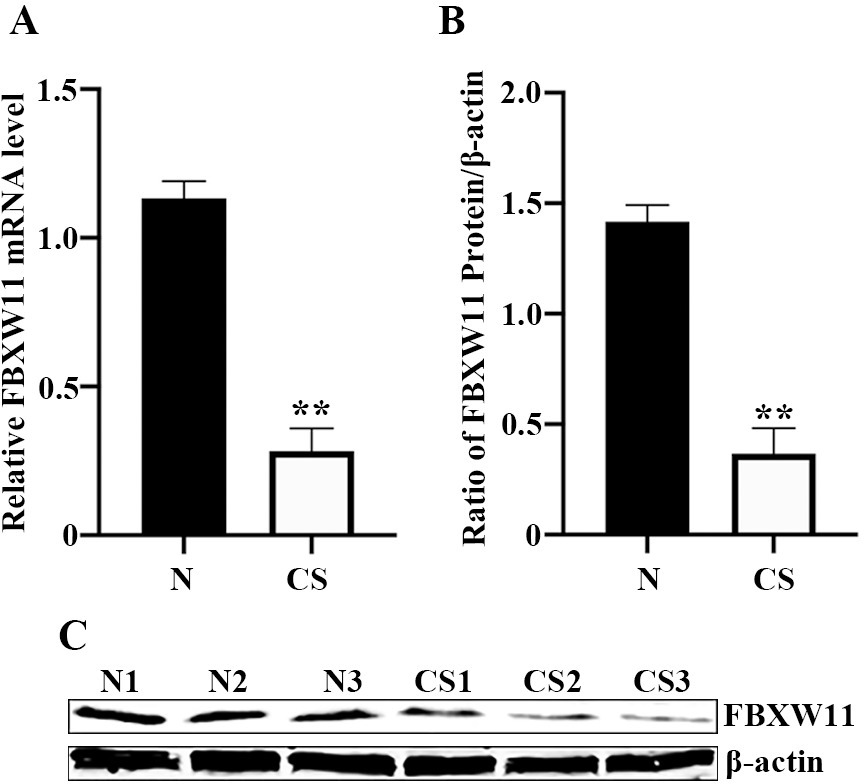
To explore whether downregulated FBXW11 expression significantly correlated with clinicopathological factors in sixty-three chondrosarcoma patients, we analyzed their relationships, and displayed that low protein expression of FBXW11 significantly correlated with the increment of histological grade (
Downregulated FBXW11 expression associated with clinicopathological factors in 63 patients with chondrosarcomas
Downregulated FBXW11 expression associated with clinicopathological factors in 63 patients with chondrosarcomas
Downregulation of FBXW11 was associated with poor prognosis in patients with chondrosarcomas. (A–B) Representative images of FBXW11 staining were presented in human cartilage tumors. Osteochondroma with high cytoplasm immunostaining of FBXW1 (A). Negative cytoplasm immunostaining of FBXW11 (B) were detected in chondrosarcoma tissues. (C) Prognostic values of FBXW11 protein expression in sixty-three patients with chondrosarcomas by Kaplan-Meier survival curves and the log-rank test. Probability of overall survival in patients with chondrosarcoma with regard to FBXW11 expression demonstrated that low FBXW11 expression remarkably correlated with poor prognosis (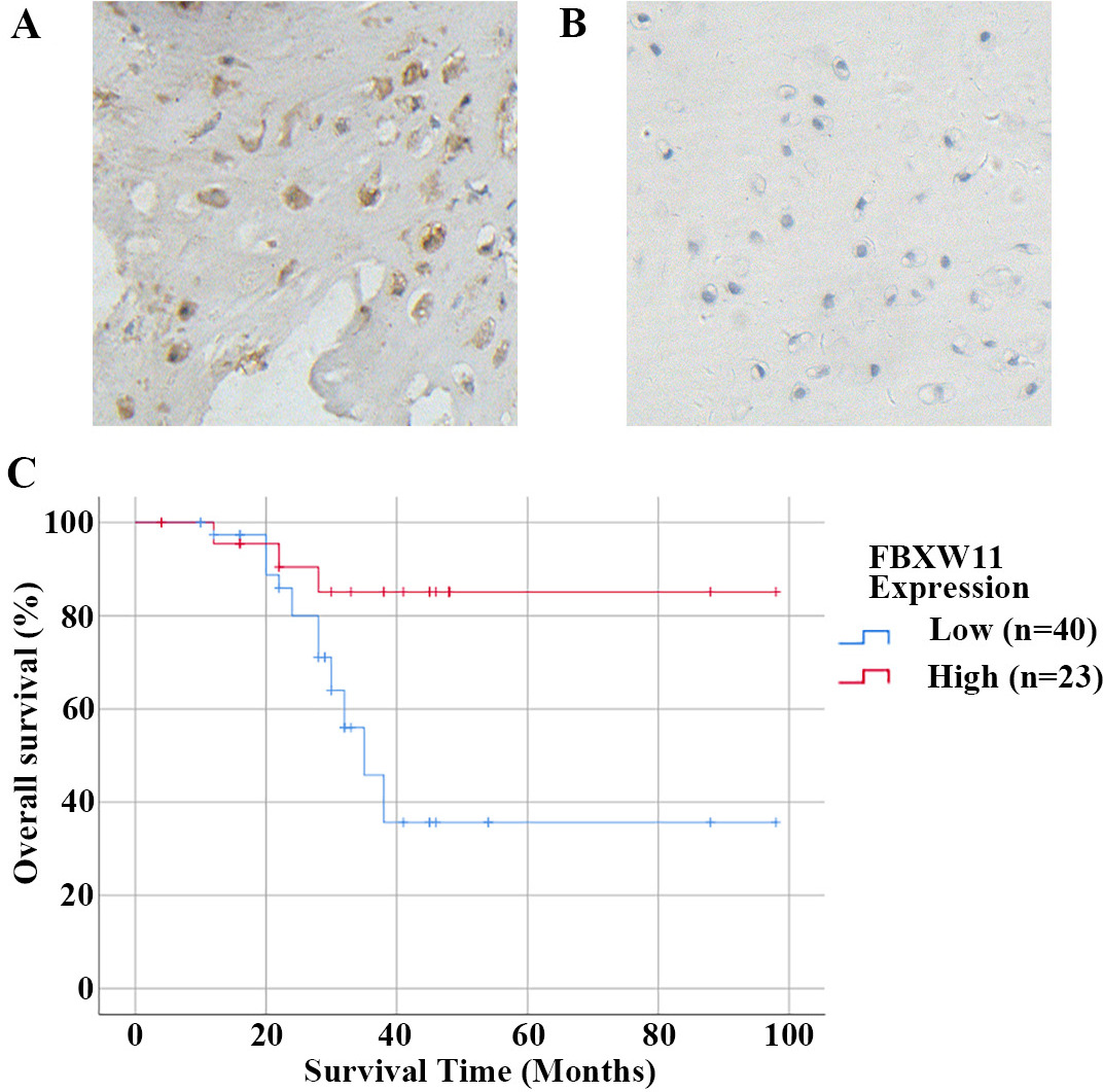
Results of Cox Regression analysis of the prognostic factors for overall survival in 63 patients with chondrosarcomas
Results of Cox Regression analysis of the prognostic factors for overall survival in 63 patients with chondrosarcomas
To identify the potential independent prognostic factors for the overall survival of chondrosarcoma patients, we had conducted Cox regression model and shown that low FBXW11 level (HR, 10.221,
Ectopic expression of FBXW11 suppressed chondrosarcoma cell growth and induced apoptosis. (A) FBXW11 was markedly upregulated by pLPCX-FBXW11. The overexpressed expression of FBXW11 was detected by Western blot from stable SW1353 cell clones with pLPCX vector or pLPCX-FBXW11 (left panel). The histograms exhibited the gray intensity analysis to the bands of Western blot (right panel, 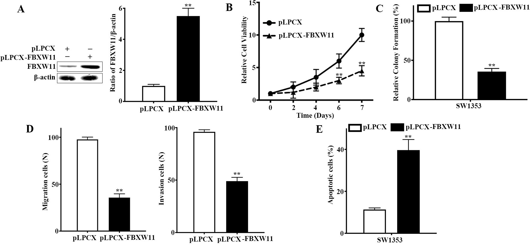
To further explore the underlying role of FBXW11 in chondrosarcoma development, we had successfully generated stable chondrosarcoma SW1353 cell clones with ectopic expression of FBXW11, as confirmed by western blot (Fig. 3A). Next, we examined the effects of restored expression of FBXW11 on chondrosarcoma cell survival, and found that restored expression of FBXW11 significantly suppressed chondrosarcoma cell viability (Fig. 3B). Our results also shown that overexpression of FBXW11 markedly inhibited SW1353 cell colony formation as displayed in Fig. 3C. As shown in Fig. 3D, the supression of migration and invasion of SW1353 cells infected with pLPCX-FBXW11 was significantly greater than that with pLPCX vector. To clarify the potential reasons for restored FBXW11 expression mediated the growth arrest, we tested apoptosis in these cells, and found that overexpression of FBXW11 significantly induced cell apoptosis (Fig. 3E). Our data unravelled that restored expression of FBXW11 remarkably suppressed chondrosarcoma cell growth, at least in part, attributed to induction of apoptosis.
Emerging data suggest dysfunction of many F-box proteins results in aberrant cellular ubiquitination and contributes to tumorigenesis and progression [11], whereas some inhibitors targeting F-box proteins have shown promising therapeutic potential [26]. Belonging to F-box protein family that recognizes and degrades substrates such as
To characterize the clinical significance of FBXW11 in chondrosarcoma, we present the first evidence that low expression of FBXW11 was significantly correlated with histological grade and MSTS stage, rather than age, gender and anatomical location (Table 2). In addition, we also found that low expression of FBXW11 had significantly unfavourable impact on the overall survival of chondrosarcoma patients (Fig. 2C). Multivariate analysis by Cox regression model had demonstrated that FBXW11 is identified as an independent prognostic factor for the overall survival of chondrosarcoma patients (Table 3). It is rather definite that the histological grading and MSTS stage are the most important predictor for clinical behavior of chondrosarcoma [34]; and the prognosis of chondrosarcoma is associated with Enneking grades, histological grades and local recurrence rather than distant metastasis, surgical approaches or the size of the tumor [35]. Notably, the histological evaluation of tumor grade is somewhat subjective because chondrosarcoma represents a collective group of cartilage-originating tumor. Thus, alternative methods, including the evaluation of DNA synthesis and content, cytogenetics and molecular markers, have been sought to assess the prognosis for patients with chondrosarcomas [34]. For other factors which affect the prognosis of chondrosarcoma, controversies arise. Therefore, we conducted this study to screen prognosis-related factors. Our findings suggest that FBXW11 can be identified as an independent prognostic biomarker for chondrosarcoma patients. Previously, many researchers reported that several molecular markers have been recognized that manifest a significant correlation with histological grade and predictive for the prognosis of chondrosarcoma patients [34, 36]. Thus, our results might help the clinician to determine the prognosis of chondrosarcoma based upon the protein level of FBXW11, indicating the potential of FBXW11 as a novel biomarker in chondrosarcoma patients. However, further investigations for the clinical value of FBXW11 in larger cohorts of chondrosarcoma are indeed needed to conduct, which validates its clinical significance and prognosis in patients with chondrosarcomas.
Accumulated data suggest that FBXW11 can exert as a potent tumor-suppressive role in tumorigenesis [18, 19, 20, 21, 29, 30]. Consistent with these results, our findings also reveal that restored expression of FBXW11 can suppress chondrosarcoma cell viability, colony proliferation, migration and invasion, and induce apoptosis in vitro (Fig. 3), supporting the evidence for its tumor-suppressive role in the pathogenesis of chondrosarcoma, at least in part, attributed to induction of apoptosis. Previous studies have documented that FBXW11 can act as an oncogene promoting melanoma development [13]. Moreover, ectopic expression of FBXW11 in lymphocytic leukemia cells can remarkably stimulate tumor proliferation, mediated by the stimulation of cell cycle progression rather than the induction of apoptosis [15]. By constract, FBXW11 can induce proteasome-mediated degradation of Mcl-1, an antiapoptotic Bcl-2 family member and overexpressed in several human cancers, and thus facilitate GSK-3
Conclusion
Our work identifies downregulation of FBXW11 significantly associated with high-grade chondrosarcoma and poor prognosis in patients with chondrosarcoma. We have unravelled that FBXW11 is recognized as an independent prognostic factor and restored FBXW11 expression can suppress chondrosarcoma growth and induce apoptosis, highlighting a novel biomarker and attractive target for against chondrosarcoma.
Data sharing statement
The datasets used and/or analyzed during this study are available from the corresponding author on reasonable request.
Author contributions
Conception: Changbao Chen, Xinlong Ma.
Interpretation or analysis of data: Hua Zhou, Xiaolin Zhang, Zhongjun Liu.
Preparation of the manuscript: Changbao Chen, Hua Zhou.
Revision for important intellectual content: Changbao Chen, Xinlong Ma.
Supervision: Xinlong Ma.
Ethics approval and consent to participate
Clinical samples such as benign cartilage tumors and chondrosarcomas were handled in accordance with the Declaration of Helsinki and the Human Tissue Act. The samples used in the present study were collected as discarded tissues after written informed consent was obtained from all patients, and the use of biological tissue material in this present project was covered by the ethical approvals from the Ethics Commission Peking University Third Hospital and Tianjin Hospital as described previously (IRB00001052-08044) [24, 25].
Consent for publication
Not applicable.
Competing interests
All authors declared that they have no competing interest in this work.
Footnotes
Acknowledgments
This work was supported by Tianjin Applied Basic Research Diversified Investment Foundation (Grant No. 21JCYBJC01100), the TianJin Youth Medicine Talents Plan, and also the National Natural Sciences Foundation of China (Grant No. 81102037).