
Abstract
BACKGROUND and OBJECTIVE:
Gastric adenocarcinoma (GAC) is one of the most common malignancies. Increasing data have indicated a correlation between soluble B7-H3 (sB7-H3) levels and tumor malignancies. In this study, we aim to investigate the level of soluble B7-H3 in serum of GAC patients. Further, we analyze the correlation between sB7-H3 level and tissue B7-H3 expression and explore the clinical evaluation value of sB7-H3 associated with pathological characteristics and prognosis of GAC patients.
METHODS:
One hundred and twenty-eight serum and tissue samples of GAC, 20 serum and tissue samples of gastritis patients and 77 serum, 5 tissue samples of healthy controls were collected. The serum levels of sB7-H3 were detected by Enzyme-linked immunosorbent assay (ELISA), while the expression of membrane B7-H3 (mB7-H3) and Ki67 were evaluated by immunohistochemistry. The correlation between sB7-H3 and mB7-H3, sB7-H3 and Ki67, sB7-H3 or mB7-H3 and clinical features were analyzed by Pearson’s Chi-square test.
RESULTS:
Both serum level of sB7-H3 and tissue B7-H3 of GAC patients were significantly higher than those of gastritis patients and healthy controls. sB7-H3 level was correlated with total B7-H3 expression in tissues (
CONCLUSIONS:
Soluble B7-H3 level may reflect the tissue B7-H3 expression on tumor cells of GAC tissues. Elevated level of sB7-H3 in serum suggests poor clinical pathological characteristics of GAC patients.
Introduction
Gastric adenocarcinoma (GAC) is one of the most fatal malignancies, and was the third leading cause of cancer death worldwide [1]. Once infiltration is present, the survival rate would reduce to 31%, even 5% when transferred to distant organs. Early detection is critical for improving the prognosis of GAC patients. The 5-year survival rate is up to 68% if GAC patients are diagnosed and treated before the spread of cancer cells [2]. Thus, a potential indicator is urgent for clinical pathological characteristics and further prognosis judgments, which may be valuable for clinical evaluation.
B7-H3 is a member of the B7 family of costimulatory molecules, which shares 20–27% amino acid identity with other B7 family ligands [3]. The mRNA of human B7-H3 has been found to be overexpressed in various human tumor tissues but relatively limited in normal tissues [4]. A vast majority of studies have demonstrated that B7-H3 could induce tumor immune escape as well as enhance tumor metastasis [5, 6]. Aberrant expression of B7-H3 was correlated with poorer prognosis in several cancers such as hepatocellular cancer, breast cancer, non-small cell lung cancer, craniopharyngioma, renal cell carcinoma and gastric cancer [7, 8, 9, 10, 11, 12]. In gastric cancer, B7-H3 was expressed in both tumor and stromal cells. Tumor B7-H3 could promote gastric tumor cell migration, invasion, radiotherapy resistance and metastasis [13, 14]. In addition, stromal B7-H3 expression was related to tumor progression and lower intratumoral CD8+ T cell density [15, 16]. Several researchers believe that B7-H3 is a potential biomarker as well as a novel and valuable target for immunotherapy. Chimeric antigen receptor (CAR) T cells and bispecific antibodies targeting B7-H3 have been developed and applied in clinical trials in patients with B7-H3 positive cancers [4, 17, 18, 19, 20, 21, 22]. Therefore, it is valuable to investigate the expression pattern of B7-H3 for judging tumor progression as well as picking suitable treatment strategies.
Soluble B7-H3 lacks the transmembrane and cytoplasmic regions and could be detected in serum and plasma in some tumors and infectious diseases [4, 23]. High levels of soluble B7-H3 have also been detected in body fluids of several tumors patients and culture supernatants of tumor cell lines, and its high level expression was significantly correlated with the invasion and metastasis of tumors [24, 25, 26]. Early in 2008, Our group discovered the existence of soluble form of B7-H3 in human serum for the first time [23]. We further implicated the diagnosis value of sB7-H3 in non-small lung cancer [27]. However, the relationship between sB7-H3 level and corresponding tissue B7-H3 expression in GAC remains unknown. In the present study, we investigated the levels of sB7-H3 in the serum by ELISA in GAC patients and evaluated its correlation with B7-H3 expressed in tumor tissues. Further, we analyzed the significance of sB7-H3 with clinical features in GAC patients.
Materials and methods
Patients and plasma samples
In this study, 128 patients diagnosed with GAC were recruited as experiment group. Ninety-seven cases matched for sex and age were recruited as control group including 20 patients diagnosed with chronic atrophic gastritis via gastroscopy and 77 healthy volunteers. All samples of patients and healthy volunteers were obtained from the First Affiliated Hospital of Soochow University (Suzhou, Jiangsu, China). Patients’ characteristics were summarized in Table 1. One hundred and twenty-eight GAC tissues were derived from surgery resection. At the same time, the clinical and pathological data including gender, age, differentiation, tumor stage, tumor depth, lymph node metastasis were retrieved. None of the patients received pre-operative chemotherapy or radiotherapy. Twenty chronic atrophic gastritis and 5 healthy control tissues which diagnosed by pathologists were derived from gastroscopy. The tissue and blood samples from the subjects were provided according to a protocol approved by the local ethics committee of Soochow University.
Characteristics of cases of GAC, gastritis and healthy control
Characteristics of cases of GAC, gastritis and healthy control
The levels of sB7-H3 in serum were measured using human B7-H3 quantikine ELISA kit (R&D systems) according to the manufacturer’s instruction. In brief, the serum samples were transferred to a 96 well plate and incubated for 2 hours at room temperature. After washed 3 times, 100
Immunohistochemistry
Immunohistochemistry was performed by the two-step EnVision method (Dako, Glostrup, Denmark). In brief, the 4
Assessment of tissue staining
All immunohistochemical studies were independently and blindly reviewed by two pathologists and scanned with a Dmetrix imaging system (Dmetrix, Tucson, AZ, USA). The staining histoscore of the slides was classified as 0–3 (0, no staining; 1,
Statistical analysis
Statistical analysis was performed using SPSS (v22.0; IBM Corporation, USA).
Ethics statement
This study was carried out in accordance with the recommendations of Clinical Research Ethics Committee of the First Affiliated Hospital of Soochow University. The protocol was approved by the Clinical Research Ethics Committee of the First Affiliated Hospital of Soochow University (approval number: 2018121). All subjects gave written informed consent in accordance with the Declaration of Helsinki.
Comparison of serum sB7-H3 in healthy controls group, gastritis group and GAC group. Error bar is Mean 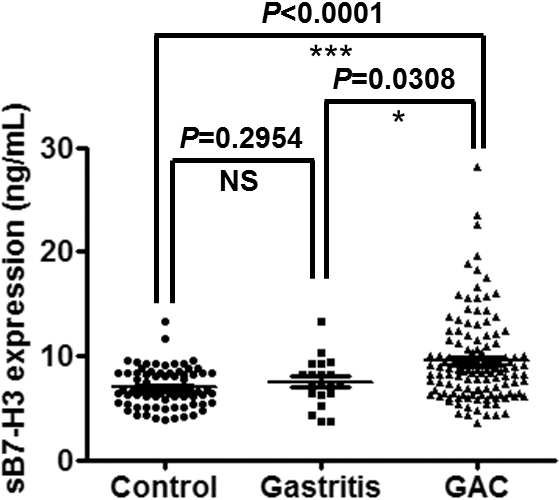
Enhancement of serum levels of sB7-H3 and mB7-H3 in GAC patients
We first investigated the levels of sB7-H3 in serum of 128 GAC patients 20 gastritis patients and 77 healthy controls (Fig. 1). The mean concentration of sB7-H3 in GAC patients was significantly higher than that in gastritis (9.524
Expression of mB7-H3 in healthy control group, gastritis group and GAC group. (A) Immunohistochemstry stain of each group. (B) Staining histoscore of each group. Error bar is Mean 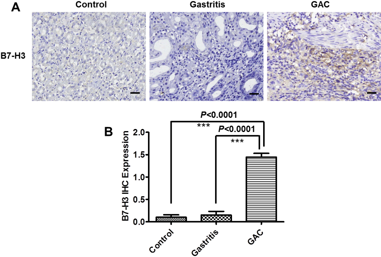
As the expression of B7-H3 in tissues is closely related to tumor progression, we then analyzed the expression of B7-H3 by immunohistochemistry in GAC patients. Our results showed that B7-H3 was either expressed in tumor cells or stromal components, or both (Fig. 3A–D). The expression of B7-H3 was detected with a frequency of 69.5% (89/128) in GAC patients. Among them, 91.0% (81/89) had stromal B7-H3 expression while 49.4% (44/89) was expressed in tumor cells as well as 41.6% (37/89) was expressed in both tumor and stromal cells according to our previous methodology [15] (Fig. 3E). In order to explore the potential source and the clinical valuable of sB7-H3 the correlation between sB7-H3 and mB7-H3 was analyzed by chi-squared test. The results showed that the levels of sB7-H3 in serum was significantly correlated with the expression of total mB7-H3 in tissues of GAC patients (
B7-H3 expression in GAC tissues. (A) B7-H3 negative expression, (B) tumor B7-H3 expression, (C) stromal B7-H3 expression and (D) both tumor and stromal B7-H3 expression, (E) Patient cases in various B7-H3 expression. The scale bar represents 20 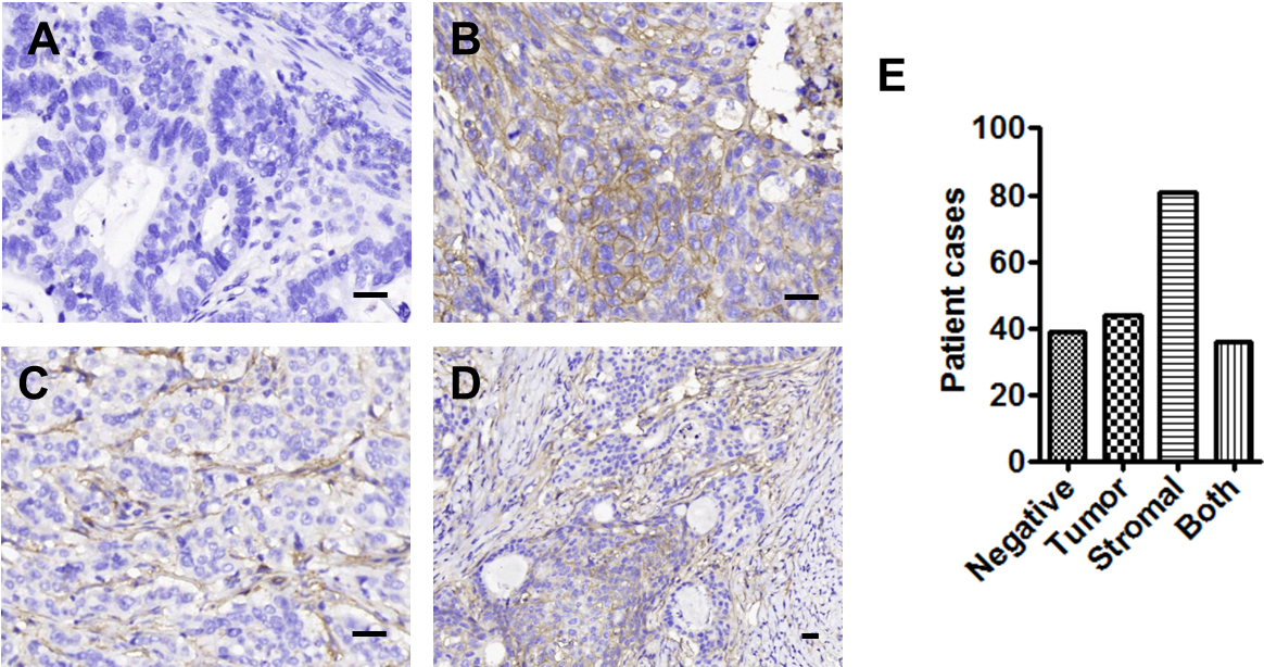
The correlation between sB7-H3 level and mB7-H3 score or Ki67 expression. (A) sB7-H3 level and total B7-H3 score. (B) sB7-H3 level and tumor B7-H3 score. (C) sB7-H3 level and stromal B7-H3 score. (D) sB7-H3 level and Ki67 expression.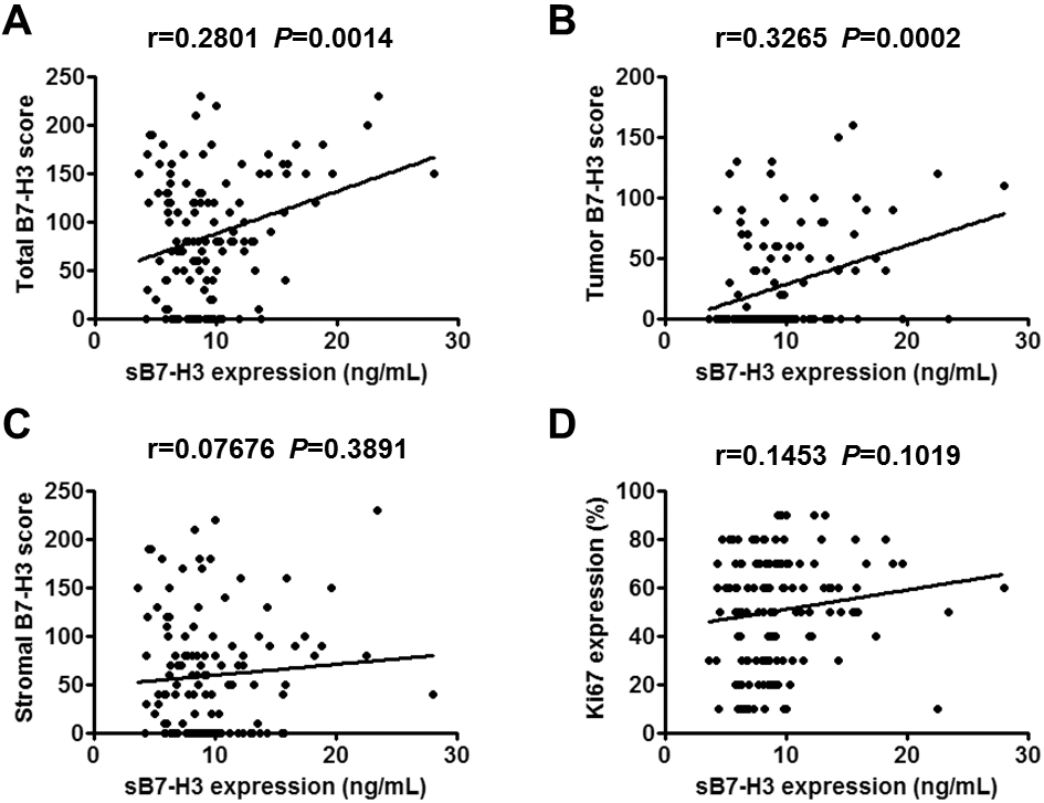
sB7-H3 level of GAC serum and various mB7-H3 expression of GAC tissue in association with cancer progression. (A) Serum sB7-H3 level (B) Total B7-H3 score (C) Tumor B7-H3 score and (D) Stromal B7-H3 score in samples with various pathological features. (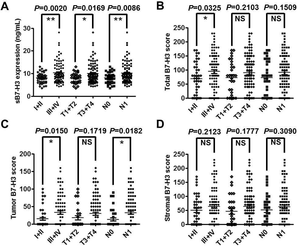
To further determine the value of increased soluble form of B7-H3 in serum of GAC patients, the correlation between sB7-H3 concentration and clinical features, including tumor grade, infiltration depth of tumor and lymph metastasis were assessed. As shown in Fig. 5A, the level of sB7-H3 in serum of GAC patients was detected to be upregulated during the neoplastic stage. The sB7-H3 levels from patients with stage III/IV or with infiltration depth T3/T4 or with lymph node metastasis were distinctly higher than that from patients with TNM stage I/II (10.32
Discussion
B7-H3, identified in 2001, is a cell surface molecule in B7 immunoglobulin super family which plays an important role in the initiation and termination of immune cell responses and cancer development. B7-H3 was overexpressed in gastric cancer and was regarded as a promising biomarker of diagnosis as well as a novel target for anti-tumor therapeutics [12, 14, 28]. Herein, we demonstrated that the soluble form of B7-H3 in serum was significantly upregulated in GAC than in gastritis and healthy controls. Furthermore, the serum levels of sB7-H3 were associated with membrane form of B7-H3 which was expressed in tumor cells but not in stromal cells. In addition, sB7-H3 levels were significantly correlated with clinical features including patients’ tumor grade, infiltration depth and lymph node metastasis. Thus, our results indicated that sB7-H3 could be a potential biomarker for clinical diagnosis and prognosis of GAC.
Prior literature have demonstrated the presence of B7-H3 in various stromal cells including endothelial cells, activated macrophages, fibroblasts, dendritic cells etc. In this study, we showed that the serum levels of sB7-H3 were positively correlated with mB7-H3 in tumor cells rather than stromal cells in GAC (
In addition, the level of sB7-H3 in serum was not statistically correlated with the expression of Ki67 but with worse prognosis, indicating that sB7-H3 had referential significance to the evaluation of tumor malignant degree but not proliferative activity of tumor cells in GAC. Increasing evidences also showed that B7-H3 could enhance tumor stem cell population and promote aggression and metastasis by targeting epithelial-mesenchymal transition in several cancers such as breast cancer, colorectal cancer and hepatocellular cancer [30, 31, 32]. Aberrant B7-H3 expression in tumor cells can also promote tumor progress through inhibiting anti-tumor immune response and several signaling pathways [4, 33, 34]. Consistent with that, sB7-H3 inhibited T cell proliferation and reduced the secretion of IL-2 and IFN-
As the overall prognosis in advanced-stage or with metastasis was poor, dynamic and timely monitor of tumor progression would improve the prognosis of patients with GAC. This study showed that sB7-H3 expression was positively correlated with TNM grade, infiltration depth and lymph node metastasis in GAC patients. Similar results were demonstrated in osteosarcoma, early-stage hepatocellular carcinoma and non-small-cell lung cancer [24, 27, 36, 37]. Soluble B7-H3 level was significantly correlated with lymph node metastasis in this study. A recent study showed that sB7-H3 promoted metastasis of pancreatic carcinoma cells through the TLR4/NF-
In general, our present findings suggested that the levels of sB7-H3 in serum were enhanced in GAC patients. Serum sB7-H3 levels were positively correlated with B7-H3 in tumor cells. We also found that the expression of sB7-H3 was correlated with tumor TNM grade, infiltration depth and lymph node metastasis. Last, sB7-H3 in serum could serve as a potential biomarker for tumor diagnosis and prognosis, as well as provide a new strategy for GAC clinical treatment.
Footnotes
Acknowledgments
This work was supported by the National Natural Science Foundation of China (grant No. 81874163, No. 81602074 and No. 31320103918) and Zhangjiagang Science and Technology Bureau Municipal Science and Technology Support Program (grant No. ZKS1948).
Author contributions
Conception: L.C., X.Z.
Interpretation or analysis of data: L.H., Y.Z., Q.S., L.C.
Preparation of the manuscript: L.H., L.C.
Revision for important intellectual content: L.C., X.Z.
Supervision: X.Z.