
Abstract
PURPOSE:
To propose MCUR1 gene as a potential biomarker for ovarian cancer prognosis.
METHODS:
The ovarian cancer patient specimen from TCGA database were analyzed using survival analysis. The immune cell infiltration ratio and checkpoints had also been investigated for different expression group of MCUR1. The function of MCUR1 as a ovarian cancer prognosis biomarker was verified in clinic.
RESULTS:
The low expression of MCUR1 was associated with the poor prognosis of ovarian cancer patients. The expressions of majority of immune cells and 6 checkpoints in low expression group of MCUR1 were significantly lower than that in high expression group of MCUR1 (
CONCLUSION:
This study has proposed a potential prognostic biomarker for ovarian cancer patients, which offers a beneficial reference for future ovarian cancer administration.
Introduction
Ovarian cancer is characterized as malignant tumors that grow on the ovary, of which 90%–95% are primary ovarian cancer [1]. Ovarian cancer represents one of the most threatening malignant tumors to the health of the women population, with a tremendous growing morbidity rate [2]. Ovarian cancer is the second most common malignancy just after breast cancer in women over the age of 40, particularly in developed countries [3]. As of 2018, ovarian cancer was around 240,000 new cases worldwide, whereas the case-to-fatality ratio is nearly three times that of breast cancer [4, 5]. Despite awareness of ovarian cancer, the curative and survival trends are far from satisfactions. There remains a big challenge for early diagnosis due to the lacking of an efficient biomarker as well as definitive screening tool with no specific early symptoms.
Calcium signaling is a key mechanism for the rapid transition of tumor microenvironmental signals into cellular responses [6]. The intracellular ubiquitous second messenger Ca
Based on the fact of unclear mechanisms underlying ovarian cancer there is still lacking an effect biomarker for prognosis of this disorder. For this purpose, in this study, using an innovative bioinformatics method combined with a clinical experimental verification, we comprehensively explored the function of MCUR1 as a potential biomarker for ovarian cancer prognosis, which offered a beneficial guidance for future ovarian cancer study.
Material and methods
Data source
The mRNA expression data information of ovarian cancer patients and corresponding control specimens were downloaded from The Cancer Genome Atlas (TCGA,
Survival analysis
The survival analysis was generated using the R language survival package and survminer (
Functional enrichment analysis
The ClusterProfiler package in R language was processed for Gene ontology (GO) analysis (including Biological Process, Molecular Function and Cellular Component) as well as Kyoto Encyclopedia of Genes and Genes (KEGG) pathway enrichment analysis [14].
Calculation of immune cell infiltration ratio
The software CIBERSORT was performed to calculate the relative proportion of 22 immune cells in ovarian cancer sample [15]. The CICERSORT software utilized the deconvolution algorithm with the preset 547 barcodes to characterize the composition of immune infiltrating cells according to the gene expression matrix.
ELISA analysis of MCUR1 for different TNM stages of ovarian cancer patients
A total of 187 ovarian cancer patients diagnosed by pathology in Tianjin Baodi Hospital from January 2018 to May 2021 were randomly selected and collected. The patients were categorized into TNM Stage I–IV using the traditional WHO classification. There was no significant characteristics difference between groups (TNM Stage I: age 63.2
The relationship between the expression of MCUR1 and the development and prognosis of ovarian cancer. (A) The expression of MCUR1 in different TNM stages of ovarian cancer patients. (B) Kaplan-Meier survival curves of ovarian cancer patients in MCUR1 high-expression group and low-expression group. The horizontal axis represents time, the vertical axis represents survival rate, and the colors indicate different groups respectively.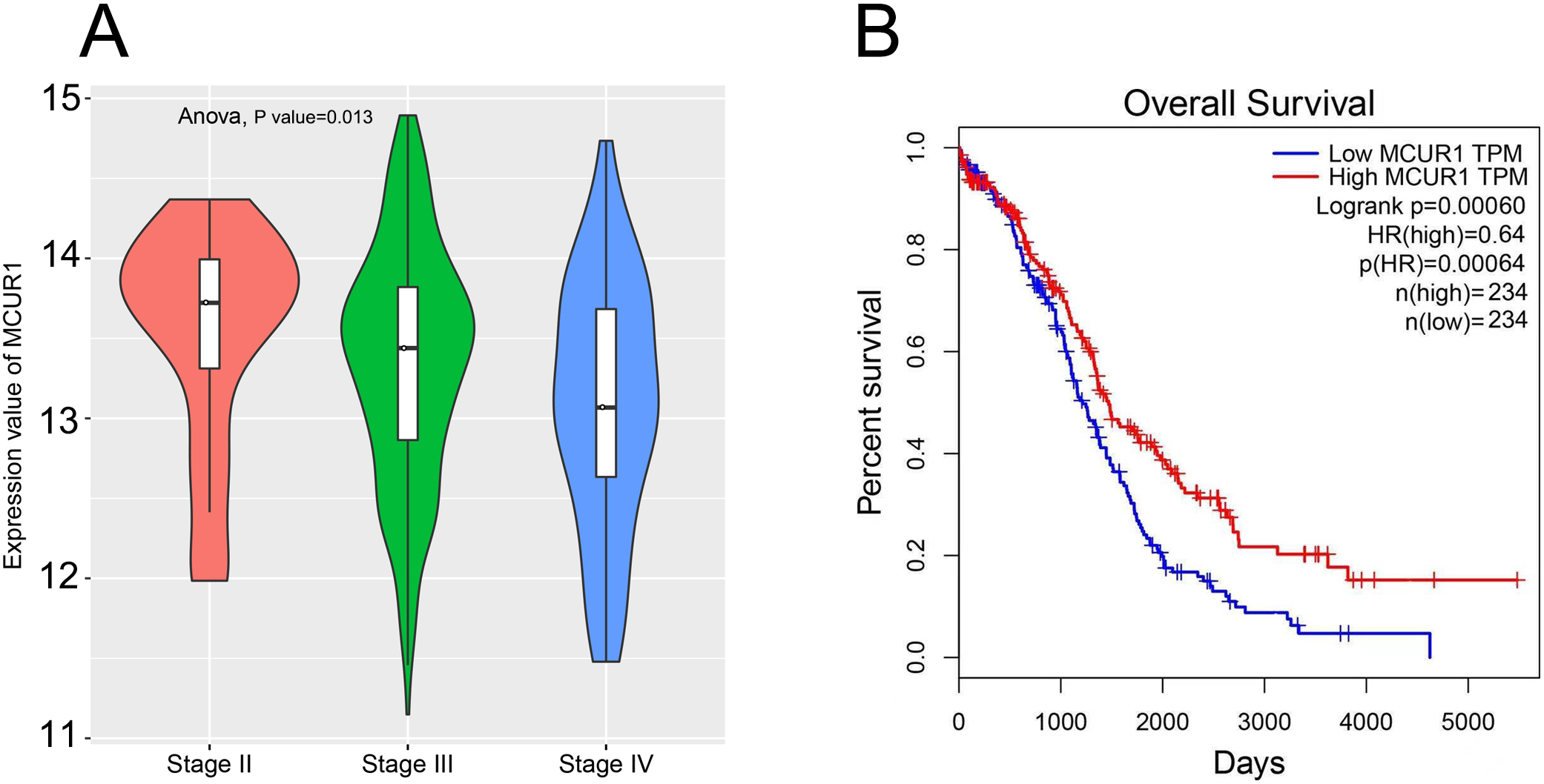
Immune infiltration status of ovarian cancer patients in the MCUR1 high and low expression group. (A) Correlation matrix of the proportions of 22 immune infiltrating cells. Red indicates positive correlation and blue indicates negative correlation. The darker the color, the greater the correlation. (B) Violin chart of immune checkpoints in the MCUR1 high and low expression groups.
MCUR1 was a primary marker for the prognosis of ovarian cancer
The dataset from TCGA was composed of different TNM stages of ovarian cancer. The gene of MCUR1 demonstrated differential expression between different TNM stages, whereas a significant decrease of MCUR1 expression level with the later stage of ovarian cancer (Fig. 1A). To further explore the relationship between the expression of MCUR1 and TNM stage, the ovarian cancer patients were divided into two groups based on the MCUR1 level. Compared with MCUR1 high expression group, patients in the low expression group showed a poor overall survival rate (Fig. 1B), suggesting that MCUR1 could be a potential marker gene for ovarian cancer prognosis.
Enrichment analysis of GO and KEGG. The top most enriched biological process entries.
The expression analysis for MCUR1. (A) The ELISA measurement of MCUR1 between different stages of ovarian cancer patients. (B) The corresponding linear graph among different stages of ovarian cancer patients.
Using CIBERSORT method combined with LM22 characteristic matrix, the infiltration of 22 immune cells of ovarian cancer patients displayed specific expression pattern between different level of MCUR1 (Fig. 2A). At the same time, we also deeply investigated the relation between MCUR1 expression level and 6 checkpoints including CTLA4, PDL1, PDL2, TIM3, LAG3 as well as TIGIT. It is noteworthy that all the checkpoints were found to be repressed in the MCUR1 low expression group (Fig. 2B), which indicated that the poor prognosis of ovarian cancer may be associated with the inhibition of immune microenvironment.
GO and KEGG enrichment analysis results
Using GO and KEGG enrichment analysis, we compared two groups of MCUR1 expression for the key molecular pathways and processes. As shown in Fig. 3, the processes of protein digestion and absorption, cytokine-cytokine receptor interaction as well as Wnt-related signaling pathways were significantly enriched in MCUR1 gene high expression group, which could be the possible molecular mechanisms underline the different stages of ovarian cancer prognosis.
External verification of MCUR1 as a primary marker for ovarian cancer prognosis
To verify the function of MCUR1 as a potential biomarker for ovarian cancer prognosis in clinic, we examined the protein concentrations of MCUR1 for different stages of ovarian cancer patients by ELSIA method. As demonstrated in Fig. 4A and B, the expression level of MCUR1 was 72.16
Discussion
Globally, ovarian cancer has became the seventh most common cancer in women and the eighth most common cause of cancer death, with five-year survival rates below 45%. The mortality of ovarian cancer is directly associated with the prognosis. It has been reported that the ovarian cancer diagnosed at stage I could have a 5-year survival rate of 90% compared with 25% in those diagnosed with metastatic process [16]. Currently, the overall improvements in the prognosis of ovarian cancer have been greatly hampered due to the lacking of effective biomarker. To address these issues, we proposed that the gene of MCUR1 could be functional as a biomarker for ovarian cancer prognosis based on a comprehensive gene expression analysis. MCUR1 refers to mitochondrial calcium uniporter regulator 1, which has been shown to facilitate the process of epithelial-mesenchymal transition (EMT) and metastasis via the mitochondrial calcium dependent ROS/Nrf2/Notch pathway in hepatocellular carcinoma [17]. The MCUR1 protein is a key regulator for intracellular calcium ions (Ca
In this study, using CIBERSORT method combined with LM22 characteristic matrix, the infiltration of 22 immune cells of ovarian cancer patients was demonstrated to be repressed in MCUR1 low expression group, corresponding to poor prognosis. At the same time, 6 checkpoints including CTLA4, PDL1, PDL2, TIM3 and LAG3 as well as TIGIT were also found to be downregulated in MCUR1 low expression group. The various types of cells from both the innate and adaptive immune system, including tumor-associated macrophages (TAMs), T cells as well as cancer-associated fibroblasts, enter into a malicious liaison mixed with multiple tumor cells to final create a tumor-promoting and immunosuppressive tumor microenvironment (TME) [23]. Ovarian cancer has been suggested to be characterized by a unique TME that enables specific and efficient metastatic routes, which subsequently impairs immune surveillance and mediates therapy resistance. Here, we hypothesized that the ovarian cancer TME was highly associated with intracellular calcium ions maintenance through MCUR1 protein, which would be a potential mechanism for ovarian cancer formation and development.
Here, we also examined the key developmental processes related to MCUR1 based on GO and KEGG analysis. Within these, the protein digestion and absorption, cytokine-cytokine receptor interaction as well as Wnt-related signaling pathways were significantly enriched. Previously, it had been shown that MCUR1 was not only a critical component of a mitochondrial uniporter channel complex required for mitochondrial Ca(2+) uptake but also played an important role in maintenance of normal cellular bioenergetics and metabolism [24]. Moreover, one study by Ren and her colleagues initiated that the survival advantage for cancer patients conferred by MCUR1-mediated mitochondrial Ca2+ uptake was mainly due to the elevated production of mitochondrial reactive oxygen species and subsequent AKT/MDM2- induced P53 degradation, following the expression level of apoptosis-related molecules and cell cycle-related molecules [25]. To this end, it would be beneficial to deeply investigate the functions of MCUR1 for these processes. Interestingly, the relations between WNT signaling and MCUR1 remain unclear yet, which deserve further study.
However, some limitations should be acknowledged in this study. First of all. even we performed a in-depth analysis for the MCUR1, the internal molecular mechanisms behind the critical gene have not been fully explored yet. Next, the external verification (ELISA measurement) of MCUR1 functions was collected from only one hospital center with restricted sample size and the results displayed a large deviation. Thus it might require more individuals with personal diversity for the final administration. In summary we systematically initiated a potential biomarker (MCUR1) for ovarian cancer prognosis in this study. The functions and related developmental processes of MCUR1 had been deeply investigated. At the same time, the applications of MCUR1 were verified in clinic. Overall, all the work here provided a novel insights in the administration and evaluating of prognosis biomarker in ovarian cancer research.
Footnotes
Acknowledgments
This research was funded by the National Natural Science Foundation of China (Grant NO. 82070687 to Bo Zhang).
Conflict of interest
The authors declare no competing interests.