
Abstract
This paper aimed at investigating AS1 expression in prostate cancer (PCa) and its effects on the proliferation and invasion of prostate cancer cells (PCCs). The prostate tissues and the matched adjacent normal prostate tissues excised and preserved during radical prostatectomy in our hospital were collected. The LncRNA NCK1-AS1 expression was detected. PCa patients were followed up for three years to analyze their prognosis. The correlation of LncRNA NCK1-AS1 expression with clinicopathological features was analyzed. Human normal prostate cells and human PCCs were selected, in which LncRNA NCK1-AS1 expression was tested to screen and then transfect the cells. Cell proliferation, invasion and migration were detected. Cell cycles and apoptosis were analyzed. Compared with the adjacent normal tissues, LncRNA NCK1-AS1 was highly expressed in the prostate cancer tissues. Its expression was remarkably different in those with different stages of TNM and with lymphatic metastasis or not. The prognosis of patients with high LncRNA NCK1-AS1 expression was remarkably poorer than that of those with low expression. Compared with the human normal prostate cells, LncRNA NCK1-AS1 expression in the human PCCs remarkably rose, with the greatest difference in 22Rv1 cells. Compared with the Blank group, cell proliferation and the number of plate cloned cells remarkably reduced in the sh-NCK1-AS1 group. Additionally, in this group, the number of invasive and migratory cells remarkably reduced; the expression of invasion-related protein E-cadherin remarkably rose but that of MMP-2 remarkably reduced; cell cycles were arrested and the expression of cycle-related proteins (CDK4, CDK6, cyclin D1) remarkably reduced; the apoptotic rate and the expression of apoptosis-related protein Bax remarkably rose. LncRNA NCK1-AS1 is highly expressed in PCa, so its down-regulation can inhibit PCCs from proliferating and reduce the number of invasive cells.
Introduction
Prostate cancer (PCa) is a common disease in the department of urinary surgery among elderly males. Across the world, its incidence ranks second and its mortality ranks sixth among all malignancies of males. So, it becomes the first malignancy that endangers the health of males. The vast majority of PCa patients are elderly men, and the median age of diagnosed patients is 66 years old [1, 2]. Localized PCa may be cured, and the treatment of widespread metastatic PCa is usually effective [3]. Aging, race and genetic factors have been already identified risk factors for PCa. Besides, exogenous factors affect the process progressing from latent to clinical PCa [4, 5]. At present, the best way to screen early PCa is still serum PSA test combined with digital rectal examination, and the pathological examination of prostate puncture biopsy remains the main method to diagnose PCa. CT examination, MRI examination and bone scanning can determine the infiltration depth of the tumor, the integrity of capsula prostatica and the invasion of regional lymph nodes. Although most PCa patients need to be confirmed by prostate puncture biopsy, some are pathologically diagnosed after electro-prostatectomy. Based on rectal digital examination, whole body bone imaging and pelvic MRI, the clinical staging of PCa is judged, and the danger level of the disease is evaluated according to the Gleason scores of puncture results and the clinical staging [6, 7, 8]. Since PCa is invasive, it can rapidly develop and invade surrounding tissues, and even progress to metastatic PCa, finally leading to a poor prognosis [9]. Therefore, early screening is crucial to the treatment and prognosis of PCa.
Long-chain non-coding RNAs (LncRNAs) are initially considered to have no biological functions and to be “noises” generated by genome transcription [10]. With the expansion of research on them, it has been found that they are involved in regulating transcriptional activation, genomic imprinting, X chromosome silencing and many other biological processes [11, 12]. Recent studies manifested that lots of LncRNAs are abnormally expressed in tumors, and this has a close correlation with their diagnosis, treatment and prognosis. In cancer biology, LncRNAs regulate the expression of oncogenes or tumor suppressor genes to promote the inhibition on tumor progression [13]. Considered as a carcinogenic LncRNA in cervical cancer, NCK1 antisense RNA1 (NCK1-AS1) is located in chr3 (q22.3) and consists of 4 exons and 3 transcripts [14].
In this research, LncRNA NCK1-AS1 expression in PCa and its effects on the proliferation and invasion of prostate cancer cells (PCCs) were mainly discussed.
Materials and methods
Tissue samples
Excised and preserved during radical prostatectomy in our hospital from January 2015 to June 2016, 116 specimens of prostate tissues and matched adjacent normal prostate tissues were collected. The patients were aged (63.8
Cell culture and transfection
Human normal prostate cells (WPMY-1) and PCC strains (PC-3, LNCaP, 22Rv1, DU145) were purchased from ATCC, America. They were placed in a RPMI1640 culture solution containing 10% fetal bovine serum (FBS), for routine subculture in a cell incubator (37
qPCR
The tissues preserved in liquid nitrogen were taken out. Total RNA was extracted from the tissues and cells (WPMY-1,PC-3, LNCaP, 22Rv1, DU145 and other group of 22Rv1) by Trizol method, with its concentration and quality detected by an UV spectrophotometer. Based on the reaction system and conditions in the instruction of reverse transcription kits (ABI, USA), the total RNA was reversely transcribed into cDNA. The relative expression of LncRNA NCK1-AS1 was detected (a 20
Western blot (WB)
Total protein was extracted from the cells (All groups of 22Rv1 cell line), and its concentration was determined by BCA protein assay. Next, 10% SDS separation gel and spacer gel were prepared. The samples were mixed with the sample loading buffer and boiled at 100
CCK-8 for cell proliferation
After transfection, 100
Plate clone formation assay
In each group, PC-3 cells were digested with trypsin and suspended in a single cell state. In each group, 4000 cells were cultured in a culture dish (60 mm in diameter) containing 10% FBS for 14 days. After being fixed and stained with 0.5% crystal violet for 15 min, colonies were cleaned for 3 times. Photos were taken under the microscope, Colonies containing 50 cells were counted. The triplicate plates were measured in each group.
Transwell for cell invasion
The diluted Matrigel (50
LncRNA NCK1-AS1 expression in PCa patients. A: LncRNA NCK1-AS1 expression in the prostate cancer tissues and the adjacent normal tissues. B: LncRNA NCK1-AS1 expression in low-risk and high-risk patients. C: LncRNA NCK1-AS1 expression in patients with different TNM stages. D: LncRNA NCK1-AS1 expression in the normal lymphatic tissues and the cancer tissues with lymphatic metastasis. E: Prognosis of PCa patients with high and low LncRNA NCK1-AS1 expression. ***Indicates 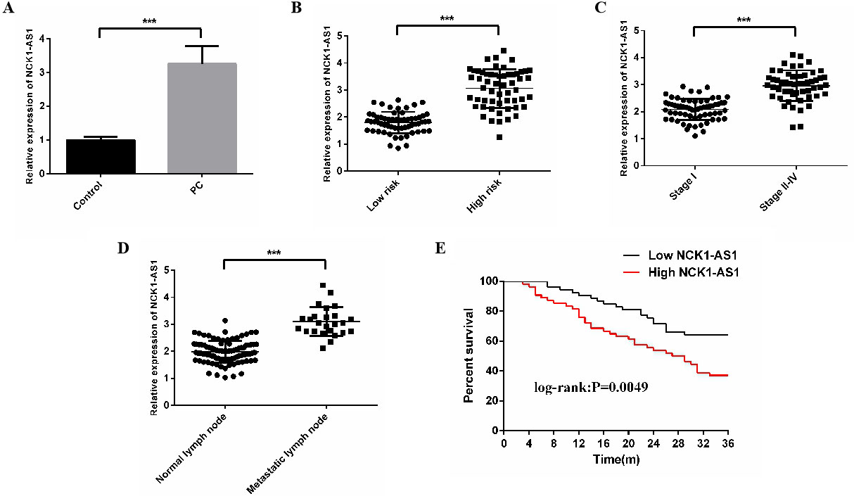
Cell cycles: All groups of the transfected 22Rv1 cell line were taken out and prepared into a single cell suspension. Then, there were inoculated on a 6-well plate at
Apoptosis: The transfected cells in each group were taken out, digested with trypsin, cleaned with pre-cooled PBS for twice, and then centrifuged. Those cells were collected, added with 1
Statistics and analysis
The data were statistically analyzed by SPSS 20.0 (Asia Analytics Formerly SPSS China) and plotted by GraphPad Prism8.0. The measurement data were expressed by mean
Results
LncRNA NCK1-AS1 expression in PCa patients
Compared with the adjacent normal tissues, LncRNA NCK1-AS1 expression rose in the prostate cancer tissues, and the difference in the expression was statistically marked between high-risk and low-risk patients. The patients were grouped by TNM staging. The expression in patients of stages II-IV was higher than that in those of stage I; the expression in cancer tissues with lymphatic metastasis was higher than that in normal lymphatic tissues (
Correlation of LncRNA NCK1-AS1 expression with clinicopathological features of PCa patients
In this part, the correlation of LncRNA NCK1-AS1 with pathological features of 116 PCa patients was statistically analyzed (Table 1). High LncRNA NCK1-AS1 was related to Gleason scores (
Correlation of LncRNA NCK1-AS1 expression with clinicopathological features of PCa patients
Correlation of LncRNA NCK1-AS1 expression with clinicopathological features of PCa patients
LncRNA NCK1-AS1 expression was compared between different PCC lines. Compared with the human normal prostate cells, LncRNA NCK1-AS1 showed high expression in the PCCs, with the greatest difference in 22Rv1 cell lines. Therefore, the 22Rv1 cell lines were selected for subsequent experiments. After cell transfection, compared with the Blank group, LncRNA NCK1-AS1 expression, cells’ proliferation, and the number of plate cloned cells remarkably reduced in the sh-NCK1-AS1 group (
LncRNA NCK1-AS1 expression in PCCs and cell proliferation. A: LncRNA NCK1-AS1 expression in PCCs. B: LncRNA NCK1-AS1 expression in 22Rv1 cells. C: Proliferation of 22Rv1 cells. D: The number of plate cloned 22Rv1 cells. Compared with the Blank group, LncRNA NCK1-AS1 expression, cells’ proliferation, and the number of plate cloned cells remarkably reduced in the sh-NCK1-AS1 group (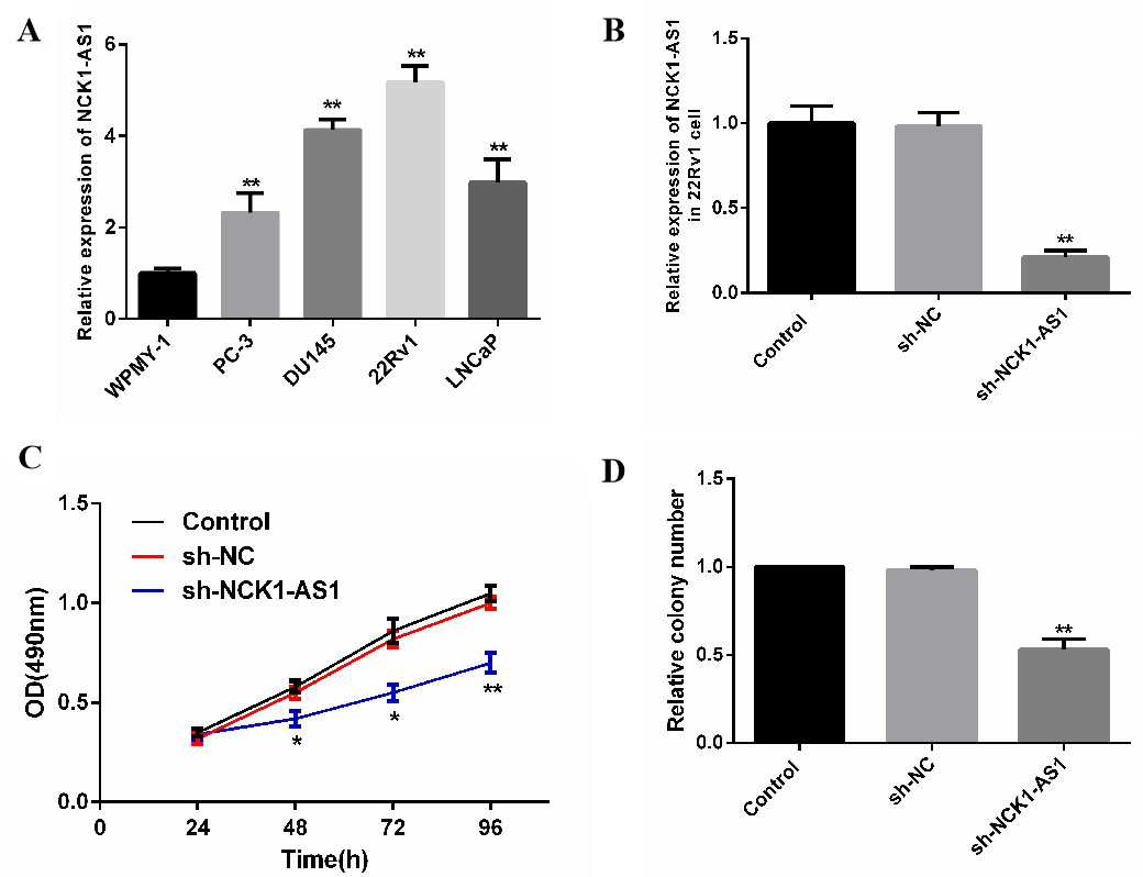
The invasion and migration of cells were compared between groups. Compared with the Blank group, the number of invasive and migratory cells and MMP-2 expression remarkably reduced, but E-cadherin expression remarkably rose in the sh-NCK1-AS1 group (
Effects of LncRNA NCK1-AS1 on invasion and migration of PCCs. A: The number of invasive cells in each group. B: The number of migratory cells in each group. C: E-cadherin expression in each group. D: MMP-2 expression in each group. Compared with the Blank group, the number of invasive and migratory cells and MMP-2 expression remarkably reduced (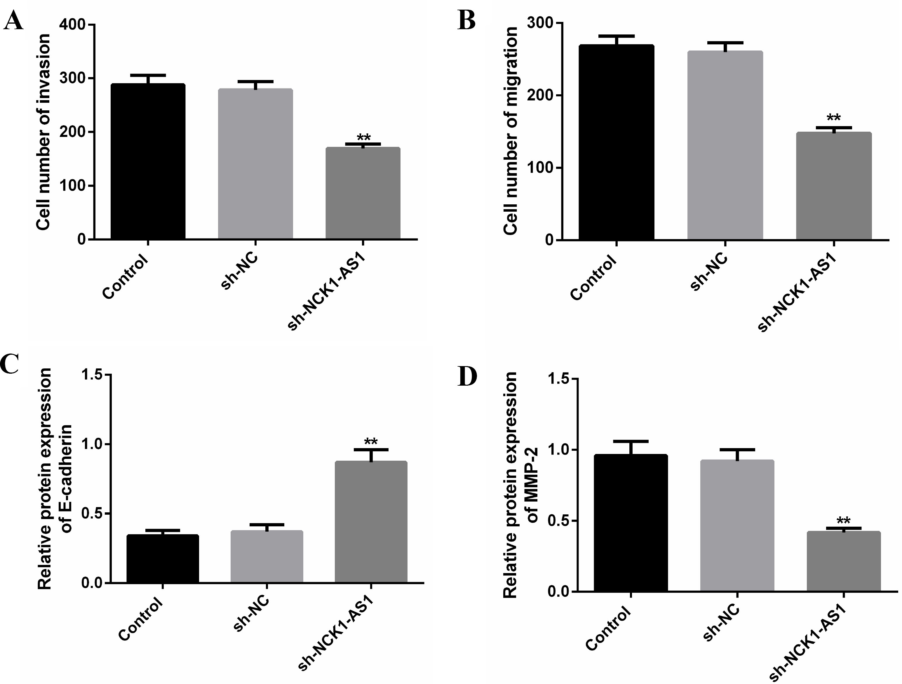
The ratio of cell cycles was compared between groups. Compared with the Blank group, the cancer cells in G0/G1 phase remarkably rose, but those in S and G2/M phases remarkably reduced in the sh-NCK1-AS1 group. This suggests that cell cycles are arrested and cell growth is inhibited. Additionally, the expression of CDK4, CDK6 and cyclin D1 remarkably reduced, but the apoptotic rate and Bax expression remarkably rose in the sh-NCK1-AS1 group (
Effects of LncRNA NCK1-AS1 on cell cycles and apoptosis of PCCs. A: Comparison of ratio of cell cycles between groups. B: Comparison of apoptotic rate between groups. C: Comparison of CDK4, CDK6 and cyclin D1 expression between groups. D: Comparison of Bax expression between groups. Compared with the Blank group, the expression of CDK4, CDK6 and cyclin D1 remarkably reduced, but the apoptotic rate and Bax expression remarkably rose in the sh-NCK1-AS1 group (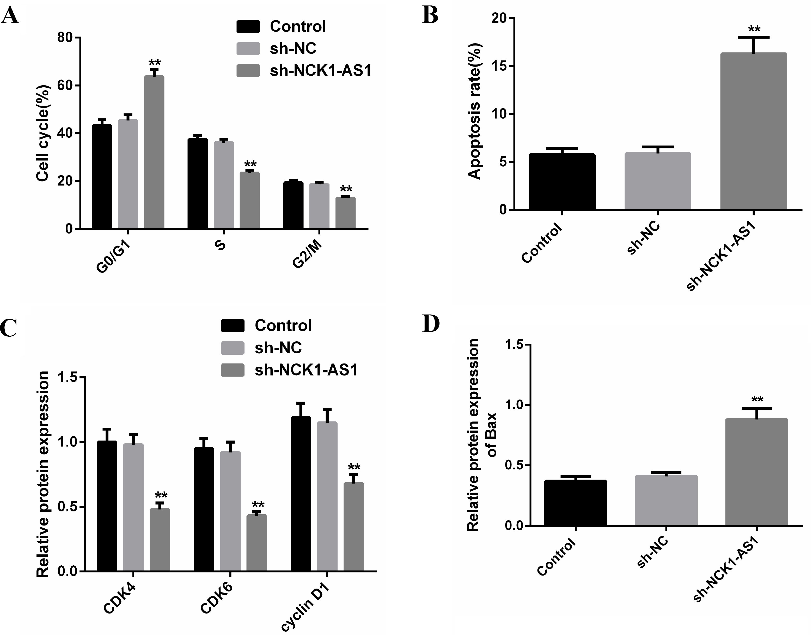
LncRNAs, which are by-products of RNA polymerase II, contain multiple stop codons or have no effective open reading frames, so they cannot encode proteins. They are involved in transcriptional regulation, epigenetic regulation and post-transcriptional regulation in the form of RNAs [16]. According to a large number of studies, they have a close correlation with tumor development and progression, especially PCa. It has been found that XIST [17], PVT1 [18], GAS5 [19] and FALEC [20] are correlated with the development and progression of PCa, so LncRNAs provide important molecular markers and therapeutic targets for disease pathogenesis, diagnosis, treatment and prognosis. However, the expression and mechanism of action of LncRNA NCK1-AS1 in the disease have not been widely reported.
In our study, LncRNA NCK1-AS1 expression in PCa and its effects on the proliferation and invasion of PCCs were explored. Compared with the adjacent normal tissues, LncRNA NCK1-AS1 was highly expressed in the prostate cancer tissues. Its expression was different in those with different stages of TNM and with lymphatic metastasis or not. The prognosis of patients with high LncRNA NCK1-AS1 expression was poorer than that of those with low expression. This indicates that LncRNA NCK1-AS1 can be used as a potential gene of PCa. Compared with the human normal prostate cells, LncRNA NCK1-AS1 expression in the human PCCs rose, with the greatest difference in 22Rv1 cells. Compared with the Blank group, cell proliferation, the number of plate cloned cells, and the expression of CDK4, CDK6 and cyclin D1 reduced in the sh-NCK1-AS1 group, in which cell cycles were arrested. Cell proliferation leads to an increase in the number of cells through cell cycles [21]. Uncontrolled cell proliferation cycle and cell dedifferentiation are important characteristics of malignancy cells. Core proteins that regulate cell cycles are cyclins, cyclin dependent kinases (CDKs) and cyclin dependent kinase inhibitors (CDIs). CDKs form various complexes with different cyclins, act on different phases of cell cycles, and determine cell cycle progression. CDIs act on the cyclin/CDK complexes, cause their inactivation, and eventually arrest cell cycles. cyclin D1, CDK4 and CDK6 are important proteins that affect the G1/S phase transition of cell cycles, and their interaction causes cells to enter S phase from G1 phase [22]. Silencing NCK1-AS1 inhibits the DNA synthesis of prostate cells, who are blocked during G1/S or G2/M transition, thereby delaying the process of cell cycles. After silencing NCK1-AS1, the number of invasive and migratory cells and MMP-2 expression reduced, but E-cadherin expression rose. As a member of the cadherin family, E-cadherin not only maintains the physical connection between cells, but also has a great effect on maintaining characteristics of epithelial cells. Its loss results in the loss of polarity and adhesive capacity between epithelial cells, presenting characteristics of non-epithelial cells. The decrease in its expression reduces the adhesive capacity between cells and makes them easy to fall off [23]. As reported by a previous study, E-cadherin expression is positively correlated with the differentiation degree of various tumors, and is involved in tumor invasion and migration [24]. Matrix metalloproteinases (MMPs) are zinc-dependent endonucleases that can promote the degradation of extracellular matrix and basement membrane. The upregulation of their expression enhances the invasion and migration of various cancer cells. As a member of the MMP family, MMP-2 has high expression in many tumors (such as PCa and breast cancer). Its high expression can promote tumor invasion and migration [25]. Besides, after NCK1-AS1 was down-regulated, the apoptotic rate and Bax expression rose. Bcl-2 family exerts a great function in the process of apoptosis. Bax is a pro-apoptotic gene in this family, and up-regulating its expression can induce tumor cell apoptosis [26].
In summary, LncRNA NCK1-AS1 is highly expressed in PCa, and silencing it can inhibit 22Rv1 cells from proliferation and invasion. Therefore, LncRNA NCK1-AS1 can be used as a potential gene of PCa, and it’s essential to clinical diagnosis, treatment and prognosis evaluation.
Author contributions
Conception: Yuxin Li, Xiaohong Zhuang, Li Zhuang, Hongjian Liu
Interpretation or analysis of data: Yuxin Li, Xiaohong Zhuang, Li Zhuang, Hongjian Liu
Preparation of the manuscript: Yuxin Li, Xiaohong Zhuang, Li Zhuang, Hongjian Liu
Revision for important intellectual content: Yuxin Li, Xiaohong Zhuang, Li Zhuang, Hongjian Liu
Supervision: Yuxin Li, Xiaohong Zhuang, Li Zhuang, Hongjian Liu
Funding
None.
Ethic approval
Informed consent in accordance with the Declaration of Helsinki of 1975, revised in 2008 and after approval from the Qingdao Women and Children’s Hospital was obtained from all patients.
Informed consent
All patients provided informed written consent for all procedures in the study.
Footnotes
Acknowledgments
None.
Conflict of interest
No author has any conflicts of interest to report.