
Abstract
BACKGROUND:
Altered cadherin expression plays a vital role in tumorigenesis, angiogenesis and tumor progression. However, the function of protocadherin 17 (PCDH17) in breast cancer remains unclear.
OBJECTIVE:
Our target is to explore PCDH17 gene expression in breast carcinoma tissues and its relation to serum angiopoietin-2 (Ang-2), carbonic anhydrase IX (CAIX) and % of circulating CD34
METHODS:
This study included Fifty female BCPs and 50 healthy females as control group. Cancerous and neighboring normal breast tissues were collected from BCPs as well as blood samples at diagnosis. PCDH17 gene expression was evaluated by RT-PCR. Serum Ang-2, CAIX levels were measured by ELISA and % CD34
RESULTS:
PCDH17 was downregulated in cancerous breast tissues and its repression was significantly correlated with advanced stage and larger tumor size. Low PCDH17 was significantly correlated with serum Ang-2, % CD34
CONCLUSIONS:
PCDH17 downregulation correlated significantly with increased angiogenic and hypoxia biomarkers. These results explore the role of PCDH17 as a tumor suppressor gene inhibiting tumor growth and proliferation.
Introduction
Breast cancer is the most prevalent malignancy among females and the world’s second major cause of cancer death among females [1]. Despite multidisciplinary treatment advances for breast cancer, thirty percent of treated patients later relapse, and more than 450 000 die every year [2].
Cadherins are transmembrane glycoproteins, which trigger calcium-dependent cell-cell adhesion. Throughout life, Cadherin-mediated adhesion controls cell growth and differentiation. These proteins are further subdivided into classical cadherins, protocadherins (PCDHs), and cadherin-related proteins [3]. Aberrant expression of protocadherins has been shown to be associated with multiple tumorigenesis [4]. Protocadherin 17 (PCDH17) gene has been located on chromosome 13q21.1 which plays important roles in the regulation of cell adhesion and signal transduction. However, its function in breast cancer remains unknown [5].
Angiogenesis, the growth of new blood vessels, is one of the cancer’s hallmarks. For the initiation of angiogenesis, tumor cells produce and release proangiogenic factors resulting in tumor growth, invasion and metastasis [6]. Numerous physiological and pathological stimuli are used to develop new blood vessels in cancer settings, where hypoxia is the key stimulus [7]. Hypoxia induced angiogenesis is primarily mediated by Vascular Endothelial Growth Factor (VEGF) through activation of the signaling pathway for prolyl-hydroxylase-hypoxia inducible factor (HIF) [8].
Angiopoietin-2 (Ang-2), a cytokine highly expressed in tumors primarily by endothelial cells as well as some malignant cells, promotes tumor angiogenesis in combination with other proangiogenic factors, particularly VEGF. In fact, Ang-2 is upregulated by tumor hypoxia [9].
Cluster of differentiation (CD) 34 is a single-chain transmembrane glycoprotein with a molecular weight of 105–120 kDa, its gene is present on the long arm of chromosome 1. CD34 is an endothelial cell-specific marker, mainly expressed on endothelial and hematopoietic progenitor cells, and closely linked with the process of angiogenesis [10, 11].
Carbonic anhydrase IX (CAIX), one of the 15 carbonic anhydrase (CA) isoforms found in humans, is a membranous metalloenzyme that facilitates the reversible hydration of carbon dioxide to bicarbonate and protons. CAIX is controlled by hypoxia inducible factor-1 alpha (HIF-1
The main objective of this work was to explore PCDH17 gene expression in breast carcinoma tissues and its relation to serum angiopoietin-2, percent of circulating CD34
Patients and methods
This study included 50 female patients newly diagnosed with breast cancer as patients group and 50 healthy females with normal mammography findings and no previous history of any kind of cancer as a control group, matched for age and menopausal status with patients group.
Patients were chosen from those presented to the Department of Cancer Management and Research and Experimental and Clinical Surgery Department, Medical Research Institute, Alexandria University. According to the Declaration of Helsinki, written consent was obtained for participation in the study and approved by the Ethical Committee of the Medical Research Institute, Alexandria University. Metastatic patients and patients undergoing chemotherapy before surgery were excluded from this study.
Patients underwent preoperative assessment, which included history taking, clinical examination to determine tumor location and the presence of swollen axillary lymph nodes. Patients were subjected to radiological examinations included mammogram, abdominal ultrasound and chest x-ray and fine needle aspiration cytology (FNAC) to diagnose the existence of malignancy. Patients underwent surgery (modified radical mastectomy or conservative surgery) followed by pathological evaluation of the tumor included tumor type, grade, tumor size, numbers of axillary lymph nodes involved, and presence or absence of vascular invasion. Assessments of estrogen, progesterone receptors (ER, PR) and Her2/neu expression were also confirmed.
Tissue samples collection
Tumor tissues and adjacent normal breast tissues were collected from patients with breast cancer at the time of surgery and frozen at
Blood samples collection
A total of 5 ml fasting venous blood sample was taken from each control participant and within a week before surgery for breast cancer patients. This sample was subdivided into two aliquots. The first aliquot was allowed to clot for 30 minutes before centrifugation, centrifuged at 3000 rpm for 10 minutes to isolate sera. Serum was stored at
Determination of serum carbonic anhydrase IX levels by ELISA
Principle
The assay is based on sandwich enzyme-linked immunosorbent technology. Anti-CAIX antibody was adsorbed onto the microwells. Human CAIX present in the sample or standard was bound to antibodies adsorbed to the microwells. A biotin conjugated anti-human CAIX was added to human CAIX captured by the first antibody. After incubation unbound biotin-conjugated anti-human CAIX antibody was removed during a wash step. Streptavidin-Horseradish peroxidase (HRP) was added and bound to the biotin-conjugated anti-human CAIX antibody. Following incubation unbound streptavidin-HRP was removed during a wash step, and substrate solution reactive with HRP was added to the wells. A coloured product was formed in proportion to the amount of human CAIX present in the sample or standard. The reaction was stopped by acid addition and absorbance was measured at 450 nm.
Determination of serum Angiopoietin-2 levels by ELISA
Principle
The assay is based on sandwich enzyme-linked immunosorbent technology. Anti-Ang-2 antibody was adsorbed onto the microwells. Human Ang-2 present in the sample or standard was bound to antibodies adsorbed to the microwells. A biotin conjugated anti-human Ang-2 was added to human Ang-2 captured by the first antibody. Following incubation unbound biotin-conjugated anti-human Ang-2 antibody was removed during a wash step. Stertavidin-Horseradish peroxidase (HRP) was added and bound to the biotin-conjugated anti-human Ang-2 antibody. After incubation unbound streptavidin-HRP was removed during a wash step, and substrate solution reactive with HRP was added to the wells. A coloured product was formed in proportion to the amount of human Ang-2 present in the sample or standard. The reaction was stopped by acid addition and absorbance was measured at 450 nm.
Determination of circulating CD34
cells by flow cytometry
Peripheral blood samples obtained from all subjects under study were used for assaying CD34 expression which is expressed on the endothelial cells and the hematopoietic progenitor cells. The level of CD34 was measured by BD FACS Canto II flow cytometer provided with FACS Diva software using relevant monoclonal antibody tagged with a fluorescent marker.
Statistical analyses
Data were supplied to the computer and analyzed using IBM SPSS software package version 20. (Armonk, New York: IBM Corp). Comparison between two independent populations were done using independent
Results
Clinico-pathological characteristics of breast cancer patients
General characterizations of breast cancer patients were represented in Table 1.
Clinico-pathological characteristics of breast cancer patients
Clinico-pathological characteristics of breast cancer patients
Abbreviations: ER: estrogen receptor, PR: progesterone receptor, Her-2/neu: human epidermal growth factor receptor-2.
Tissue protocadherin 17 gene expression, angiopoietin-2, the percentage of circulating CD34
Protocadherin 17 gene expression in adjacent normal breast tissues and breast cancer tissues.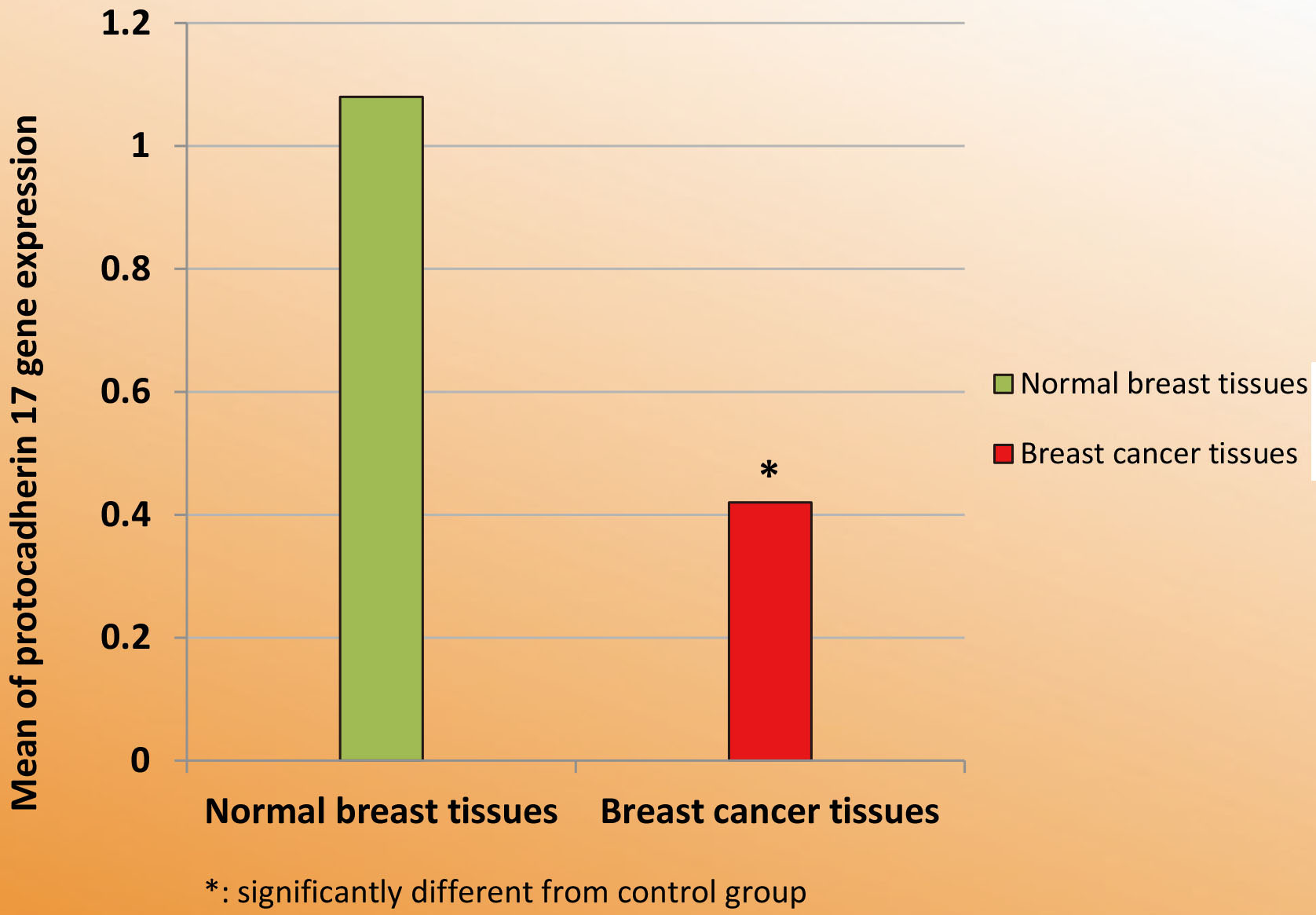
Values of relative quantification of PCDH17 gene expression were ranged from 0.47–2.4 with a mean value 1.08 in normal adjacent breast tissues. In breast cancer tissues, it ranged from 0.0–1.57 with a mean value 0.42 (Table 2, Fig. 1). Statistical analysis of this result revealed that PCDH17 gene expression in BC tissues was significantly lower than that in normal breast tissues (
Serum Angiopoietin-2
Range and mean
Serum angiopoietin-2 (pg/ml) in control group and breast cancer patients group.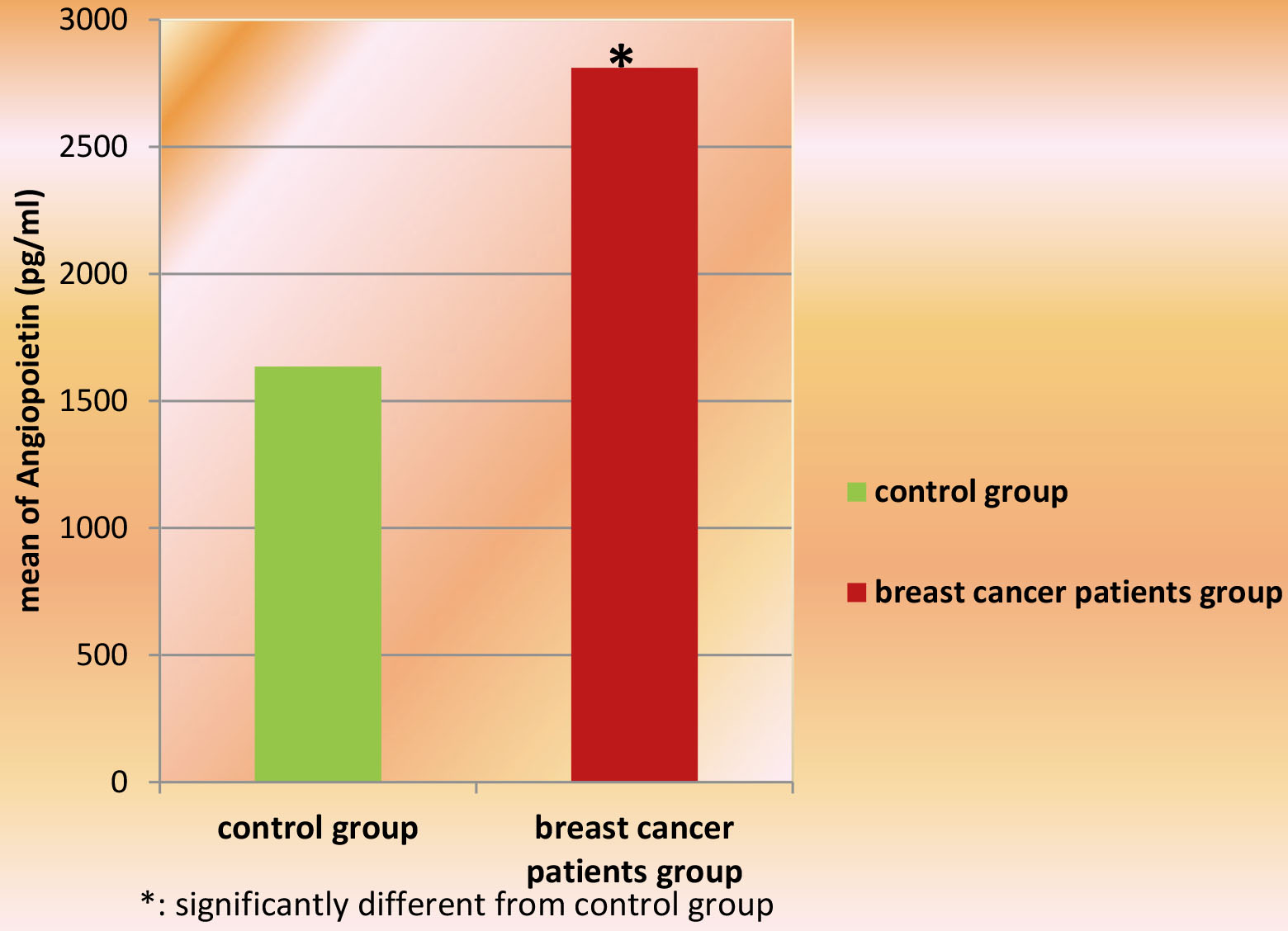
Range and mean of the percent (%) of CD34
Percent of CD34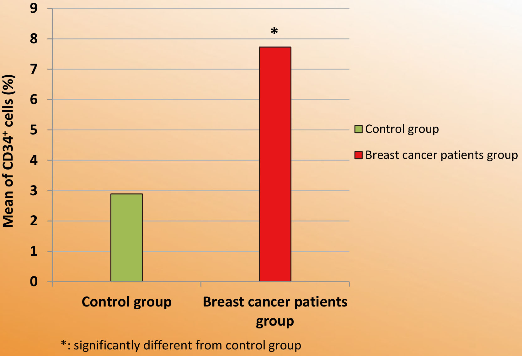
Range and mean
Serum CAIX (pg/ml) in control group and breast cancer patients group.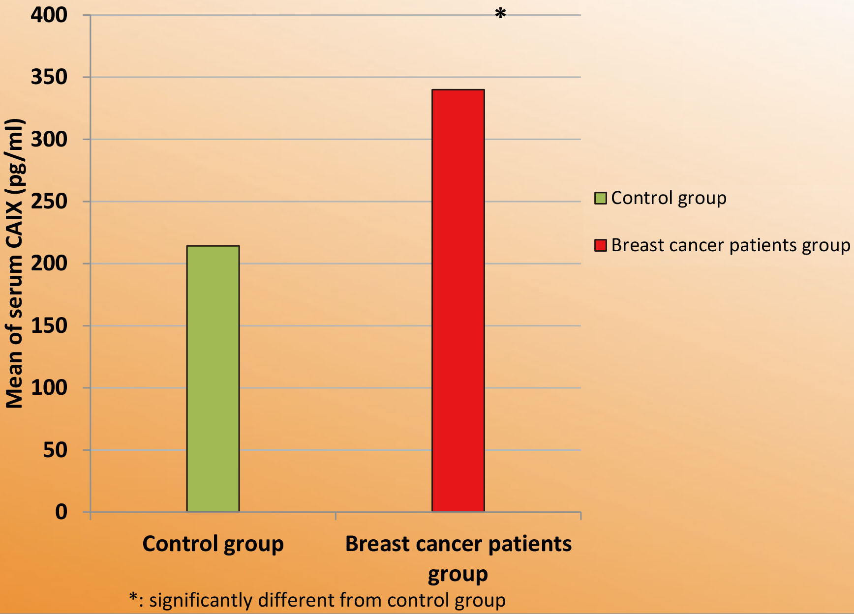
Correlations between studied biochemical parameters and clinicopathological characteristic in patients with breast cancer
r: Pearson coefficient.
According to Table 3, downregulation of PCDH17 gene was associated with increased serum CAIX and Ang-2 and higher percentage of CD34
Regarding serum Ang-2, it was significantly positively correlated with clinical stage, vascular invasion, axillary lymph node involvement, serum CAIX and the percentage of CD34
With respect to the percentage of CD34
Diagnostic performance of the studied parameters
Diagnostic performance of the studied parameters
With regards to serum CAIX, it was significantly positively correlated with clinical stage, tumor size, axillary lymph node involvement, serum Ang-2 and the percentage of CD34
The ROC curve analysis was used to assess the diagnostic values of the studied markers. The higher area under the Roc curve (AUC) corresponds to a better diagnostic test.
Graphical presentation of the Roc curves for serum CAIX, angiopoietin-2, CD34 and tissue PCDH17 in breast cancer patients.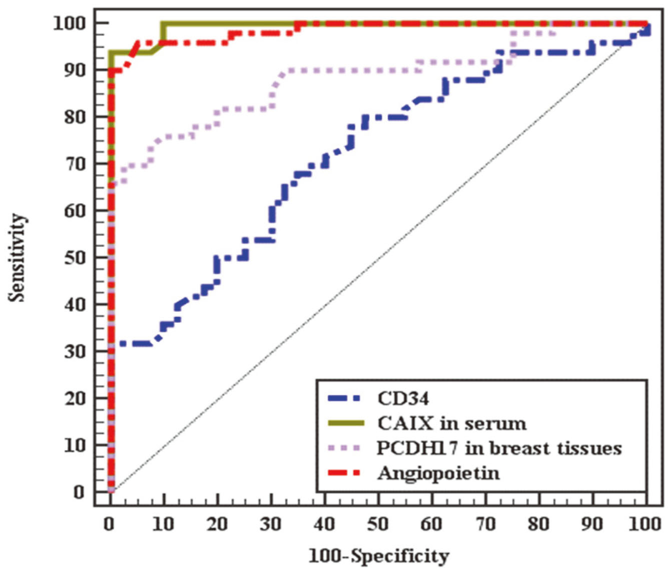
Table 4 and Fig. 5 showed that serum CAIX showed significant AUC (0.994,
Protocadherin 17 gene has been located on chromosome 13q21.1 and plays essential functions in the regulation of cell adhesion and signal transduction. However, little is known about its role in cancer. Our results showed that the expression of PCDH17 gene in breast cancer tissues was significantly lower than that in adjacent normal breast tissues. Suggesting that silencing of protocadherin 17 might encourage tumorigenesis. This finding agrees with previous studies recognized PCDH17 as a tumor suppressor gene (TSG) and is often down regulated [14, 15, 16, 17]. Several mechanisms have been proposed explaining PCDH17 downregulation including promoter methylation, mutation of PCDH17 gene, histones modifications, absence of transcriptional activators or the existence of transcriptional repressor that could contribute to gene silencing especially in primary tumors leading to loss of its tumor suppressive activity and initiating carcinogenesis. Hyper methylation of its promoter has been found in different cancer types including esophageal, renal, and digestive carcinoma [17, 18, 19].
Subsequently, we examined the relationship between PCDH17 expression and clinicopathologic parameters. Interestingly, PCDH17 downregulation significantly correlated with larger tumor size and advanced stage, which are risk factors for breast cancer progression and poor outcome. These results explore the role of PCDH17 as a tumor suppressor gene by inhibiting tumor growth and proliferation. These findings agree with Lin et al. who found that PCDH17 methylation is correlated with advanced pathological stage and poor clinical outcome in prostate cancer patients [20].
Furthermore, we examined the correlation between PCDH17 expression and angiogenesis and hypoxia biomarkers. We found that downregulation of PCDH17 is correlated significantly with increased angiogenesis biomarkers Ang-2 and CD34. In addition, decreased PCDH17 expression is also correlated with increased hypoxia marker CAIX. These findings suggest that PCDH17 act as an endogenous inhibitor of angiogenesis. He et al. [16] stably transfected the PCDH17 gene into CNE1 cells and found that ectopic expression of PCDH17 inhibits angiogenesis and VEGF secretion. He concluded that PCDH17 inhibits the growth, migration, and invasion of nasopharyngeal carcinoma cells and was involved in the cell cycle, apoptosis, angiogenesis, and in vivo tumor formation.
Hypoxia was found to be linked with malignant initiation, progression, increasing the occurrence of metastasis and therapy resistance in many cancer types [21]. Hypoxia is an efficient angiogenesis-driving force which represents a compensable mechanism against the ischemia of the tissue [22]. Hypoxia stimulates CAIX expression, a transmembrane zinc metalloenzyme. In addition to its role in regulating pH, it has function in cell adhesion, growth and tumor cell survival under normoxia and hypoxia [23, 24]. In addition to the cell-associated membrane protein, a soluble isoform of CAIX is released by proteolytic cleavage and can be detected in the serum [25].
Based on the result of the current study, the statistical analysis of the data indicates that serum CAIX was significantly higher in breast cancer patients than healthy control subjects. The elevated levels of serum CAIX may be attributed to its expression and secretion from breast cancer cells and stromal cells [26]. The present result is in line with the study done by Takcova et al. [27] who found that serum CAIX levels determined by ELISA were significantly higher in clear cell renal cell carcinoma patients (CCRCC) than in non CCRCC. Similarly, Finkelmeier et al. [28] reported elevated CAIX level in hepatocellular carcinoma (HCC) patients compared to healthy controls. Moreover, our results demonstrated that high levels of CAIX were positively significantly correlated with clinical stages of patients and axillary lymph node involvement. This finding indicated that CAIX may be considered as a biological marker reflecting unfavorable prognosis of BC patients. This correlation was proven in previous study carried out by Finkelmeier et al. [28] who reported that high serum level of CAIX in HCC patients was correlated with higher clinical stage. Similarly, the concentration of this parameter in serum of BCPs was significantly positively correlated with tumor size. Which hypothesize that elevated levels of serum CAIX function to promote cell growth of tumor cells. This hypothesis is coincided with that reported by Takcova et al. [27].
CD34 is a highly glycosylated transmembrane protein molecule. CD34 is especially expressed in vascular endothelial cells and closely linked with the process of angiogenesis. Furthermore, it is an indicator of tumor neovascularization during the growth of a tumor [10, 11]. The current study demonstrated that the percentage of CD34
Importantly, it has been shown that VEGF and Ang-2 function synergistically to cause endothelial destabilization, enhance vascular branching and boost angiogenesis [32]. The present study showed that serum Ang-2 was highly elevated in BCPs in comparison to control group. Moreover, we found that higher serum Ang-2 levels were significantly positively correlated with clinical stage, axillary lymph node involvement, and vascular invasion, suggesting that Ang-2 might be involved in breast cancer metastasis. Accumulating evidence has shown a link between increased expression of Ang-2 and invasive metastatic phenotypes of different kinds of human cancers including breast cancers [33, 34]. For example, Sallinen et al. [35] found that Ang-2 levels were significantly elevated in serum samples of patients with ovarian carcinoma compared with healthy controls and correlated with decreased patients’ survival. Li et al. [36] showed that the levels of serum Ang-2 were significantly increased in patients with breast cancer compared with healthy controls and significantly positively correlated with histological grade, lymph node involvement, and clinical stage.
In the current study, a significant positive correlation was found between angiogenesis biomarkers Ang-2 and CD34 with serum CAIX which highlights the role of hypoxia in stimulating angiogenic markers. For a tumor to grow, there is necessary to stimulate vasculogenesis and/or neoangiogenesis. This ability of tumor cell encompasses a multistep process, called the ‘angiogenic switch’, a phenomenon occurs when the balance of pro-angiogenic factors outweighs anti-angiogenic factors. HIF can promote vascularization process through induction of the expression of a large number of pro-angiogenic factors, including VEGF [37, 38].
The significant evaluation of serum CAIX, serum Ang-2, % of CD34
Conclusion
PCDH17 gene is downregulated in breast carcinoma tissues and its silencing correlated significantly with advanced stage and larger tumor size. Downregulation of PCDH17 is correlated significantly with increased angiogenic and hypoxia biomarkers. These results explore the role of PCDH17 as a tumor suppressor gene inhibiting tumor growth and proliferation. Serum CAIX, serum Ang-2 and percent of circulating CD34 Serum CAIX and Ang-2 may act as non-invasive biomarkers for BC screening and diagnosis with high sensitivity and specificity.
Recommendations
Further large-scale studies are warranted to investigate the new opportunity for PCDH17 targeted therapy to prevent angiogenesis and to suppress invasion and metastasis.
Author contributions
Sanaa A. El-Benhawy: Conception, Preparation of the Manuscript and interpretation of data.
Samia A. Ebeid: Supervision and interpretation of data.
Nadia A. Abd El Moneim: Supervision and revision for important intellectual content.
Rabie R. Abdel Wahed: Supervision and revision for important intellectual content.
Amal R.R. Arab: Conception, Preparation of the Manuscript and analysis of data.
Footnotes
Acknowledgments
All authors have made significant contributions to this work.
Conflict of interest
No conflict of interest is declared.