
Abstract
BACKGROUND:
Non-small cell lung cancer (NSCLC) is the major subtype of lung cancer, imposing a huge disease burden worldwide. MicroRNA-1303 (miR-1303) has been demonstrated to be involved in several diseases, including cancers. In this study, we aimed to investigate the role of miR-1303 in NSCLC.
METHODS:
We quantified the expression levels of miR-1303 in NSCLC tissues and cells using the qRT-PCR assay. Then the association between miR-1303 expression and clinical characteristics of patients was analyzed using the
RESULTS:
The expression of miR-1303 was upregulated in NSCLC tissue samples and cells. The upregulation of miR-1303 was associated with TNM stage and lymph node metastasis. The survival time of NSCLC patients with high expression of miR-1303 was shorter than those with low expression. The functional analyses revealed that overexpression of miR-1303 in H1299 and A549 cells promoted cell proliferation, migration, and invasion.
CONCLUSION:
These results suggest that miR-1303 may be a potential prognostic biomarker for NSCLC and be involved in the progression of NSCLC.
Introduction
Lung cancer, an aggressive tumor with high morbidity and mortality, remains the leading cause of cancer-related deaths around the world [1]. Histologically, non-small cell lung cancer (NSCLC) and small cell lung cancer (SCLC) are the two major types of lung cancer, in which NSCLC comprises more than 80% of all lung cancer cases [2]. The environmental tobacco smoke exposure, family history of lung cancer, and air pollution are the main risk factors for the initiation and progression of NSCLC [3, 4]. In addition, metastasis is the main factor leading to disease progression and poor prognosis of NSCLC patients [5]. In recent years, although available therapies (such as surgery, chemotherapy, and radiotherapy) for the treatment of NSCLC have been developed, the 5-year overall survival rate of patients is still less than 20%, which was not significantly improved [6]. Therefore, searching for more molecules and genes associated with the development of NSCLC is particularly important for the treatment of NSCLC.
MicroRNAs (miRNAs) are small (appreciate 21 nucleotides long), non-coding, single-stranded RNAs that negatively regulate gene expression by binding the 3’-UTR of target mRNAs [7, 8]. Abnormal expression of miRNAs has been reported in various types of diseases, including cancers, where they may function as oncogenes or suppressors [9, 10]. Moreover, a number of studies have revealed that aberrant expression of miRNAs might function as diagnostic and/or prognostic biomarkers in various tumors [11, 12]. Importantly, miRNAs play crucial roles in various biological processes, including cellular growth and proliferation, differentiation, metastasis, and apoptosis [13, 14]. In NSCLC, miR-187 [15], miR-593-5p [16], and miR-141 [17] have been reported aberrantly expressed, and acted an oncogenic or a suppressive role to regulate cell proliferation, invasion, and metastasis. Previous researches have also indicated that miR-1303 expression was involved in the development of several diseases [18, 19], including cancers, such as hepatocellular carcinoma [20] and esophageal squamous cell carcinoma [21]. A miRNA expression profiling study revealed that numerous miRNAs were differentially expressed in NSCLC tissues versus adjacent normal tissues, including miR-1303 [22]. However, the clinical significance and functional role of miR-1303 in NSCLC remain largely unknown.
In this study, we detected the miR-1303 expression levels in NSCLC tissues and adjacent normal lung tissues, as well as NSCLC cell lines and normal epithelial cells. Based on its expression and survival information, we explored the clinical prognostic value of miR-1303 in NSCLC. Furthermore, we investigated the functional role of miR-1303 in NSCLC. Our results suggest that miR-1303 might play an oncogenic role and be a potential prognostic biomarker and therapeutic target in NSCLC.
Materials and methods
Patients and tissue samples
The present study was approved by the Ethical Review Committee of Liaocheng People’s Hospital. All the NSCLC patients enrolled in the study signed written informed consent before tissue collection. The patients were recruited based on the following inclusion criteria: (1) All the patients did not undergo preoperative therapies (chemotherapy or radiotherapy). (2) All the cases were pathologically diagnosed as NSCLC. (3) The patients who were at early or middle stages, and those at advanced stages, whose lesion is confined to one side of the thorax and can be completely removed, or with solitary metastasis. A total of 113 paired NSCLC tissue samples and adjacent non-tumor tissue samples (approximately 5 cm away from the edge of the lung tumor) were collected from patients who received surgical resection at Liaocheng People’s Hospital during the study. These tissue specimens were immediately frozen in liquid nitrogen after resection until RNA extraction. The clinical medical records of NSCLC patients were collected and corresponding clinical characteristics were recorded in Table 1. The TNM stage of the tumors was confirmed following the 7
Cell culture and transfection
Human NSCLC cell lines (H1299, A549, NCI-H1650, HCC827) and immortalized bronchial epithelial cell BEAS-2B were purchased from the cell bank of Chinese Academy Sciences (Shanghai, China). All cells were cultured in DMEM medium (Gibco, NY, USA) supplemented with 10% FBS (Gibco, Grand Island, NY, USA) in a humidified incubator containing 5% at 37
miR-1303 is upregulated in NSCLC tissue samples and cells. A. Relative expression of miR-1303 in NSCLC tissue samples and corresponding normal tissue samples. B. Expression of miR-1303 in four NSCLC cell lines and normal epithelial cell BEAS-2B. 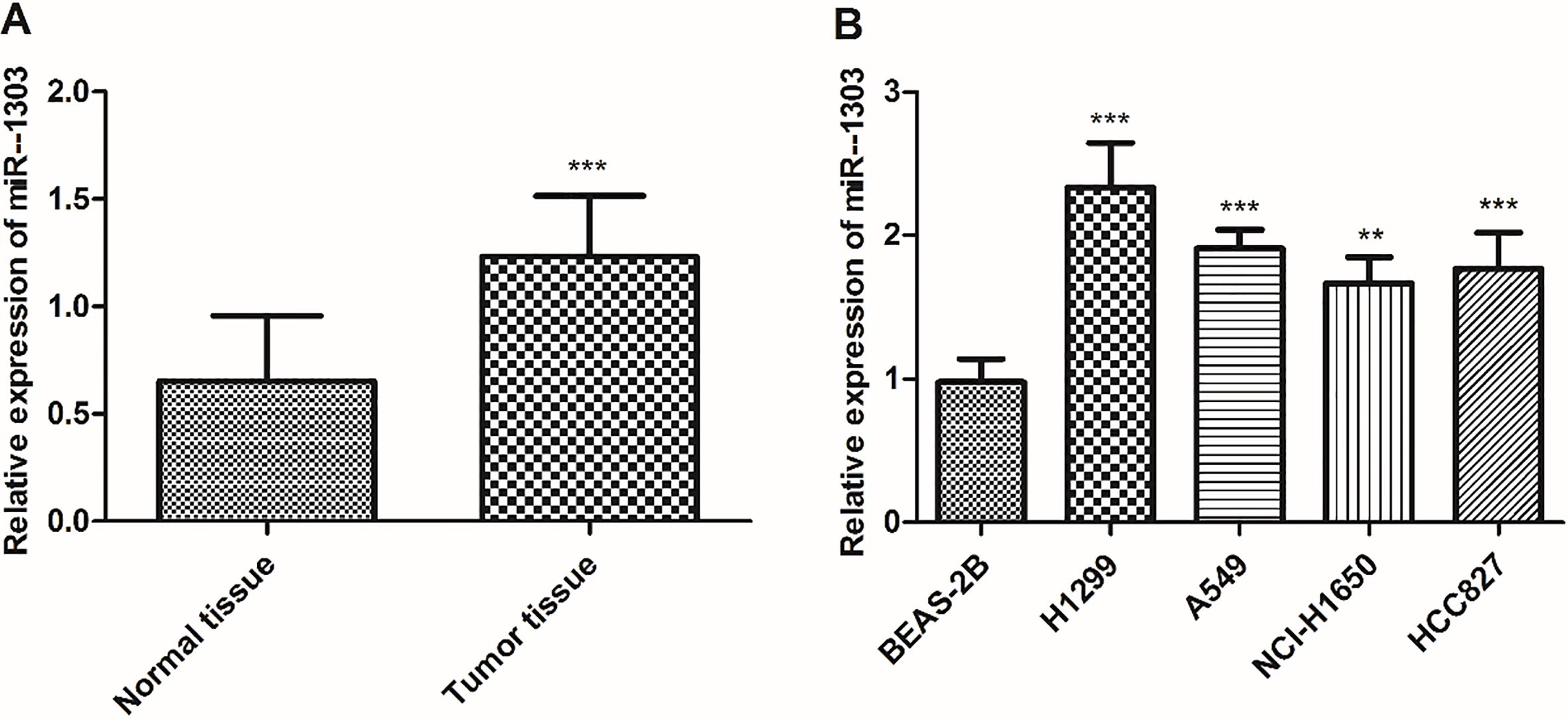
Before transfection, NSCLC cells were seeded into 6-well plates and incubated overnight. The miR-1303 mimics, mimic negative control (mimic NC), miR-1303 inhibitors, inhibitor NC were chemically synthesized by GenePharma Co., Ltd. (Shanghai, China). Then the cells were transfected with miR-1303 mimic, miR-1303 inhibitor, or their corresponding NCs using Lipofectamine 3000 (Invitrogen, Carlsbad, CA, USA) following the manufacturer’s protocol. After 24 h transfection, further experiments were performed.
The total RNA was extracted from patients’ tissue specimens and cultured cells using TRIzol reagent (Thermo Fisher Scientific, Waltham, MA, USA) according to the manufacturer’s instructions. Then RNA was reverse transcribed to cDNA using the TaqMan miRNA reverse transcription kit (Applied Biosystems, Thermo Fisher Scientific) according to the manufacturer’s instructions. Subsequently, the qRT-PCR assay was carried out using SYBR Premix Ex Taq II (TaKaRa, Dalian, China) on a 7500 Real-Time PCR System (Applied Biosystems, USA). The expression levels of miR-1303 were quantified using the 2
Cell proliferation assay
The cell proliferation of A549 and H1299 cells was assessed using cell counting kit-8 (CCK-8; Dojindo, Tokyo, Japan). Briefly, transfected cells were seeded into 96-well plates (4
Cell migration and invasion assays
The migratory and invasive capabilities of A549 and H1299 cells were detected using a 24-well Transwell chamber (8
Correlation of the miR-1303 expression with clinical characteristics in NSCLC
Correlation of the miR-1303 expression with clinical characteristics in NSCLC
All the experiments were repeated at least three times, and all the data in the present study were expressed as mean
Results
miR-1303 is upregulated in NSCLC tissues and cells
First, the expression of miR-1303 in NSCLC tissue specimens was measured using the qRT-PCR assay. As shown in Fig. 1A, miR-1303 was significantly upregulated in NSCLC tissue specimens compared with adjacent normal tissues (
Kaplan-Meier method was used to analyze the 5-year survival rate of NSCLC patients with high and low expression of miR-1303. Patients with high miR-1303 expression exhibit a shorter overall survival rate than those with low miR-1303 expression (log-rank test 
Based on the relative expression of miR-1303 in NSCLC tissues, we also analyzed the relationship between miR-1303 expression and clinical characteristics of NSCLC patients, and 113 NSCLC patients were separated into low miR-1303 expression group (
Increased expression of miR-1303 associates with prognosis in NSCLC
To determine whether the miR-1303 expression has the clinical prognostic significance in NSCLC, the Kaplan-Meier curve was conducted using the survival information of patients. As shown in Fig. 2, patients with high miR-1303 expression levels had a shorter overall survival time, whereas those with low miR-1303 expression exhibited longer survival time (log-rank test,
Overexpression of miR-1303 promotes cell proliferation, while inhibition of miR-1303 suppresses cell proliferation of H1299 and A549 cells, compared with untreated cells. A. miR-1303 expression was determined by qRT-PCR. miR-1303 expression in H1299 and A549 cells transfected with miR-1303 mimics or miR-1303 inhibitors. B. The CCK-8 assay was used to detect the effects of miR-1303 mimics or inhibitors on the cell proliferation of H1299 and A549 cells. 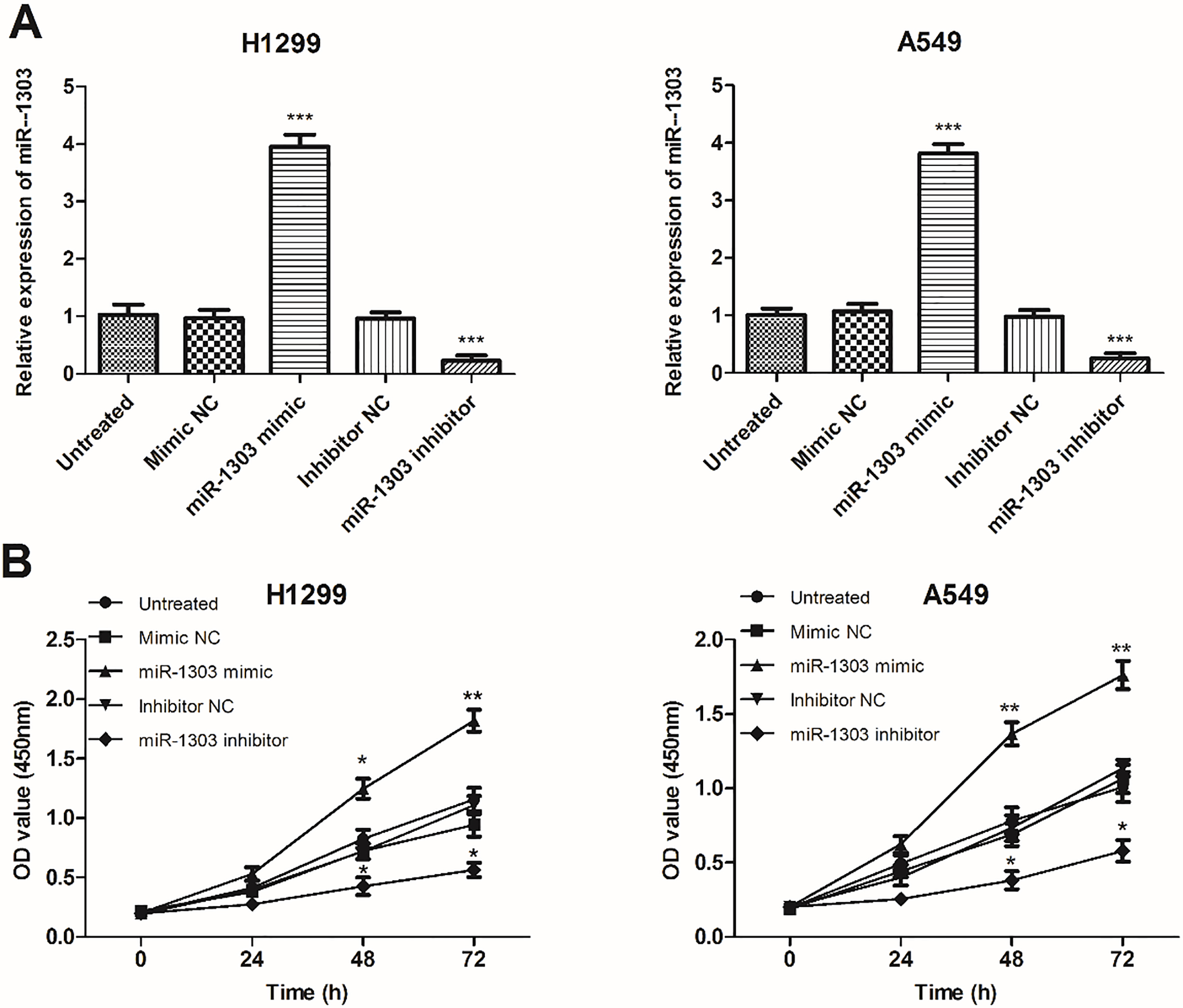
Multivariate Cox analysis of clinical characteristics in relation to overall survival
To study whether miR-1303 expression would affect the progression of NSCLC, subsequently, we explored the effects of miR-1303 in NSCLC by upregulating or downregulating miR-1303 expression in A549 and H1299 cells. Then qRT-PCR assay was used to confirm transfection efficiency. As expected, the expression of miR-1303 was significantly overexpressed after transfection of miR-1303 mimic in H1299 and A549 cells, while the expression of miR-1303 was significantly downregulated after transfection of miR-1303 inhibitor in H1299 and A549 cells (
Upregulation of miR-1303 promotes cell migration and invasion, while knockdown of miR-1303 inhibits cell migration and invasion of H1299 and A549 cells, compared with untreated cells. A. Transwell migration assay was used to detect the effects of miR-1303 on the cell migration of H1299 and A549 cells. B. Transwell invasion assay was used to detect the effects of miR-1303 on the cell invasion of H1299 and A549 cells. 
The development of NSCLC is a series of complex, multi-step and multi-factor process. In recent years, miRNAs have been reported that associated with the development, occurrence, and metastasis of various types of cancers [9, 23]. Moreover, miRNAs have gained growing research and considerable attention as therapeutic targets for the treatment of diseases, including cancers [24]. Thus, identifying novel markers as possible prognostic biomarkers or therapeutic targets may be helpful for improving the treatment of NSCLC.
miR-1303, located on 5q33.2, is an important miRNA that has been indicated to be aberrantly expressed in several types of diseases, including cancers, and involved in the development of several types of cancers [25, 26]. In the present study, we examined the expression of miR-1303 in NSCLC tissue samples and cell lines. The results showed that the expression of miR-1303 was significantly upregulated in NSCLC tissue samples and all the NSCLC cells, compared with corresponding normal tissue samples and BEAS-2B cells, respectively. The results suggest that miR-1303 may be a tumor suppressor in NSCLC. These results are consistent with a number of previous studies. For instance, miR-1303 was significantly overexpressed in gastric cancer tissues and cell lines [27].
Aberrantly expression of miRNAs was involved in the growth and development of cancers, which indicated the clinical potential of miRNAs as diagnostic and/or prognostic biomarkers [28, 29, 30]. For instance, miR-133b is downregulated in NSCLC, is involved in NSCLC metastasis, and may function as a biomarker for the diagnosis and prognosis of NSCLC [31]. A previous study used the metastasis-free survival rates information for analysis and demonstrated that miR-1303 was associated with metastasis-free survival in inflammatory breast cancer [26]. In this study, we used the overall survival information of NSCLC patients and further explored the potential clinical significance of miR-1303 in NSCLC. The results indicated that patients with high miR-1303 expression levels exhibited a shorter overall survival rate compared with those with low miR-1303 expression levels. Multivariate Cox regression assay revealed that miR-1303 expression was an independent prognostic predictor for NSCLC. The above results suggest that miR-1303 expression may be a novel prognostic biomarker for NSCLC.
To investigate the functional role of miR-1303 in NSCLC, gain/loss-of-function experiments were carried out to upregulate or downregulate the expression of miR-1303 in H1299 and A549 cells. The results indicated that overexpression of miR-1303 in NSCLC cells promoted the proliferative, migratory, and invasive capabilities of NSCLC cells, while downregulation of miR-1303 inhibited the proliferative, migratory, and invasive capabilities of NSCLC cells, compared with untreated cells. Previously, miR-1303 has been demonstrated to promote the proliferation, migration, and invasion of prostate cancer cells through activating the Wnt/
All in all, there are some limitations to the present study. The sample size in the present study is limited and the clinical prognostic significance of miR-1303 still needs to be confirmed using large scale samples. In addition, another limitation of this study is that the precise molecular mechanism of miR-1303 needs to be confirmed in future studies.
Taken together, our study demonstrated miR-1303 is upregulated in NSCLC tissues and cells, and its overexpression of miR-1303 promotes cell proliferation, migration, and invasion. NSCLC patients with high miR-1303 expression have a shorter overall survival rate than those with low miR-1303 expression. Thus, our results provide a novel insight into the prognosis and treatment of NSCLC, and miR-1303 may be associated with prognostic overall survival rates, indicating the great potential of miR-1303 as a therapeutic target for the treatment of NSCLC.
Footnotes
Conflict of interest
The authors declare that they have no conflict of interest.
Supplementary data
The expression of miR-1303 in NSCLC patients according to TNM stage
TNM stage
Cases No.
miR-1303 expression in tissue
I
39
1.144
0.277
II
35
1.176
0.301
III
36
1.368
0.216
IV
3
1.429
0.230