
Abstract
MicroRNAs (miRNAs) have been demonstrated that play a critical role in tumorigenesis. The aim of this study is to identify the functional role of miR-378 in cholangiocarcinoma (CCA). Quantitative real-time polymerase chain reaction (qRT-PCR) was used to measure the expression levels of miR-378 in human CCA tissue samples and CCA cell lines. The receiver operating characteristic (ROC) curve was established, and the area under the ROC curve (AUC) was calculated to estimate the capacity of miR-378 in distinguishing CCA patients with different TNM stages. Kaplan-Meier survival analysis and Cox regression assay were performed to explore the prognostic value of miR-378. Cell proliferation capacity was assessed by MTT assay. Cell migration and invasion were identified by Transwell assays. miR-378 was significantly elevated in CCA tissues when compared with adjacent normal tissues, and in CCA cell lines compared to HIBEC cells. And we found that the expression of miR-378 was significantly associated with TNM stage (
Introduction
Cholangiocarcinoma (CCA) is a rare malignant tumor originating in the epithelium of bile ducts prone to the lymph node metastasis [1]. It is the most frequent primary hepatic malignancy, which is classified as intrahepatic, extrahepatic or perihilar according to anatomical location [2, 3]. The incidence and mortality rate of CCA is increasing in recent years [4, 5]. Due to early metastasis and advanced stages diagnosis, the survival rate and prognosis of CCA patients are dismal. Surgical resection is perhaps the only potentially curative treatment, because of almost all types of CCA are resistant to chemotherapies [6]. However, the disease is difficult to be controlled given that CCA patients are usually diagnosed at an advanced stage. Despite advances in treatment, the 5-year survival rate of CCA patients is still unsatisfactory. Therefore, there is a crucial need to explore novel cancer-related biomarkers for early diagnostic and novel therapeutic applications to improve the poor prognosis and therapeutic efficacy.
MircroRNAs (miRNAs) are a class of small, endogenous, non-coding RNAs that regulate target genes expression at the post-transcriptional level by binding to the 3’ untranslated region (3’ UTR) [7]. Previous studies have shown that miRNAs were closely involved in the regulation of diverse physiological and pathological functions, including cell proliferation, cell differentiation, cell migration and invasion, cell apoptosis, and tumor metastasis [8, 9, 10, 11]. Dysregulated miRNA expression has been reported in several human diseases, including cancers [12, 13, 14, 15]. The expression of miR-378 was also found to be dysregulated in many diseases and tumors, such as ovarian cancer, liver cancer, breast cancer and so on [16, 17, 18]. miR-378-5p, a member of the miR-378 family, was found upregulated and influenced SULT2A1 expression in primary sclerosing cholangitis [19]. However, the underlying clinical significance of miR-378 in CCA is still unclear.
In the present study, we investigated the expression pattern of miR-378 in CCA tissues and cell lines. The prognostic impact of miR-378 expression was also tested. Then the effects of miR-378 on biological behavior were investigated. The aim of this study is to investigate the clinical role of miR-378 in CCA.
Materials and methods
Patients and tissue samples
The corresponding resected CCA tissues and adjacent normal tissues (all were confirmed by at least two pathologists) were collected from 120 CCA patients diagnosed in Yidu Central Hospital of Weifang from May 2008 to April 2012. All the patients did not receive adjuvant treatment prior to surgical resection. All the tissues and adjacent noncancerous tissues were snap frozen in liquid nitrogen subsequent stored at
Association between miR-378 expression and clinicopathological variables of CCA patients
Association between miR-378 expression and clinicopathological variables of CCA patients
The human CCA cell lines (QBC939 and RBE), and normal human intrahepatic biliary epithelial cell line (HIBEC) were purchased from Cell Bank of Type Culture Collection (CTCC), Chinese Academy of Sciences (Shanghai, China). All cells were cultured in RPMI-1640 medium with 10% fetal bovine serum (FBS) and were incubated at 37
RNA isolation and qRT-PCR analysis
Total RNA was isolated from the tissues and cell using TRIzol reagent (Invitrogen, Carlsbad, CA, USA) following the manufacturer’s instructions. And complementary DNA (cDNA) synthesis was performed using Transcriptor First Strand cDNA Synthesis Kit (Roche, Vilvoord, Brussel, Belgium). The relative miR-378 expression was analyzed with qRT-PCR on the 7300 Real-Time PCR System (Applied Biosystems, Foster City, USA) using SYBR Green PCR Master Mix (Applied Biosystems). The U6 gene was used as the endogenous control. The primers in this reaction referred to the study by Zeng et al. [20]. The relative expression levels were calculated with the 2
Expression of miR-378 was determined by qRT-PCR. A. The expression of miR-378 in CCA tissues was significantly higher than that in normal tissues (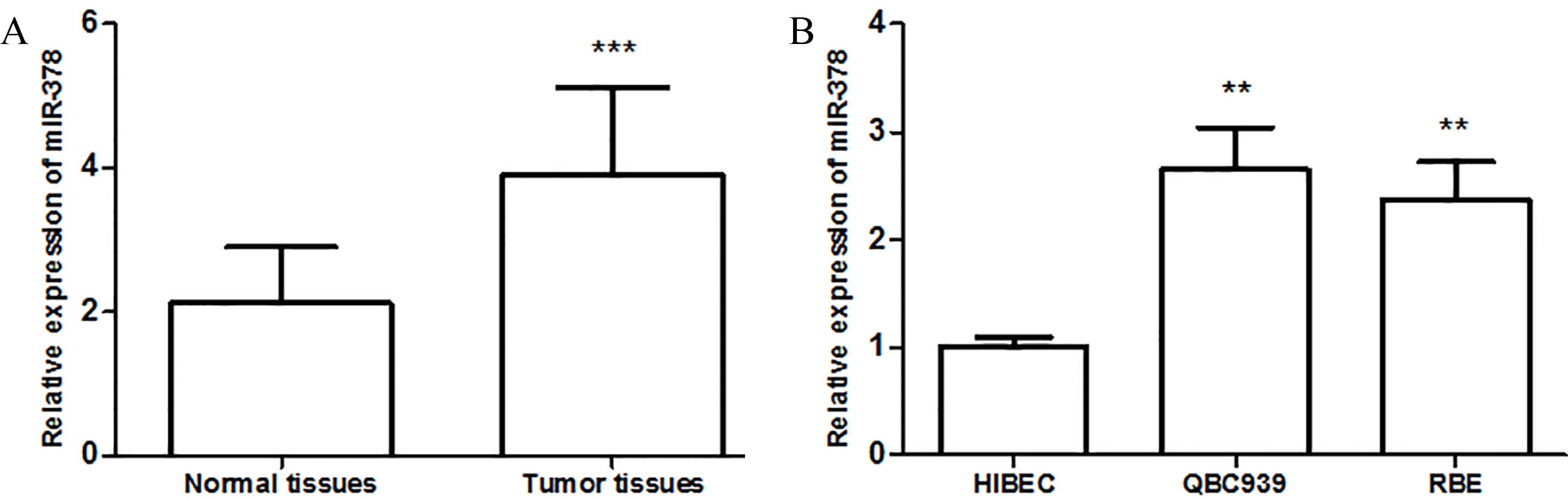
The viability of CCA cells was measured with the colorimetric 3-(4,5-dimethylthiazol-2-yl)-2, 5-diphenyltetra-zolium bromide (MTT) assay. After the transfection was stabilized, QBC939 and RBE cells were plated in 96-well plates (5
Migration and invasion assays
Cell migration and invasion studies were performed using the Transwell assays (Corning, Tewksbury, MA, USA, 8.0
Statistical analysis
All statistical analyses were performed using the SPSS 21.0 software (SPSS, Inc., Chicago, IL, USA) and presented by GraphPad Prism software (GraphPad Software, Inc. La Jolla, CA, USA). Data were presented as the mean
Results
Expression of miR-378 in the CCA tissues and cell lines
To explore the expression pattern of miR-378 in CCA, we first determined the expression levels of miR-378 in CCA tissues and adjacent normal tissues using qRT-PCR. As shown in Fig. 1A, the expression level of miR-378 in CCA tissues was significantly higher than that in normal tissues (
The capacity of miR-378 in distinguishing CCA patients with different stages. A. Relative expression levels of miR-378 in CCA patients with different TNM stages (***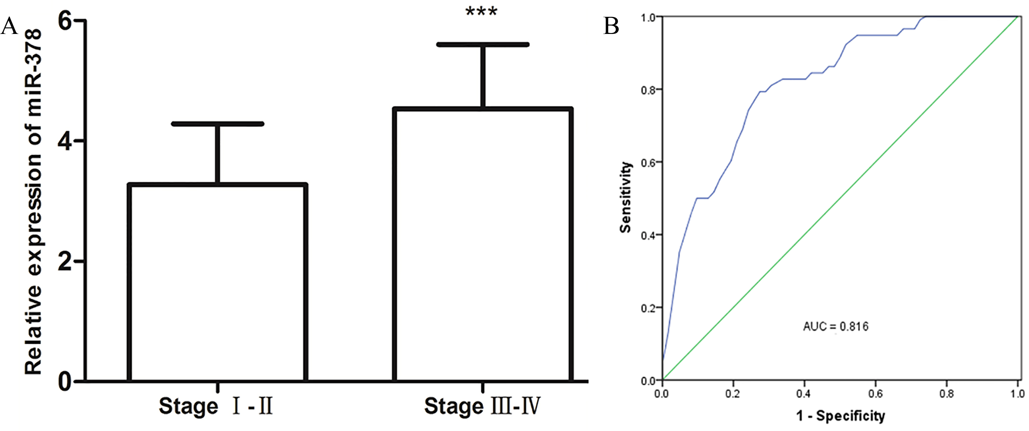
To measure the association between miR-378 expression and clinicopathological characteristics, the CCA patients were divided into two groups according to the mean value of miR-378 expression levels (3.896), including low-expression group (
Kaplan-Meier survival curves of patients with CCA based on miR-378 expression status. Patients with the high expression group have a significantly shorter overall survival rate than those in low expression group.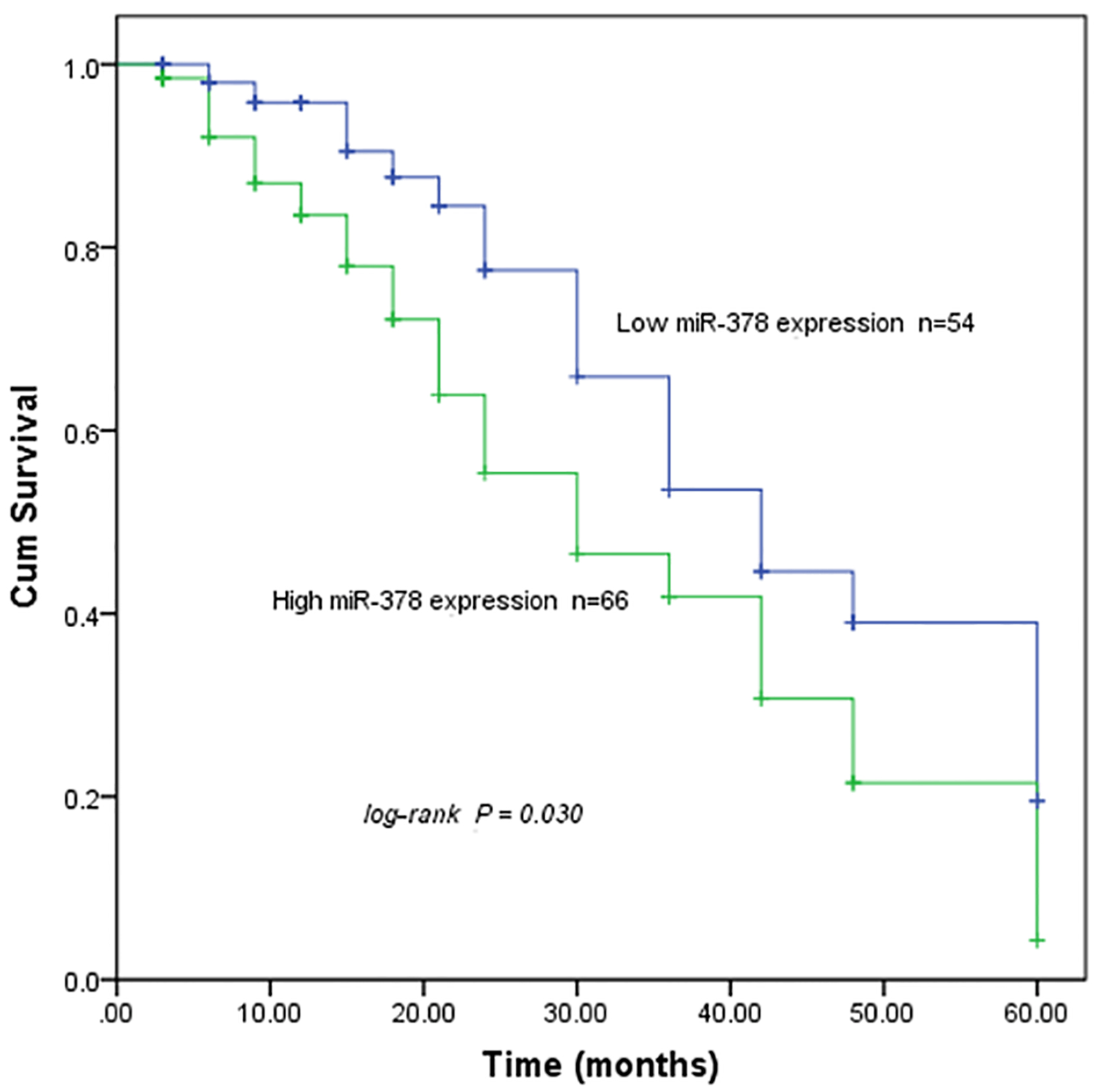
Univariate and Multivariate Cox analysis of clinical parameters in relation to overall survival
–, indicated no related data.
Effects of miR-378 on the proliferation of QBC939 and RBE cell line. A. qRT-PCR analysis of miR-378 in QBC939 and RBE cells. The expression of miR-378 was increased in cells transfected with miR-378 mimic, and down-regulated in cells transfected with miR-378 inhibitor. B. Cell proliferation was enhanced in the QBC939 and RBE cells transfected with miR-378 mimic but was inhibited by downregulation of miR-378 (*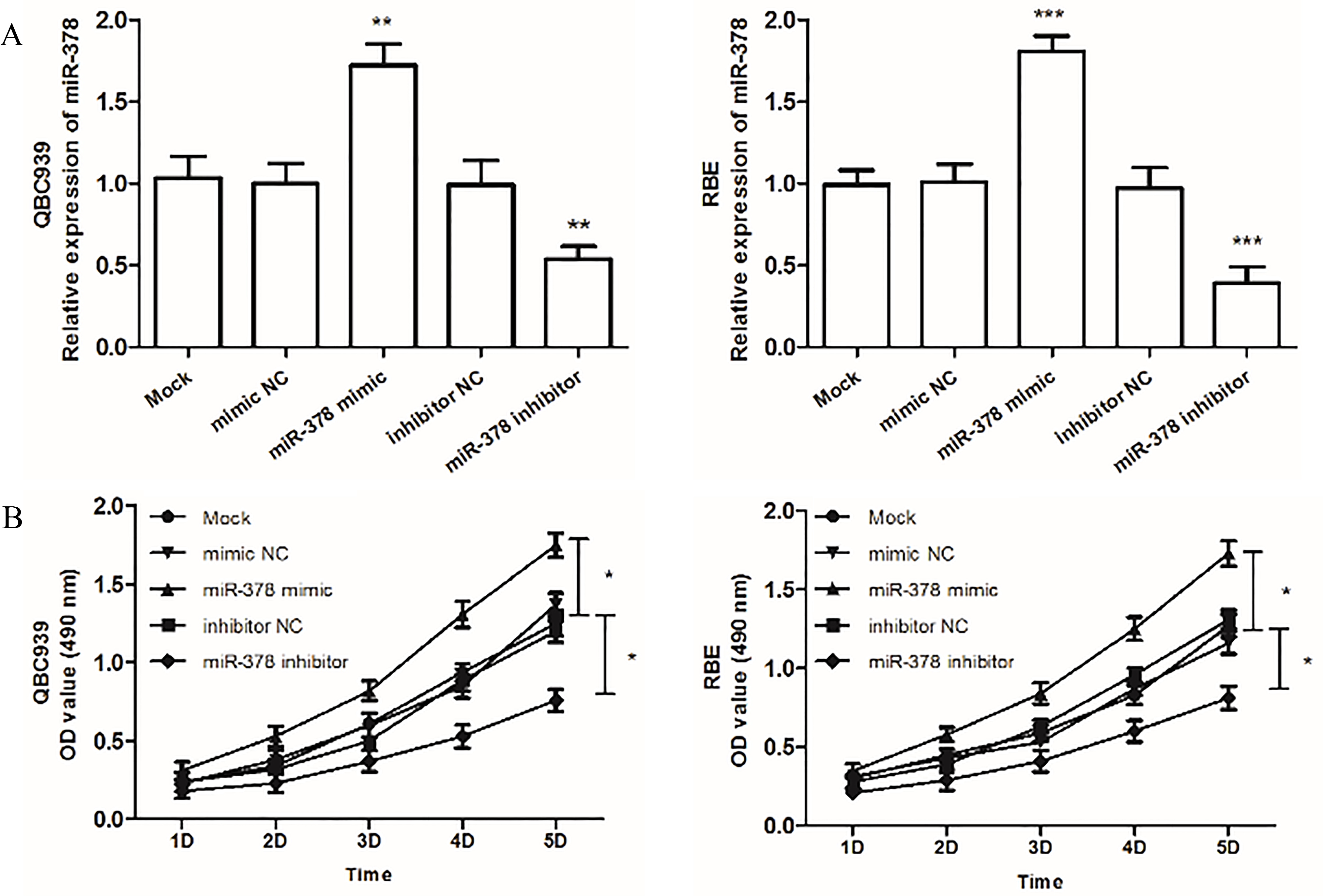
The effects of miR-378 on the migration and invasion abilities of QBC939 and RBE cells. A. The results of migration analysis.B. Overexpression of miR-378 by miR-378 mimic enhanced the cell migration, whereas downregulated miR-378 expression by miR-378 inhibitor suppressed the cell migration. C. The results of invasion analysis. D. Cell invasion was promoted by overexpression of miR-378 but was inhibited by downregulation of miR-378 in both cell lines (*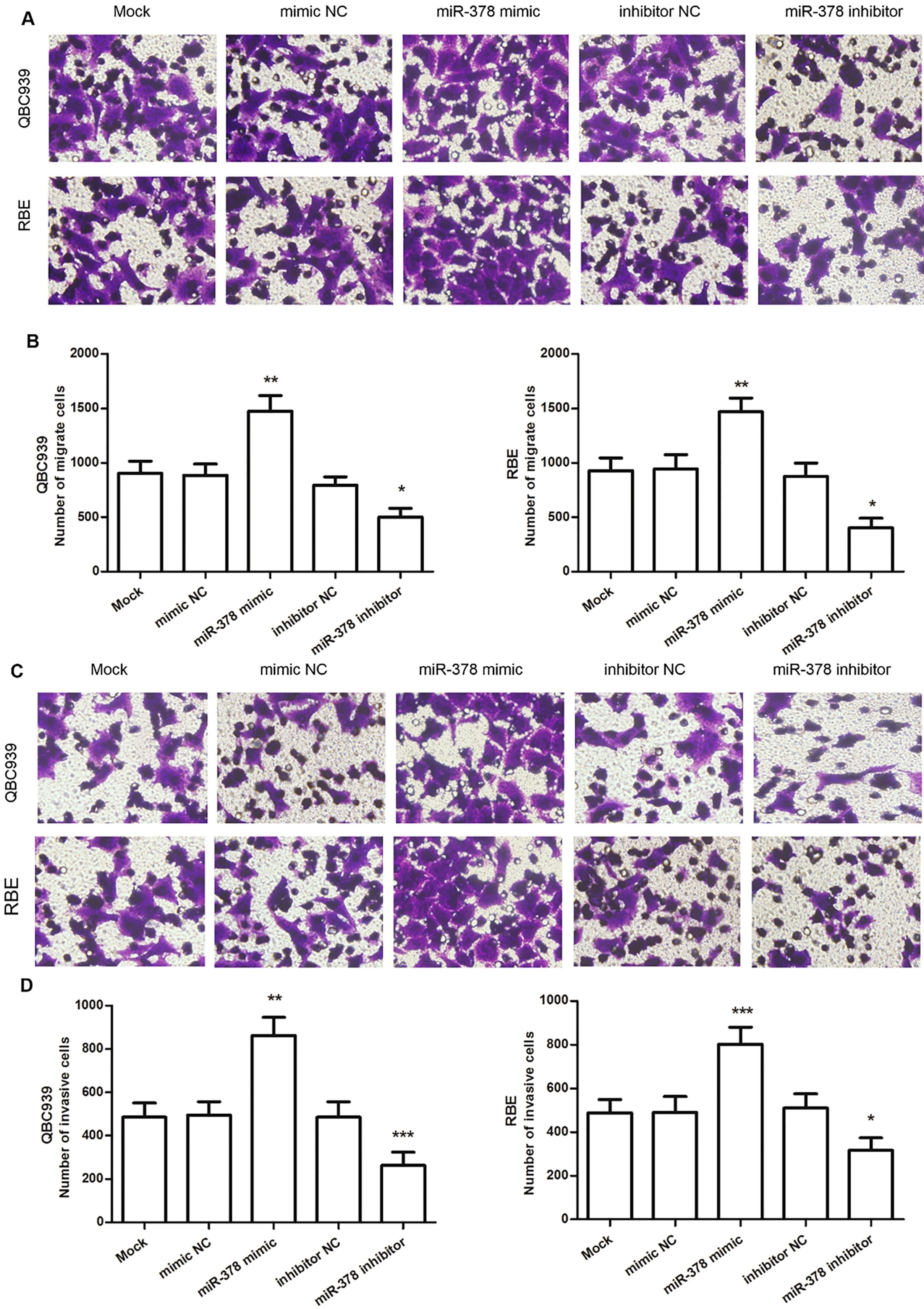
Since the expression of miR-378 was significantly associated TNM stage, it may have capacity in distinguishing patients with different stages. The TNM stages of CCA patients were confirmed according to the American Joint Committee on Cancer (AJCC) edition staging system [21]. To investigate the capacity of miR-378 in distinguishing patients with different stages, CCA patients were divided into two groups, including low TNM stage (I and II) group and high (III and IV) group. The expression levels of miR-378 were 3.21
Correlations of miR-378 expression and survival of CCA patients
To determine the prognostic value of the miR-378 expression in CCA, we used the Kaplan-Meier survival analysis. As shown in Fig. 3, the overall survival of patients with high miR-378 expression was significantly worse than those with low miR-378 expression (log-rank test:
In vitro effect of miR-378 on CCA cell proliferation, migration, and invasion
To investigate the functional significance of miR-378 in CCA, the QBC939 and RBE cells were transfected with miR-378 mimic or miR-378 inhibitor, and the viability of the cells was detected with an MTT assay. The qRT-PCR results indicated that the expression level of miR-378 in cells transfected with miR-378 mimic was significantly higher, and in those transfected with miR-378 inhibitor was decreased, compared to those transfected with negative control or mock (
In addition to cell proliferation, the effects of miR-378 on CCA cell migration and invasion were also examined with a Transwell assay. As presented in Fig. 5, compared with the mock group, transfection with a miR-378 mimic increased the migration and invasion ability in the QBC939 and RBE cells, whereas transfection with a miR-378 inhibitor reduced the migration and invasion ability in QBC939 and RBE cells (all
Discussion
CCA has few distinguishable symptoms, although MRI and computed tomography (CT) are common methods used in the diagnosis of CCA, the patients are diagnosed at the advanced tumor stage, resulting to poor outcomes and a relatively low survival rate [22, 23]. Currently, numerous studies have devoted to exploring specific markers with diagnosis or/and prognosis in CCA [24, 25, 26]. For instance, Lee et al. indicated that HMGA2 expression and lymph node metastasis were associated with shorter patient survival and were an independent marker of poor prognosis in intrahepatic CCA [26]. Watanabe et al. indicated the EMT inducer FOXC2 expression contributed to a shorter cancer-specific survival in extrahepatic cholangiocarcinoma (EHCC) patients and might be a promising molecular target for regulating EHCC metastasis [27]. As novel biomarkers, the potential rules of miRNAs in cancers that have broad prospects has also been identified [28, 29, 30, 31]. These studies suggest that the progress of cancer-related biomarkers may be useful for improving the accuracy of diagnosis and prognosis, as well as help predict treatment strategies for cancer patients.
Numerous studies indicated miR-378 function as an oncogene in various cancers [16, 17, 18]. However, interestingly, in human colon cancer, miR-378 was downregulated in the cancer tissues and cell lines, indicating that miR-378 may act as a tumor suppressor to inhibit the proliferation, migration, and invasion of colon cancer cells by targeting SDAD1 [20]. Similarly, miR-378 was also found that serves as a tumor suppressor and played an important role in inhibiting tumor migration and invasion, such as in glioma, colorectal cancer [32, 33]. In the current study, which aimed to explore the functional significance of miR-378 in CCA, we demonstrated that miR-378 was upregulated in CCA tissues when compared with adjacent normal tissues, and in cancer cell lines compared with HIBEC cells. The data showed that miR-378 may act as an oncogene in CCA. Considering the multiple functions of miR-378 during the development of different cancers, we indicated that miR-378 may act as either an oncogene or a tumor suppressor gene depending on the cancer type. Moreover, the expression levels of miR-378 were significantly correlated with TNM stage and lymph node metastasis, which suggest miR-378 is involved in the development of CCA. Since the expression of miR-378 was significantly associated with TNM stage of patients, ROC curve analysis results showed miR-378 has the ability to distinguish CCA patients with high TNM stages from those with low TNM stages, indicating high miR-378 is significantly correlated with the advanced TNM stages.
To further investigate the prognostic role of miR-378 in CCA patients, Kaplan-Meier and Cox regression analysis were used. Firstly, we found that the levels of miR-378 were positively correlated with the TNM stage and lymph node metastasis, which showed miR-378 expression was associated with the progression of CCA. Then in the Kaplan-Meier survival analysis, the patients with higher miR-378 expression level had a significantly poorer prognosis than those with low miR-378 expression level (
Moreover, we performed cells experiments to explore the biological function of miR-378 in CCA cells. The results showed that miR-378 mimic promoted cell proliferation, migration, and invasion, whereas the miR-378 inhibitor could inhibit proliferation, migration, and invasion of CCA cells. Various studies have indicated the effects of miR-378 on biological behaviors during cancer progression, and the molecular pathways regulated by miR-378 [34, 35]. For instance, a study by Sun et al. demonstrated that miR-378 functioned as an onco-miRNA by targeting the ST7L/Wnt/
In conclusion, our present work showed that miR-378 expression was increased in CCA and was tightly associated with cancer cell proliferation, migration, and invasion. Our study provides miR-378 may be a prognostic biomarker for therapeutic strategies in the treatment of CCA.
Footnotes
Conflict of interest
The authors declare that they have no competing interests.