
Abstract
BACKGROUND:
Ovarian cancer (OC) is one of the most common malignancy worldwide. Emerging evidences have demonstrated that microRNAs (miRNAs) play an important role in regulating the initiation and development of OC.
OBJECTIVE:
The present study was to explore the clinical significance of serum exosomal miR-484 in OC.
METHODS:
A total of 113 OC patients and 60 healthy volunteers were enrolled in this study. Quantitative real-time polymerase chain reaction (qRT-PCR) was performed to measure serum exosomal miR-484 levels in blood samples.
RESULTS:
Our results showed that serum exosomal miR-484 levels were significantly lower in OC patients. Serum exosomal miR-484 was able to discriminate OC cases from controls, with an area under the receiver-operating characteristics (ROC) curve (AUC) of 0.821. Combination of serum exosomal miR-484 with CA-125 showed an elevated AUC of 0.912 in identifying OC patients from controls. Moreover, decreased serum exosomal miR-484 expression was significantly associated with aggressive clinical variables as well as shorter overall survival and progression-free survival. The OC patients with simultaneously low serum exosomal miR-484 expression and high serum CA-125 levels tended to suffer the worst clinical outcomes. The multivariate analysis confirmed that low serum exosomal miR-484 level was an independent indicator.
CONCLUSIONS:
Collectively, serum exosomal miR-484 could serve as a reliable and non-invasive marker for predicting the prognosis of OC.
Introduction
Ovarian cancer (OC) is the most lethal gynecologic malignancy around the world [1]. More than 50, 000 new cases were diagnosed annually in China [2]. Surgery combined with chemotherapy remain the standard therapeutic options for patients with OC [3]. Although great advance for the treatment of OC has been achieved in the past decades, the 5-year overall survival rate remains at 45% [4]. Most cases diagnosed at the late stage is the major reason accounting for the unfavorable prognosis of OC. Therefore, it is crucial to investigate some non-invasive and reliable biomarkers for early detection of this malignancy.
MicroRNAs (miRNAs) are a class of endogenous tiny non-coding RNAs with 21–25 nucleotides in length. These short molecules can negatively regulate gene expression by binding to the 3’-untranslated regions (3’-UTR) of the target mRNAs [5, 6]. MiRNAs have been demonstrated to be actively involved in regulating many biological processes including, but not limited to, proliferation, migration, survival, apoptosis and differentiation [7]. Emerging evidence has revealed that abnormal expression of miRNAs plays a pivotal role in many pathological processes including cancer [8]. Exosomes are small membrane vesicles and carry many kinds of biological molecules, including proteins, RNAs, DNAs and lipids. The exosomal miRNAs can be stably detected and serve as useful diagnostic and prognostic biomarkers [9, 10]. For instance, serum exosomal miR-1307 and miR-375 levels were significantly higher in OC patients, and strongly associated with aggressive clinical variables of OC [11]. Likewise, increased serum exosomal miR-200b and miR-200c levels were found in epithelial ovarian cancer (EOC) patients, and correlated with lymph node metastasis and advanced clinical stage [12]. The data indicated that these serum exosomal miRNAs played an oncogenic role in tumorigenesis of OC.
The gene encoding miR-484 locates at chromosome 16p13 and it seems to be highly conserved in vertebrates [13]. Aberrant expression of miR-484 has been reported in various types of cancer such as cervical cancer, lung cancer and colorectal cancer [15, 16, 17]. However, the clinical value of serum exosomal miR-484 in OC remains poorly understood. In this study, we aimed to detect the expression level of serum exosomal miR-484 in OC, and analyzed its diagnostic and prognostic value.
Materials and methods
Patients and samples
All blood samples were obtained from 113 OC patients treated at First Affiliated Hospital of Yangtze University. The study was approved by the Ethics Committee of First Affiliated Hospital of Yangtze University, and written informed consent was collected from all participants. None of the patients received any therapy prior to the treatment. Tumors were staged according to the 2014 Federation of Obstetrics and Gynecology (FIGO) classification. Of all OC patients, 35 subjects were diagnosed with lymph node metastasis while 78 subjects were without lymph node metastasis, 64 subjects were in I/II FIGO stage and 49 subjects were in III/IV FIGO stage. Moreover, 60 healthy volunteers were recruited as controls. Venous blood was withdrawn from each participant and centrifugation at 3000 g for 10 min, then the supernatants were collected and stored at
Exosome isolation
Isolation of exosomes was performed with ExoQuick Exosome Precipitation Solution kit (System Biosciences, Mountain View, CA, USA) based on the manufacturers’ protocols. Briefly, the serum was thawed on ice and centrifuged at 2000 g for 30 min to remove cell debris. Subsequently, the supernatant was mixed with ExoQuick reagent, followed by incubation for 30 min at 4
Total RNA isolation and quantitative real-time polymerase chain reaction (qRT-PCR)
Exosomal RNA was isolated using the miRNeasy serum/plasma kit (Qiagen, Hilden, Germany). In the RNA isolation step, a total of 2
(A) The expression level of serum exosomal miR-484 was dramatically downregulated in OC patients in comparison with the healthy controls. Compared to the respective controls, serum exosomal miR-484 levels were significantly reduced in OC patients with poorer histological grades (B), with lymph node metastasis (C) or at the advanced FIGO stage (D).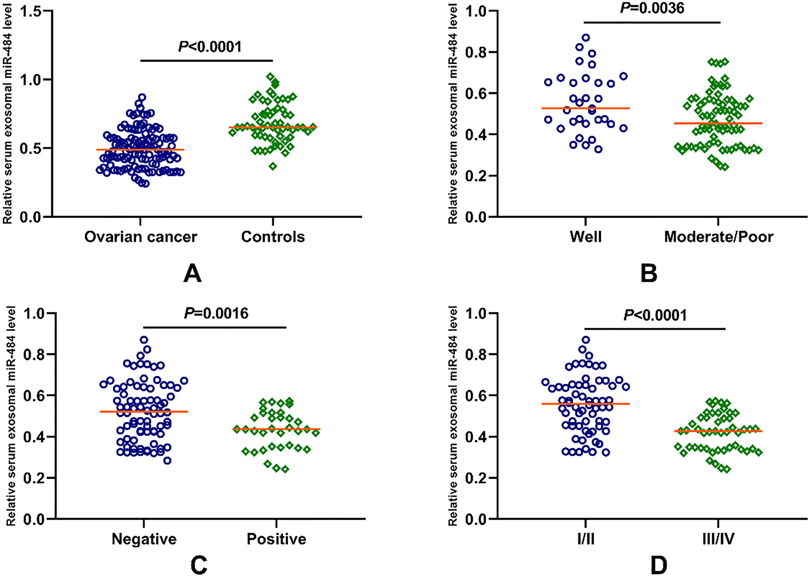
GraphPad Prism software (GraphPad Software, San Diego, CA, USA) and MedCalc 12.7.0 (MedCalc, Ostend, Belgium) were used for all statistical analyses. The Mann-Whitney U test was performed to compare the differences in serum exosomal miR-484 expression between two groups. The Chi-square test was used to evaluate the correlations between serum exosomal miR-484 expression and clinical variables. Receiver operating characteristic (ROC) curve was constructed and the area under the ROC curve (AUC) was computed to assess the diagnostic power of serum exosomal miR-484 and serum CA-125. The Kaplan-Meier method and log-rank test was used to plot the survival curves. A multivariate Cox proportional hazard model was performed to determine the risk ratios (RR) for overall survival (OS) and progression free survival (PFS). OS was defined as the time between the date of surgical treatment and the date of death or last follow- up. PFS was defined as the time between the date of surgical treatment and the date of recurrence or last follow-up. A
Results
Serum exosomal miR-484 levels decreased in OC
The serum exosomal miR-484 levels in OC patients and normal controls were detected by qRT-PCR. The relative expression levels of exosomal miR-484 in the serum samples were determined by comparing with the levels of cel-miR-39 spike in control. It was observed that serum exosomal miR-484 levels were significantly lower in 113 OC cases (median value
(A) The diagnostic accuracy of serum CA-125 for discriminating serous OC, non-serous OC or OC patients from healthy controls. (B) The diagnostic accuracy of serum exosomal miR-484 for discriminating serous OC, non-serous OC or OC patients from healthy controls. (C) The diagnostic accuracy of combining serum CA-125 and serum exosomal miR-484 for identifying OC patients from healthy volunteers.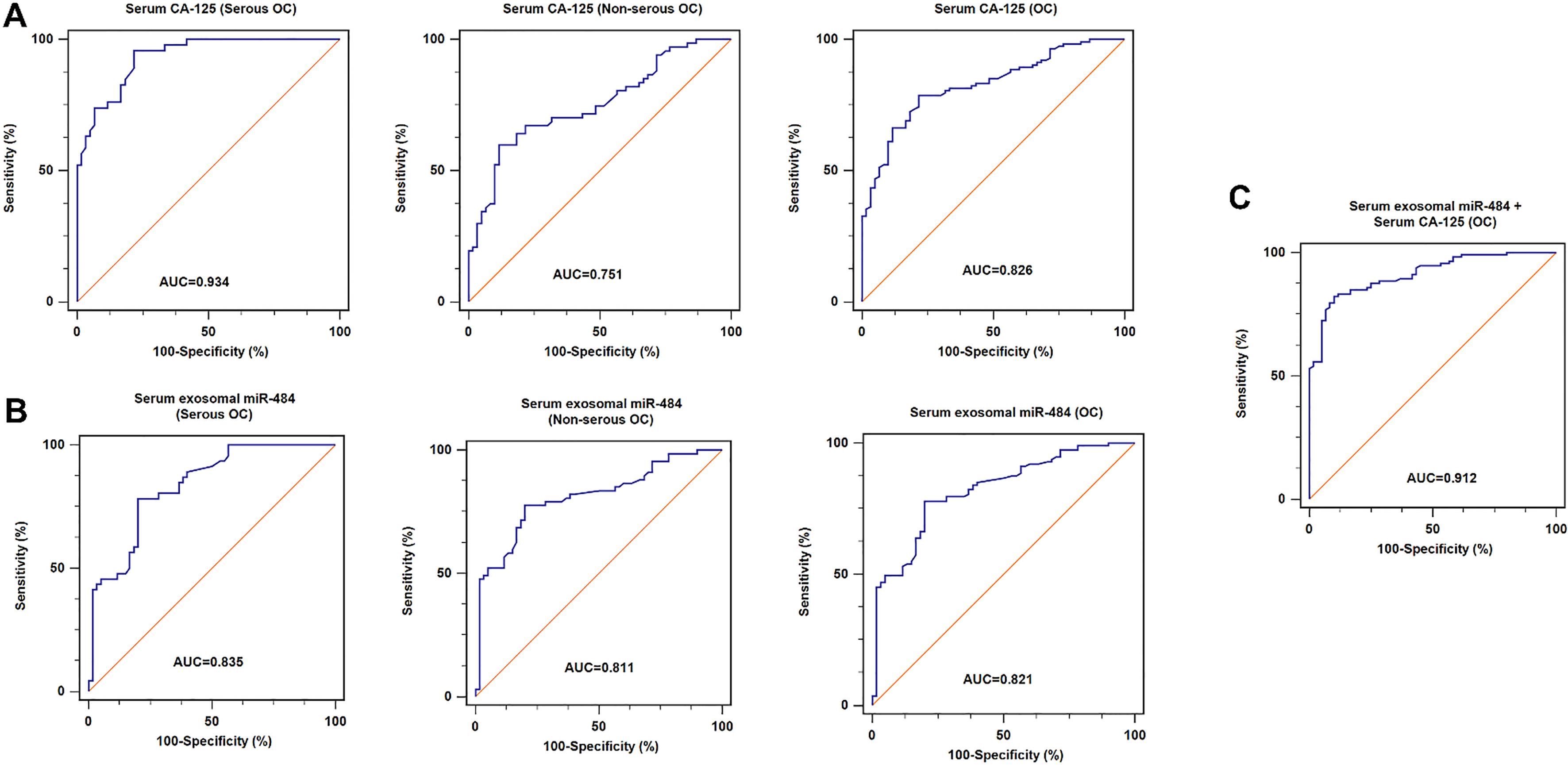
We first performed the ROC analyses for evaluating the diagnostic accuracy of serum CA-125 or serum exosomal miR-484 for the serous, non-serous OC and OC. Our results showed that serum CA-125 could well discriminate patients with serous OC from healthy controls with an AUC value of 0.934 (sensitivity
Association between serum exosomal miR-484 expression and clinical features
The association between serum exosomal miR-484 expression and patient’s clinicopathological variables was analyzed. As shown in Table 1, lower serum exosomal miR-484 expression was closely correlated with poorer histological grade (
Clinicopathological correlation of serum exosomal miR-484 expression in OC patients
Clinicopathological correlation of serum exosomal miR-484 expression in OC patients
Kaplan-Meier method and log-rank test were used to evaluate the association between serum exosomal miR-484 expression and survival. The OS (
(A) and (B) The OS and PFS were significantly lower in low serum exosomal miR-484 expression group than in high serum exosomal miR-484 expression group. (C) and (D) OC patients in the high serum CA-125 group suffered a significantly shorter OS and PFS than those in the low serum CA-125 group. (E) and (F) Statistically significant differences were found in both OS and PFS among the four compared groups.
We have also determined the association between serum CA-125 levels and the OS/PFS of patients with OC as well as the cumulative prognostic power of CA-125/miR-484 index. A total of 59 cases were in the low serum CA-125 group (
Multivariate analysis of prognostic factors in all OC patients
In the multivariate Cox proportional hazard model, it was demonstrated that histological grade (RR
Serum exosomal miRNAs are promising biomarkers for diagnosis, prognosis and treatment monitoring of OC. The potential clinical utility of serum exosomal miR-484 as a reliable biomarker in OC was assessed in this study. We found that serum exosomal miR-484 expression was downregulated in OC subjects compared to normal controls. In addition, serum exosomal miR-484 showed good performance to differentiate OC cases from healthy controls. When serum exosomal miR-484 and CA-125 were combined, ROC analysis yielded an increased AUC of 0.912 in discriminating OC cases from controls. Moreover, low serum exosomal miR-484 expression was strongly associated with poorer histological grade, positive lymph node metastasis and advanced FIGO stage. Furthermore, OC patients with the lower serum exosomal miR-484 expression had shorter OS and PFS. The OC patients with simultaneously low serum exosomal miR-484 expression and high serum CA-125 levels tended to suffer the worst survival outcomes. Finally, the multivariate analysis showed that serum exosomal miR-484 expression was an independent risk factor for OS and PFS. Consistent with our findings, downregulation of miR-484 was closely associated with enhanced chemoresistance of OC cells [14].
There were two possible reasons for the poor performance of serum CA-125 in our study. Firstly, previous studies have shown that serum CA-125 might be more sensitive for detecting serous OC than other types of OC [15, 16]. Only 42.48% (48/113) of patients were diagnosed as serous type OC in our study, which might account for the poor overall performance of CA-125. In addition, the sample size was relatively small in our study. Increasing the sample size might improve the diagnostic power of serum CA-125 in OC. Surprisingly, it seems that the OC patients in the low serum exosomal miR-484
Except for OC, several previous studies have shown that miR-484 acted as a tumor suppressor in other types of cancers. For instance, the expression level of miR-484 was reduced in cervical cancer tissues and cell lines. In addition, ectopic expression of miR-484 suppressed the oncogenic activities of cervical cancer cells by targeting ZEB1 and SMAD2 [17]. MiR-484 was the most downregulated miRNA in colorectal cancer (CRC) with microsatellite instability. Both in vitro and in vivo studies showed that overexpression of miR-484 inhibited the proliferation of cancer cells and tumor growth [18]. Lu et al. reported that the expression level of serum miR-484 was dramatically reduced in CRC patients at the early stage [19]. Similar findings were also found in patients with gastric cancer, the expression level of miR-484 was remarkably reduced in gastric cancer tissues [20].
However, miR-484 had also been reported to be functioned as an oncogene in some types of cancers. For instance, miR-484 enhanced glioma tumor-initiating properties in vitro and in vivo. In addition, high miR-484 expression was closely associated with unfavorable clinical outcome in patients with glioma [21]. The expression level of miR-484 was overexpressed in non-small-cell lung cancer (NSCLC) tissues and cell lines. In addition, upregulation of miR-484 promoted the proliferation and migration capacity of lung cancer cells, and opposite results were observed when miR-484 was downregulated. Moreover, Apaf-1 was demonstrated to be a direct target of miR-484, suggesting that miR-484 acted as an oncomiR in NSCLC [22]. These data indicated that the concrete role of miR-484 in tumorigenesis might be closely associated with the tumor type and microenvironment.
Conclusions
To the best of our knowledge, this is the first report describing the clinical significance of serum exosomal miR-484 in OC. We have demonstrated that serum exosomal miR-484 expression was significantly lower in OC patients compared to that in healthy individuals. Downregulation of serum exosomal miR-484 was closely correlated with worse clinical variables and poor prognosis. Therefore, serum exosomal miR-484 might serve as a promising diagnostic and prognostic biomarker for OC. The relatively small sample size was the main limitation of the current study, and further studies with larger scale of OC samples are required to validate our findings.
Footnotes
Acknowledgments
This study was partly supported by the Grant from First Affiliated Hospital of Yangtze University.
Conflict of interest
None.