Anaplastic thyroid cancer/ATC is highly lethal malignancy without reliable chemotherapeutic drug. Resveratrol possesses anti-ATC activities but encounters resistance in some cases due to certain unknown reason(s).
OBJECTIVE:
Because signal transducer and activator of transcription/STAT3 signaling is critical for ATC cell survival and the main molecular target of resveratrol, its roles in determining the fates of resveratrol-treated ATC cells were investigated here.
METHODS:
Human THJ-11T, THJ-16 and THJ-21T ATC cell lines were treated by 100 M resveratrol and their growth, statuses of STAT3 signaling and STAT3-related gene expression were examined. The relevance of STAT3 activation with resveratrol resistance was elucidated using STAT selective inhibitor AG490. Leukemia inhibitory factor/LIF expression and phosphorylated-STAT3/p-STAT3 nuclear translocation in ATC tissues were immunohistochemically analyzed.
RESULTS:
Resveratrol inhibited proliferation, p-STAT3 nuclear translocation as well as LIF and STAT3 expression of THJ-16T and THJ-21T but not THJ-21T cells which showed LIF upregulation and more frequent p-STAT3 nuclear translocation. AG490 significantly prevent p-STAT3 nuclear translocation, and reversed the resveratrol tolerance of THJ-11T cells. Immonohistochemical staining revealed 14.3% (4/28) of LIF and 3.6% (1/28) of p-STAT3 detection in noncancerous ATC-surrounding tissues, which increased to 89.5% (17/19) and 52.6% (10/19) respectively among ATC specimens. The correlative analysis indicated the relevance of LIF expression and STAT3 activation ( 0.825; 0.01).
CONCLUSIONS:
The status of STAT3 activation and LIF expression are closely correlated with the therapeutic effect of resveratrol on ATCs. Frequent LIF upregulation and STAT3 activation are the unfavorable factors of ATCs and the potential targets of anti-ATC therapy.
Thyroid cancer is the most common endocrine malignancy and its incidence keeps increasing [1]. According to the standards of World Health Organization, thyroid cancer can be divided into differentiated and undifferentiated thyroid cancer [2]. Differentiated thyroid cancer has a favorable prognosis and long survival period, while ATC, though accounts for less than 2% of the total thyroid cancer cases, has the highest mortality rate and the patients usually die within one year of diagnosis [3]. Surgery and adjuvant chemotherapy are the standard management of ATCs, but the prognosis is very poor because of the highly aggressive growth and metastasis tendency of the tumors [4, 5, 6]. All-trans retinoic acid has been used to promote ATC cell differentiation for improving iodine uptake and therefore radiosensitivity, while the outcomes are not promising and even show the opposite effects as we have reported recently [7]. Therefore, it would be of clinical values to explore safer and more reliable agent(s) against ATCs.
Resveratrol, a polyphenolic compound commonly found in natural plants, has multiple biological activities, including anti-inflammatory and anti-cancer effects [8]. More importantly, resveratrol in the anticancer dose has little toxic effects on normal cells [9]. Our previous results show that all of three ATC cell lines (THJ-11T, THJ-16T and THJ-21T) so far checked are resistant to RA treatment [10], but two of them (THJ-16T and THJ-21T) are sensitive to resveratrol, suggesting the better therapeutic effects of resveratrol on ATCs. However, THJ-11T cells are not sensitive to resveratrol. This suggests the necessity to investigate the underlying reason(s) leading to differential response of ATC cells to resveratrol for the personalized anti-ATC therapy.
STAT3 pathway is known to play an important role in cancer formation and progression [11, 12]. It regulates cancer stem-cell properties in anaplastic thyroid cancer cells and confers on drug resistance of anaplastic thyroid cancer [13]. STAT3 pathway can be activated by certain cytokines including LIF that has been known to play promoting roles in thyroid carcinogenesis [14, 15]. Because STAT3 pathway is the main molecular target of resveratrol [16, 17], we speculate that the statuses of STAT3 signaling and its upstream stimulators such as LIF may be related to the resveratrol sensitivity of ATC cells. Our study thus aims to address this issue using the ATC cell lines with different resveratrol sensitivity.
Materials and methods
Thyroid cancer cell lines and treatment
Human anaplastic thyroid cancer THJ-11T, THJ-16T and THJ-21T cell lines [18] were kindly provided by Dr. Liu, Institute of Cancer Stem Cell, Dalian Medical University. They were cultured in 1640 medium with L-glutamine (Hyclone, Logan, UT, USA), supplemented with 10% fetal bovine serum (Gibco, Grand island, NY, USA) for THJ-11T and THJ-21T, and with 5% fetal bovine serum for THJ-16T. An amount of 5 10/mL cells were plated onto culture dishes (Nunc A/S, Roskilde, Denmark) at 37C and 5% CO for 24 h before the experiments were performed. For haematoxylin and eosin (H/E), immunocytochemical staining and flow cytometry analyses (Becton Dickinson, San Jose, CA, USA), dozens of cell-bearing coverslips were prepared under the same experimental conditions using coverslip-preparation dishes (Jet Biofile Tech. Inc., Guangzhou, China; China invention patent No. ZL200610047607.8) for multiple experimental purposes. Resveratrol (Sigma-Aldrich, St. Louis, MO, USA) were dissolved in dimethylsulfoxide (DMSO; Sigma-Aldrich) and diluted with culture medium to 100 M as the working concentrations just before use. The experimental groups were set in triplicate, and the experiments were repeated three times to establish a definite conclusion.
Evaluation of cell proliferation and death
To elucidate the response of ATC cells to resveratrol treatment, HE staining, viable/nonviable cell counting (Automated Cell Counter, Bio-Rad, Singapore), 3-[4,5-Dimethylthiazol-2-yl]-2,5-diphenyl-tetrazolium bromide (MTT) cell proliferation assay and a modification of the terminal deoxynucleotide transferase (TdT)-mediated dUTP-biotin nick end-labeling method (TUNEL; Roche Inc., Basel, Switzerland) were performed on coverslips bearing THJ-11T, THJ-16T and THJ-21T cells without and with resveratrol treatment by the methods described elsewhere [10].
Western blot analyses
Western blotting was performed using antibodies against -actin (Santa Cruz, USA; 1:3000), p-STAT3 (Santa Cruz, USA; 1:500), STAT3 (Santa Cruz, USA; 1:500), LIF (Santa Cruz, USA; 1:200), NF-B (Zhongshan-Jinqiao, Inc., Beijing, China; 1:500), survivin (Bioss. Inc., Beijing, China; 1:500). Total cellular proteins were prepared from the cells under different culture conditions by the method described previously [20]. For Western blot analyses, the sample proteins (20 g/well) were separated by electrophoresis in 10% sodium dodecylsulfate-polyacrylamide gel electrophoresis, transferred to polyvinylidene difluoride membrane (Amersham, Buckinghamshire, UK). The membrane was blocked with 5% skimmed milk in TBS-T (10 mM Tris-Cl, pH 8.0, 150 mM NaCl, and 0.5% Tween 20) at 4C overnight, rinsed three times (10 minutes each time) with TBS-T, followed by 3 hours of in cubation at room temperature with the first antibodies in appropriate concentrations followed by 1 hour of incubation with horseradish peroxidase/ HRP-conjugated anti-mouse or anti-rabbit IgG (Zymed Lab, Inc). The bound antibody was detected using the enhanced chemiluminescence system (Roche GmbH, Mannheim, Germany). After removing the labeling signal by incubation with stripping buffer (62.5 mM Tris-HCl, pH 6.7, 100 mM 2-mercaptoethanol, 2% SDS) at 55C for 30 minutes, the membrane was reprobed with other antibodies one by one by the same experimental procedures until all of the parameters were examined.
Immunocytochemical staining
Immunocytochemical staining was performed on the coverslips obtained from each of the experimental groups. The antibodies against p-STAT3 (Santa Cruz. Inc., USA; 1:80), LIF (Santa Cruz. Inc., USA): 1:80, NF-B (Zhongshan-Jinqiao, Inc., Beijing, China; 1:100), survivin (Bioss. Inc., Beijing, China; 1:100). Briefly, the coverslips were washed with phosphate-buffered solution (PBS, pH 7.4), incubated for 10 minutes in 3% HO and then with the appropriately diluted first antibody at 37C for 60 minutes in a humid chamber, followed by the treatments with reagent A containing polymer enhancer for 20 minutes and with reagent B containing polymerized HRP anti-mouse/rabbit IgG for 30 minutes (Zymed Lab, Inc, San Francisco, CA, USA). Color reaction was developed using 3,3’-diaminobenzidine tetrahydrochloride.
Inhibition of STAT3 activation with AG490
AG490 (Sigma-Aldrich St. Louis, MO, USA) is a JAK2-specific inhibitor and therefore selectively inhibits STAT3 phosphorylation. This reagent was dissolved in DMSO to a stock concentration of 80 mM and was diluted to the final concentration of 80 M with conventional culture medium just before use [19]. Four experimental groups were set as follows: Group 1, normal culture supplemented with 0.2% DMSO; Group 2, treatment with 100 M resveratrol; Group 3, treatment with 80 M AG490; Group 4, treatment with 100 M resveratrol and 80 M AG490.
Tissue microarray based immunohistochemical staining
The archived paraffin blocks of 28 cases of noncancerous tumor-surrounding thyroid tissues and 19 anaplastic thyroid cancers were used to construct the tissue microarrays in the densities of 56 spots/cm by the method described elsewhere [11]. The microarrays prepared were sectioned to 5-m thickness for immunohistochemical profiling of p-STAT3 and LIF expression and intracellular distribution, respectively. The antibodies used were the same as that used in immunocytochemistry. The tissue microarray sections lacking incubation with individual primary antibodies were used as background controls. The staining results were evaluated separately by two investigators, with the intensity of immunolabeling scored as negative (), weakly positive (), moderately positive () or strongly positive ().
Statistical analyses
Each experiment was conducted for three times, and the data obtained were analyzed together. The results of the MTT cell proliferation assay and cell counting were evaluated with ANOVA and the independent-samples -test. The bar graphs present the mean standard deviation (SD) of separate experiments ( 8). When required, -values are provided in the figures and their legends.
Results
Differential resveratrol sensitivities of ATC cells
Human THJ-11T, THJ-16T and THJ-21T ATC cell lines were treated with 100 M resveratrol for 48 hours, respectively. HE morphological staining (Fig. 1A) showed that resveratrol-treated THJ-16T and THJ-21T cells show extensive cell death, while no distinct morphological alteration could be observed in THJ-11T cell population under the same experimental condition (Fig. 1B). In accordance, TUNEL fluorescent apoptotic cell labeling revealed abundance of TUNEL-positive cells in resveratrol-treated THJ-16T and THJ-21T rather than THJ-11T cell populations (Insets in Fig. 1A).
Suppressed STAT3 signaling in resveratrol-sensitive THJ-16T and THJ-21T but not in resveratrol-resistant THJ-11T cells. Cellular morphology (H/E) of resveratrol-treated THJ-11T, THJ-16T and THJ-21T cells. N, normally cultured cells; Res, 100
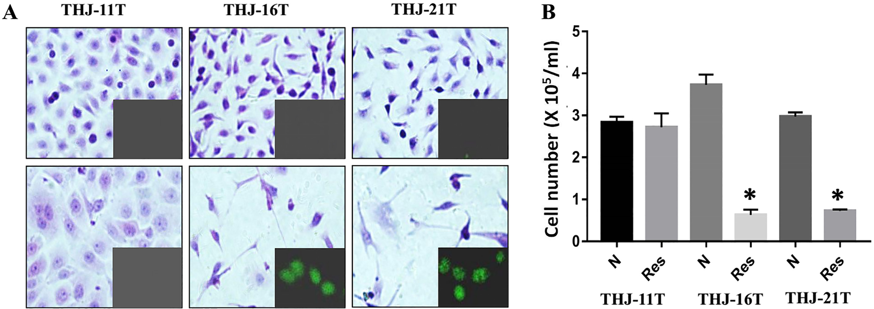
 M resveratrol treated cells. The corresponding immunofluorescent TUNEL images are shown in the insets. The total cell numbers of the three ATC cell lines without (N) and with 48 hour 100 M resveratrol treatment (Res). , 0.01 in comparison with Group-N.
Evaluation of NF-B expression and STAT3 signaling of THJ-11T, THJ-16T and THJ-21T cells without (N) and with 48 hour 100 M resveratrol treatment (Res) by Western blotting (A) and immunocytochemical staining (B).
Different NF-B and STAT3 statuses in resveratrol-sensitive ATC cells
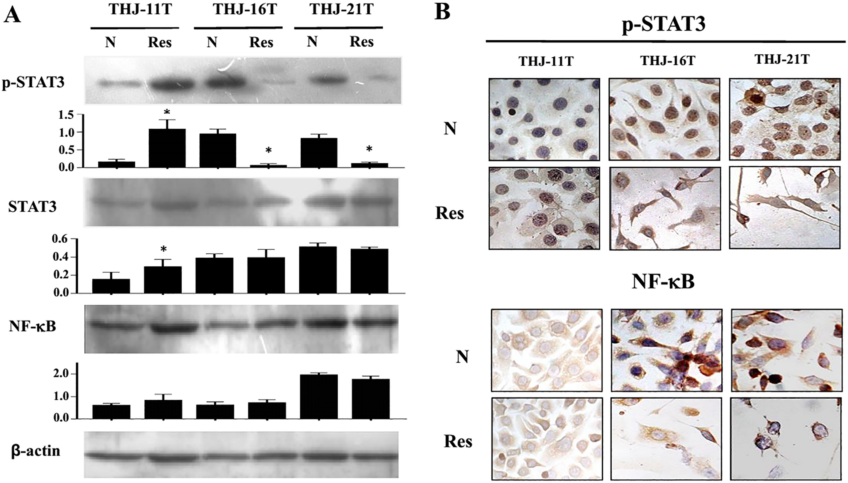
The results of Western blotting revealed that the levels of p-STAT3 but not NF-B/p65 in THJ-16T and THJ-21T cells were 15.4 folds and 10.2 folds decreased after 48 hours of 100 M resveratrol treatment; in contrast, the NF-B/p65 and STAT3 levels in resveratrol-treated THJ-11T cells kept stable (Fig. 2A). Immunocytochemical staining showed that the nuclear translocation of NF-B/p65 and p-STAT3 in THJ-16T and THJ-21T cells decreased significantly after resveratrol treatment; the frequency of nuclear translocation of p-STAT3 in resveratrol treated THJ-11T cells increased slightly but the distribution pattern of NF-B/p65 remained unchanged (Fig. 2B).
Negative correlation of LIF expression with resveratrol sensitivity
LIF is the main activator of NF-B and STAT3 signaling pathways. Western blotting results showed that the LIF expression was significantly down-regulated in THJ-16T (1.9 folds) and THJ-21T (2.42 folds) but was elevated (1.65 folds) in THJ-11T cells after 48 hours of resveratrol treatment (Fig. 3A). Similarly, the results of immunocytochemical staining showed the distinct decrease of LIF immunolabeling in resveratrol-sensitive THJ-16T and THJ-21T cells and the increased LIF staining in THJ-11T cells (Fig. 3B).
Evaluation of LIF expression in THJ-11T, THJ-16T and THJ-21T cells without (N) and with 48 hour 100 M resveratrol treatment (Res) by Western blotting (A) and immunocytochemical staining (B).
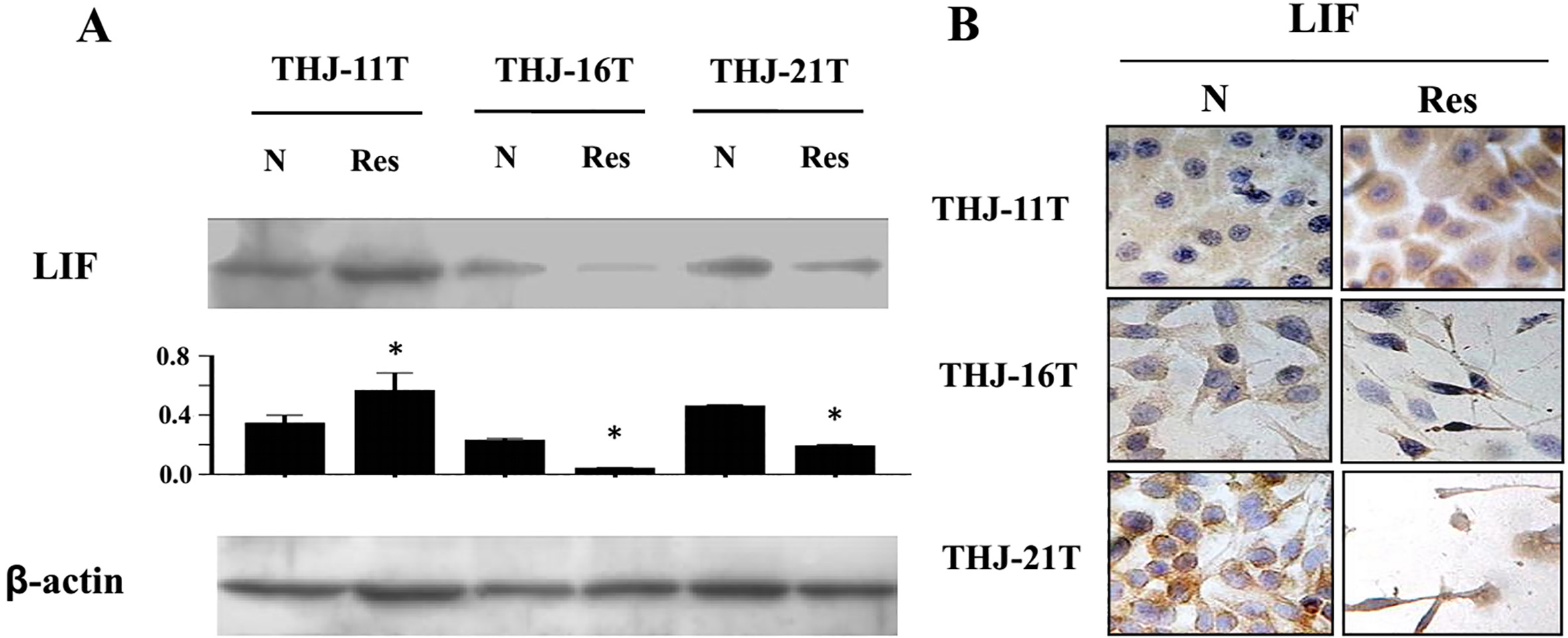
AG490 suppressed STAT3 activation and improved resveratrol sensitivity of THJ-11T cells. Immunocytochemical evaluation of p-STAT3, LIF, NF-B and survivin expression of THJ-11T cells cultured under normal condition (N), treated with 100 M resveratrol (Res), 80 M AG490 (AG490) and 100 M resveratrol and 80 M AG490 combination (Res&AG490) for 48 hours. Arrows indicate the portions shown in the insets with higher magnification (X 80). H/E morphological staining of THJ-11T cells. N, normally cultured cells; Res, 100 M resveratrol treated cells; AG490, 80 M AG490 treated cells; AG490&Res, 100 M resveratrol and 80 M AG490 treated cells. Arrows indicate the portions shown in the insets with higher magnification (X 80). MTT assay performed on THJ-11T cells under N, Res, AG490 and AG490&Res experimental conditions. , 0.01 in comparison with the Control and the Res groups; , 0.01 in comparison with the Control, the Res and the AG490 groups.
AG490-enhanced resveratrol sensitivity of THJ-11T Cells
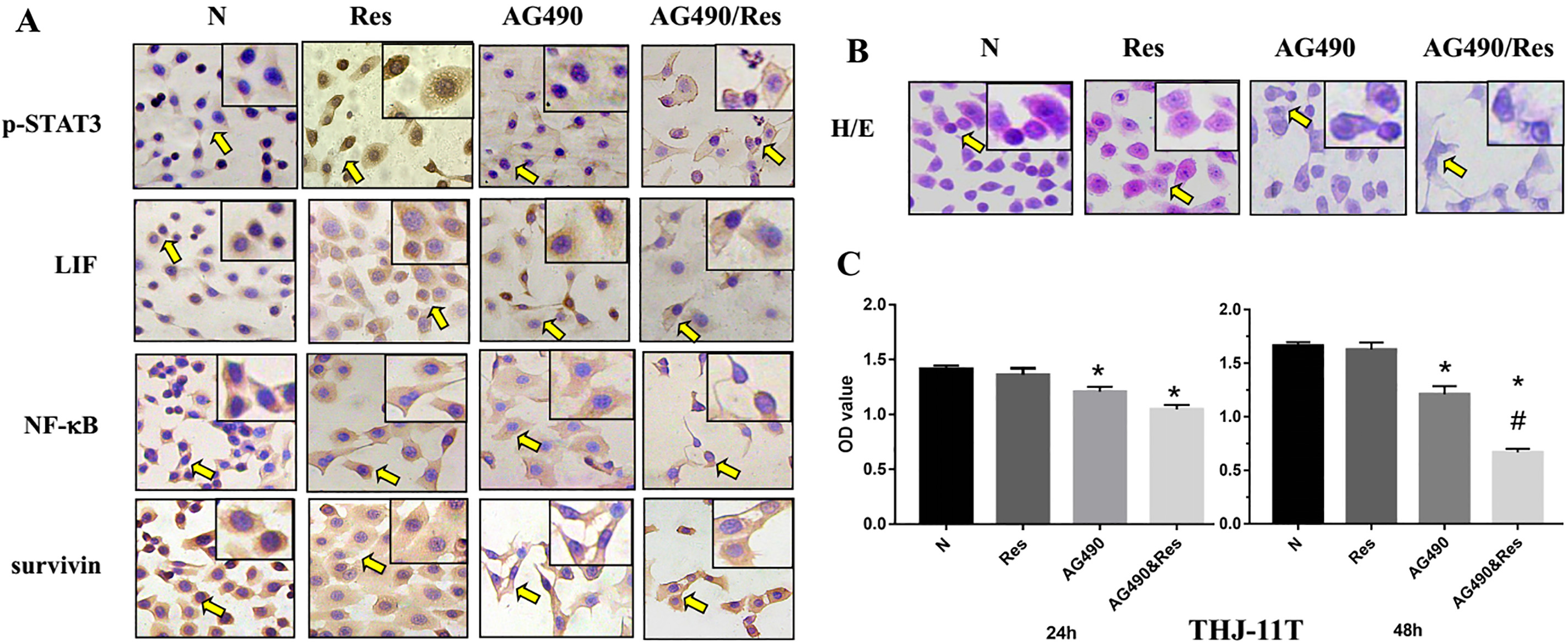
To investigate the relationship between STAT3 pathway status and resveratrol sensitivity in ATC cells, 80 M AG490 and its combination with 100 M resveratrol were used to treat THJ-11T cells, respectively. Immunocytochemical staining showed that the nuclear translocation of p-STAT3 and LIF expression increased after 48 hours of resveratrol treatment. Remarkable reduction of p-STAT3 nuclear translocation was observed in THJ-11T cells treated either by 80 M AG490 only or by 80 M AG490 and 100 M resveratrol combination (Fig. 4A). The changes of NF-B/p65 nuclear translocation as well as LIF and survivin expression in THJ-11T cells were not distinct irrespective to the above two treatments (Fig. 4A). HE staining showed cell number reduction (Fig. 4B) in both AG490- and AG490/Res-treated populations; MTT assay (Fig. 4C) demonstrated that the optical density (OD) value was 27.2% and 25.6% decreased ( 0.01) in AG490-treated cells and 59.8% and 58.9% decreased in AG490/Res-treated ones ( 0.01) in comparison with that of their normally cultured (N) and Res-treated counterparts.
Correlative analysis of p-STAT3 nuclear translocation and LIF expression in noncancerous thyroid tissues and the poorly differentiated/anaplastic thyroid cancers
LIF
p-STAT3
N
24 (85.7%)
3 (10.7%)
0
#
0.825 ( 0.01)
0
1 (3.6%)
0
0
0
0
ATC
0
0
0
0
2 (10.5%)
7 (36.8%)
0
0
10 (52.6%)
#, Significant difference comparing with the data of nancancerous thyriod tissues ( 0.05).
Tissue microarray-based immunohistochemical profiling of p-STAT3 nuclear transloication and LIF expression in anaplastic thyroid cancers and their noncancerous counterparts. A. Immunohistochemical illustration (X 20) of p-STAT3 distribution patterns and LIF level in a case of anaplastic thyroid cancer (ATC) in the form of and its noncancerous tumor-surrounding tissue (N) in . Arrows indicate the potions shown in the insets with higher magnification (X 60). B. The frequencies of p-STAT3 nuclear translocation and LIF expression in noncancerous tumor-surrounding tissues (Noncancerous) and anaplastic thyroid cancers (ATC). The intensity of immunolabeling was scored as negative (), weakly positive (), moderately positive () or strongly positive ().
Frequent LIF upregulation and p-STAT3 nuclear translocation in ATC tissues
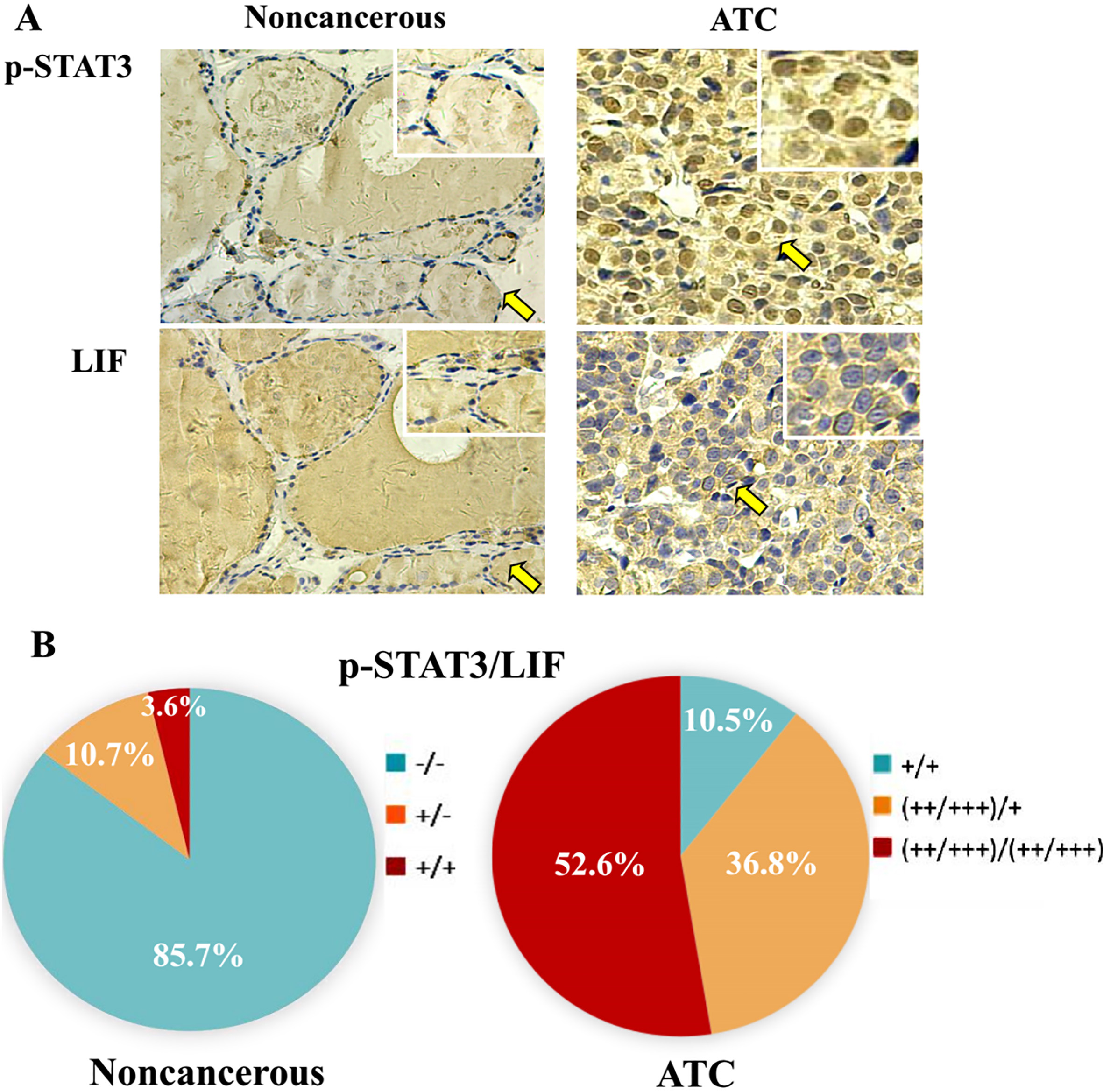
LIF-oriented immunohistochemical staining (Table 1 and Fig. 5) showed that 85.6% (24/28) of noncancerous thyroid tissues is negative in LIF expression and the remaining 14.4% (4/28) expressed LIF at a low level (); while all of 19 ATC cases was positive in LIF expression, of which 89.5% (17/19) was strongly positive (). The nuclear translocation of p-STAT3 was hardly observed in the nuclei of the noncancerous thyroid epithelail cells except one case (3.6%); intranuclear p-STAT3 labeling was observed in all of the 19 ATC cases studied (100%) and 10 of them (52.6%) was strongly positive ().
Correlation of LIF expression with STAT3 activation in ATC tissues
The positive rates of LIF and p-STAT3 in the epithelial cells of the noncancerous thyroid tissues adjacent to cancer were 14.4% and 3.6% respectively, although strong background was observed in the lumens of thyroid follicles; the positive rates of LIF and p-STAT3 in ATC tissues were 100% with higher levels of their production. The correlation analysis (Pearson) indicated the close correlation of LIF expression with the p-STAT3 nuclear translocation by showing 0.825 ( 0.01) of the correlation coefficient (Table 1 and Fig. 5).
Discussion
Anaplastic thyroid cancer is the most lethal thyroid malignancy without effective chemotherapeutic remedy [4]. Resveratrol, a natural-occuring polyphenol compound, has been known as a potential chemopreventive or chemotherapeutic drug because of its extremely low toxicity to normal tissues [8], multifaceted biological activities [20] and broad anticancer spectrum [9, 21, 22]. Our recent results show that resveratrol effectively reduces the risk of carcinogen-induced rat thyroid cancer formation [23], inhibits the growth of retinoic acid-resistantg human ATC cell lines via increasing oxidative stress [24] and reverses retinoic acid-resistance of ATC cells by erasing methylation in CRABP2 promoter region [25]. Nevertheless, there are still some ATC cell lines such as THJ-11T are not sensitive to resveratrol, suggesting the necessity to investigate the internal factors leading to their resveratrol resistance for personalized therapy.
Resveratrol exerts multiple biological effects on cancer cells, of which inhibition of STAT3 activation is considered as a critical molecular event [16, 26, 27]. The activated STAT3 signaling pathway is also important for ATC cells because it regulates stem cell attributes of ATC [13] and inhibition of STAT3 activation delays the thyroid cancer formation in obese mice [28]. STAT3 pathway can be activated by IL-6-like cytokines and mediates drug resistance of thyroid cancers [2]. Given the above evidence, we supposed that the status of STAT3 signaling might be related with the anti-ATC efficacy of resveratrol. In view of the above reasons, the statuses of STAT3 pathway in resveratrol-sensitive THJ-16T and THJ-21T and -resistant THJ-11T ATC cells before and after drug treatment were analyzed. The results showed that after 48 hours after 100 M resveratrol treatment, p-STAT3 levels in THJ-16T and THJ-21T cells were significantly decreased accompanied with the reduced frequencies of p-STAT3 nuclear translocation. These results suggest the ability of resveratrol to block STAT3 signaling via inhibiting the phosphorylation of STAT3 protein in THJ-16T and THJ-21T cells and the inactivated STAT3 signaling results in suppressed cell proliferation and apoptosis. Unlike the situation in THJ-16T and THJ-21T cells, STAT3 expression and p-STAT3 level were lower in normally cultured THJ-11T cells, indicating their lesser importance in the maintenance of THJ-11T cells. Interestingly, STAT3 signaling activity is increased in resveratrol-treated THJ-11T cells in terms of upregulated STAT3 expression, increased p-STAT3 level as well as nuclear translocation. STAT3 activation has been known as an important cell survival machinary, which leads to drug resistance of cancer cells [30]. Therefore, selective inhibition of STAT3 signal transduction become one of the main strategies of anti-cancer therapy. This point is further confirmed by the data from resveratrol-treated THJ-16T and THJ-21T cells. However, few reports are available concerning resveratrol-promoted STAT3 expression and activation in cancer cells resistant to it. Our results demonstrate that the responding manners of STAT3 to resveratrol are related with resveratrol’s anti-ATC efficacy. These findings indicate that the activation of STAT3 signaling in resveratrol-treated THJ-11T cells may be a protective response to prevent cell death and, therefore, result in drug resistance.
AG490, a synthetic derivative of benzomethylene malonitrile, has been used as a specific inhibitor of JAK/STAT3 [31]. AG490 can inhibit the proliferation of human medullary thyroid cancers and improve their radiosensitivity [32]. To elucidate the relevance of the activated STAT3 signaling with the resveratrol resistance, THJ-11T cells were treated by 80 M AG490 alone or combined with 100 M resveratrol, respectively. The results showed that the inhibitory rate of 80 M AG490 on the growth of THJ-11T cells was 27.2%, which significantly increased to 59.8% when 80 M AG490 and 100 M resveratrol were used together, further confirming the importance of STAT3 signaling in maintaining the survival of ATC cells. We therefore consider that the activated STAT3 signaling would be a key factor to rescue ATC cells from resveratrol treatment and the blockage of its activation by AG490 or other STAT3 inhibitor such as pyrimethamine can sensitize THJ-11T cells to resveratrol. Because the expression and distribution of NF-B/p65 in three ATC cell lines remain stable irrespective to resveratrol treatment, its involvement in determining the anti-ATC efficacy of resveratrol can be largely ruled out.
Because of the critical roles of STAT3 signaling in determing the response of ATC cells to resveratrol, it is necessary to investigate the upstream activator of STAT3 in THJ-11T cells before and after resveratrol treatment. Leukemia inhibitory factor/LIF is a famous member in IL-6 family, which specifically binds to gp130 receptor and activates STAT3 signaling pathway [33, 34]. We therefore checked the in vitro and in vivo expression patterns of LIF in ATCs and analyzed its correlation with activation of STAT3 pathway. In similar patterns with STAT3 alteration, 100 M resveratrol significantly decreased LIF expression in THJ-16T and THJ-21T, while LIF in THJ-11T cells was upregulated upon resveratrol treatment. In accordance with the in vitro findings, tissue-microarray based immunohistochemical staining revealed that STAT3 nuclear translocation and upregulated LIF expression were closely correlated and frequently observed in ATC rather than the noncancerous thyroid tissues. These results suggest that LIF may act as an upstream survival factor for resveratrol-treated THJ-11T cells via triggering STAT3 activation.
Taken together, our in vitro and in vivo analyses demonstrate that the status of STAT3 signaling in resveratrol-treated ATC cells is a critical element in determining the therapeutic outcome. Frequent LIF expression and its close correlation with STAT3 activation are evidenced in ATC cells and surgical specimens, which suggest a poor prognosis in ATC patients. In this context, LIF and/or IL-6 targeting therapy may improve the outcome of anti-ATC chemotherapy.
Footnotes
Acknowledgments
The authors express gratitude to Dr. Quentin Liu at the Institute of Cancer Stem Cell, Dalian Medical University, for providing the three human anaplastic thyroid cancer cell lines. This work was supported by the grants from the National Natural Science Foundation of China (No. 81272786 and 81450016 to Dr. J Liu), the Program Fund for Liaoning Provincial Department of Education Key Laboratory (LF2017002 to Dr. J Liu), and the fund from Natural Science Foundation of Liaoning Province (No. 2019-ZD-0650 to Dr. PN Li).
References
1.
La VecchiaC.MalvezziM.BosettiC.GaravelloW.BertuccioP.LeviF. and NegriE., Thyroid cancer mortality and incidence: a global overview, Int J Cancer136(9) (2015), 2187–2195.
2.
HedingerC.WilliamsE.D. and SobinL.H., The WHO histological classification of thyroid tumors: a commentary on the second edition, Cancer63(5) (1989), 908–911.
3.
SmallridgeR.C.AinK.B.AsaS.L.BibleK.C.BrierleyJ.D.BurmanK.D.KebebewE.LeeN.Y.NikiforovY.E.RosenthalM.S.ShahM.H.ShahaA.R.TuttleR.M., T. American Thyroid Association Anaplastic Thyroid Cancer Guidelines, American Thyroid Association guidelines for management of patients with anaplastic thyroid cancer, Thyroid22(11) (2012), 1104–1139.
O’NeillJ.P. and ShahaA.R., Anaplastic thyroid cancer, Oral Oncol49(7) (2013), 702–706.
6.
HaddadR.I.LydiattW.M.BallD.W.BusaidyN.L.ByrdD.CallenderG.DicksonP.DuhQ.Y.EhyaH.HaymartM.HohC.HuntJ.P.IagaruA.KandeelF.KoppP.LamonicaD.M.McCaffreyJ.C.MoleyJ.F.ParksL.RaeburnC.D.RidgeJ.A.RingelM.D.ScheriR.P.ShahJ.P.SmallridgeR.C.SturgeonC.WangT.N.WirthL.J.HoffmannK.G. and HughesM., Anaplastic thyroid carcinoma, Version 2.2015, J Natl Compr Canc Netw13(9) (2015), 1140–1150.
7.
LanL.BasourakosS.CuiD.ZuoX.DengW.HuoL.ChenH.ZhangG.DengL.ShiB. and LuoY., ATRA increases iodine uptake and inhibits the proliferation and invasiveness of human anaplastic thyroid carcinoma SW1736 cells: involvement of beta-catenin phosphorylation inhibition, Oncol Lett14(6) (2017), 7733–7738.
8.
BorrielloA.BencivengaD.CaldarelliI.TramontanoA.BorgiaA.ZappiaV. and Della RagioneF., Resveratrol: from basic studies to bedside, Cancer Treat Res159 (2014), 167–184.
9.
RaufA.ImranM.ButtM.S.NadeemM.PetersD.G. and MubarakM.S., Resveratrol as an anti-cancer agent: a review, Crit Rev Food Sci Nutr58(9) (2018), 1428–1447.
10.
LiY.T.TianX.T.WuM.L.ZhengX.KongQ.Y.ChengX.X.ZhuG.W.LiuJ. and LiH., Resveratrol suppresses the growth and enhances retinoic acid sensitivity of anaplastic thyroid cancer cells, Int J Mol Sci19(4) (2018).
11.
LiC.LiH.ZhangP.YuL.J.HuangT.M.SongX.KongQ.Y.DongJ.L.LiP.N. and LiuJ., SHP2, SOCS3 and PIAS3 expression patterns in medulloblastomas: relevance to STAT3 activation and resveratrol-suppressed STAT3 signaling, Nutrients9(1) (2016).
12.
SlatteryM.L.LundgreenA.KadlubarS.A.BondurantK.L. and WolffR.K., JAK/STAT/SOCS-signaling pathway and colon and rectal cancer, Mol Carcinog52(2) (2013), 155–166.
13.
ShiraiwaK.MatsuseM.NakazawaY.OgiT.SuzukiK.SaenkoV.XuS.UmezawaK.YamashitaS.TsukamotoK. and MitsutakeN., JAK/STAT3 and NF-kappaB signaling pathways regulate cancer stem-cell properties in anaplastic thyroid cancer cells, Thyroid29(5) (2019), 674–682.
14.
ParkJ.I.StrockC.J.BallD.W. and NelkinB.D., Interleukin-1beta can mediate growth arrest and differentiation via the leukemia inhibitory factor/JAK/STAT pathway in medullary thyroid carcinoma cells, Cytokine29(3) (2005), 125–134.
15.
LumachiF.BassoS.M. and OrlandoR., Cytokines, thyroid diseases and thyroid cancer, Cytokine50(3) (2010), 229–233.
16.
YuL.J.WuM.L.LiH.ChenX.Y.WangQ.SunY.KongQ.Y. and LiuJ., Inhibition of STAT3 expression and signaling in resveratrol-differentiated medulloblastoma cells, Neoplasia10(7) (2008), 736–744.
17.
ZhongL.X.ZhangY.WuM.L.LiuY.N.ZhangP.ChenX.Y.KongQ.Y.LiuJ. and LiH., Resveratrol and STAT inhibitor enhance autophagy in ovarian cancer cells, Cell Death Discov2 (2016), 15071.
18.
MarlowL.A.D’InnocenziJ.ZhangY.RohlS.D.CooperS.J.SeboT.GrantC.McIverB.KasperbauerJ.L.WadsworthJ.T.CaslerJ.D.KennedyP.W.HighsmithW.E.ClarkO.MilosevicD.NetzelB.CradicK.AroraS.BeaudryC.GrebeS.K.SilverbergM.L.AzorsaD.O.SmallridgeR.C. and CoplandJ.A., Detailed molecular fingerprinting of four new anaplastic thyroid carcinoma cell lines and their use for verification of RhoB as a molecular therapeutic target, J Clin Endocrinol Metab95(12) (2010), 5338-5347.
19.
ZhangP.LiH.YangB.YangF.ZhangL.L.KongQ.Y.ChenX.Y.WuM.L. and LiuJ., Biological significance and therapeutic implication of resveratrol-inhibited Wnt, Notch and STAT3 signaling in cervical cancer cells, Genes Cancer5(5-6) (2014), 154–164.
20.
ShuX.H.WangL.L.LiH.SongX.ShiS.GuJ.Y.WuM.L.ChenX.Y.KongQ.Y. and LiuJ., Diffusion efficiency and bioavailability of resveratrol administered to rat rrain by different routes: therapeutic implications, Neurotherapeutics12(2) (2015), 491–501.
21.
KimS.E.ShinS.H.LeeJ.Y.KimC.H.ChungI.K.KangH.M.ParkH.R.ParkB.S. and KimI.R., Resveratrol induces mitochondrial apoptosis and inhibits epithelial-mesenchymal transition in oral squamous cell carcinoma cells, Nutr Cancer70(1) (2018), 125–135.
22.
LiY.G.XiaH.J.TaoJ.P.XinP.LiuM.Y.LiJ.B.ZhuW. and WeiM., GRIM19mediated Stat3 activation is a determinant for resveratrolinduced proliferation and cytotoxicity in cervical tumorderived cell lines, Mol Med Rep11(2) (2015), 1272–1277.
23.
ZhengX.JiaB.SongX.KongQ.Y.WuM.L.QiuZ.W.LiH. and LiuJ., Preventive potential of resveratrol in carcinogen-induced rat thyroid tumorigenesis, Nutrients10(3) (2018).
24.
ZhengX.JiaB.TianX.T.SongX.WuM.L.KongQ.Y.LiH. and LiuJ., Correlation of reactive oxygen species levels with resveratrol sensitivities of anaplastic thyroid cancer cells, Oxidative Medicine and Cellular Longevity2018 (2018), 1.
25.
LiuX.LiH.WuM.L.WuJ.SunY.ZhangK.L. and LiuJ., Resveratrol reverses retinoic acid resistance of anaplastic thyroid ancer cells via demethylating CRABP2 gene, Frontiers in Endocrinology10 (2019), 734.
26.
KhanM.W.SaadallaA.EwidaA.H.Al-KatranjiK.Al-SaoudiG.GiacconeZ.T.GounariF.ZhangM.FrankD.A. and KhazaieK., The STAT3 inhibitor pyrimethamine displays anti-cancer and immune stimulatory effects in murine models of breast cancer, Cancer Immunol Immunother67(1) (2018), 13–23.
27.
ZhongL.X.LiH.WuM.L.LiuX.Y.ZhongM.J.ChenX.Y.LiuJ. and ZhangY., Inhibition of STAT3 signaling as critical molecular event in resveratrol-suppressed ovarian cancer cells, J Ovarian Res8 (2015), 25.
28.
ParkJ.W.HanC.R.ZhaoL.WillinghamM.C. and ChengS.Y., Inhibition of STAT3 activity delays obesity-induced thyroid carcinogenesis in a mouse model, Endocr Relat Cancer23(1) (2016), 53–63.
29.
NotarangeloT.SisinniL.TrinoS.CaliceG.SimeonV. and LandriscinaM., IL6/STAT3 axis mediates resistance to BRAF inhibitors in thyroid carcinoma cells, Cancer Lett433 (2018), 147–155.
30.
WangX.M.LiuY.FanY.X.LiuZ.YuanQ.L.JiaM.GengZ.S.GuL. and LuX.B., LncRNA PTCSC3 affects drug resistance of anaplastic thyroid cancer through STAT3/INO80 pathway, Cancer Biol Ther19(7) (2018), 590–597.
31.
Caceres-CortesJ.R., A potent anti-carcinoma and anti-acute myeloblastic leukemia agent, AG490, Anticancer Agents Med Chem8(7) (2008), 717–722.
32.
LiJ.GanS.LuoC.LiuS. and PengZ., AG490 inhibits the proliferation of human medullary thyroid carcinoma TT cells and increases their radiosensitivity, Xi Bao Yu Fen Zi Mian Yi Xue Za Zhi31(6) (2015), 753–757.
33.
HuangS.LiuQ.LiaoQ.WuQ.SunB.YangZ.HuX.TanM. and LiL., Interleukin-6/signal transducer and activator of transcription 3 promotes prostate cancer resistance to androgen deprivation therapy via regulating pituitary tumor transforming gene 1 expression, Cancer Sci109(3) (2018), 678–687.
34.
GrivennikovS.I.GretenF.R. and KarinM., Immunity, inflammation, and cancer, Cell140(6) (2010), 883–899.