
Abstract
BACKGROUND:
Lung cancer is the main cancer-related deaths worldwide. In this study, we explored the clinical prognostic significance and functional role of miR-939-3p in lung cancer.
METHODS:
We analyzed the expression of miR-939-3p in lung cancer tissues and cells by qRT-PCR. The prognostic significance of miR-939-3p was investigated using the Kaplan-Meier survival and Cox regression analyses. The CCK-8 assay was used to determine the role of miR-939-3p in cell proliferation. Transwell assays were used to determine the effects of miR-939-3p on cell migration and invasion abilities.
RESULTS:
The expression of miR-939-3p was upregulated in cancer tissues and cell lines compared with adjacent normal tissues and normal cells, respectively. The upregulated miR-939-3p was significantly associated with lymph node metastasis, TNM stage and poor prognosis of lung cancer patients. After the transfection of miR-939 mimic, overexpression of miR-939-3p promoted lung cancer cell proliferation, migration, and invasion.
CONCLUSION:
These findings suggested that miR-939-3p acts as an oncogene and promotes cell proliferation, migration, and invasion in lung cancer. miR-939-3p may be a potential independent prognostic biomarker in lung cancer.
Introduction
Lung cancer is one of the most commonly diagnosed cancer with high incidence and high mortality rate worldwide [1]. Among lung cancer, approximately 85% is non-small cell lung cancer (NSCLC), which is associated with high mortality as the majority of patients have already reached an advanced stage at the time of initial diagnosis [2, 3, 4]. Although only about 15% of lung cancer cases are small cell lung cancer (SCLC), it has a higher degree of malignancy, faster progress and shorter over-survival compared to the NSCLC [5, 6]. Despite advances in standard treatments, such as surgery, radiotherapy, and chemotherapy, the 5-year overall survival rate of lung cancer patients is still relatively low. Thus, identifying novel molecules markers and new therapeutic strategies is still crucial.
MicroRNAs (miRNAs), a class of small noncoding (19–25 nucleotides) RNAs, serve as negative regulators of genes by binding to 3’-UTR of the target genes [7, 8]. Aberrant expression of miRNAs has been reported leading to altered biological function, such as cell growth, proliferation, differentiation, and migration [9, 10, 11]. Increasing evidence indicated that abnormal expression of miRNAs plays an important role in the pathological processes in various diseases, including cancers [12, 13, 14]. Identifying the role of miRNAs in diseases may provide a perspective therapeutics for the management of cancers or other diseases [14]. miR-939 has been reported dysregulated in several types of cancers, such as hepatocellular carcinoma [15], pancreatic cancer [16], and colon cancer [17]. miR-939-3p is found upregulated in non-small cell lung cancer and circulating miR-939 may have potential as a diagnostic biomarker for adenocarcinoma [18, 19]. However, the expression pattern of miR-939-3p in lung cancer tissues and functional role of miR-939-3p has not been thoroughly investigated in lung cancer.
In the present study, we examined the expression of miR-939-3p in lung cancer tissues and cell lines. We also analyzed the association between miR-939-3p expression and prognosis of lung cancer patients. In addition, the functional role of miR-939-3p in lung cancer was investigated.
Materials and methods
Patients and samples
A total of 118 pairs of lung cancer tissue specimens and adjacent normal tissue specimens were collected from lung cancer patients who underwent surgical resection between March 2011 and January 2013 at our hospital. All the patients did not receive any preoperative therapies, such as radiotherapy and chemotherapy. These tissue specimens were immediately put into liquid nitrogen and stored at
Cell culture and cell transfection
Human lung cancer cell lines (A549, H1299, HCC827, and H1650), as well as a non-cancerous MRC-5 cell line, were purchased from the Cell Source of the Academy of Sciences (Shanghai, China). All cell lines were cultured in DMEM medium (Invitrogen; Thermo Fisher Scientific, Inc., Waltham, MA, USA) containing 10% FBS (Invitrogen; Thermo Fisher Scientific, Inc.) and cultivated in a humidified incubator (Thermo Fisher Scientific, Inc.) at 37
Before transfection, lung cancer cells were seeded into a 6-well plate at 30–50% confluence. miR-939-3p mimic, mimic negative control (mimic NC), miR-939-3p inhibitor or inhibitor NC was transfected into lung cancer cells for miR-939-3p overexpression or inhibition. The transfection of cells was performed using Lipofectamine 3000 Reagent (Invitrogen; Thermo Fisher Scientific, Inc.) following the manufacturer’s protocol.
RNA extraction and qRT-PCR analysis
The total RNA was extracted from lung cancer tissues and cultured cells using TRIzol reagent (Invitrogen) according to the manufacturer’s instructions. Reverse transcription was performed using AMV first strand cDNA synthesis Kit (Sangon Biotech Co., Ltd., Shanghai, China). The expression levels of miR-939-3p were quantified using an SYBR Premix Ex Taq II kit (Takara, Dalian, China) in an ABI 7900HT Sequence Detection System (Applied Biosystems, Foster City, CA, USA) and normalized to U6. Relative expression of miR-939-3p was quantified with the 2
Cell proliferation assay
Cell proliferation assay was assessed by cell counting kit-8 (CCK-8, Dojindo, Kumamoto, Japan) following the manufacturer’s introduction. Lung cancer cells were incubated in 96-well plates (1
Relative expression of miR-939-3p in lung cancer tissues and cell lines was measured by qRT-PCR. A. Expression of miR-939-3p is increased in lung cancer tissues compared with normal tissues. B. Relative expression of miR-939-3p is higher in four lung cancer cell lines compared with normal cell line MRC-5. 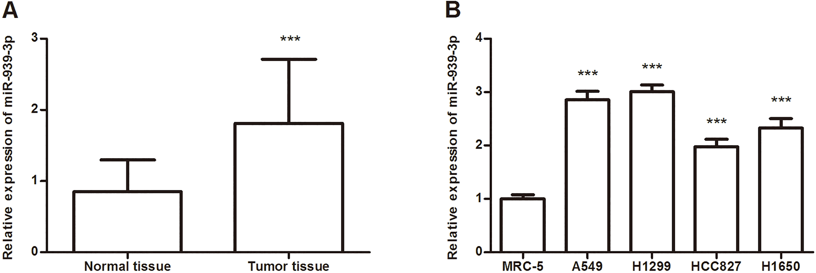
Cell migration and invasion assays were carried out using a 24-well Transwell system (Corning, NY, USA). For cell migration assay, transfected cells (1
Statistical analysis
Statistical analysis was performed using SPSS 20.0 software (SPSS, Inc., Chicago, IL, USA) and GraphPad Prism 5.0 software (GraphPad Software, Inc., Chicago, USA). All values were presented as mean
Results
Expression of miR-939-3p is increased in lung cancer
The expression of miR-939-3p in 118 paired human lung cancer tissues and adjacent normal tissues was analyzed using qRT-PCR, which revealed that miR-939-3p expression was increased in lung cancer tissues compared with normal tissues (
Correlations between lung cancer patients’ clinicopathological characteristics and miR-939-3p expression
The mean value of miR-939-3p level in lung cancer tissues was used as the cutoff point to divide lung cancer patients into low miR-939-3p expression group (
Correlation between miR-939-3p expression levels and clinical features in lung cancer patients
Correlation between miR-939-3p expression levels and clinical features in lung cancer patients
Kaplan-Meier method was used to evaluate the 5-year survival rate of patients with high and low expression of miR-939-3p.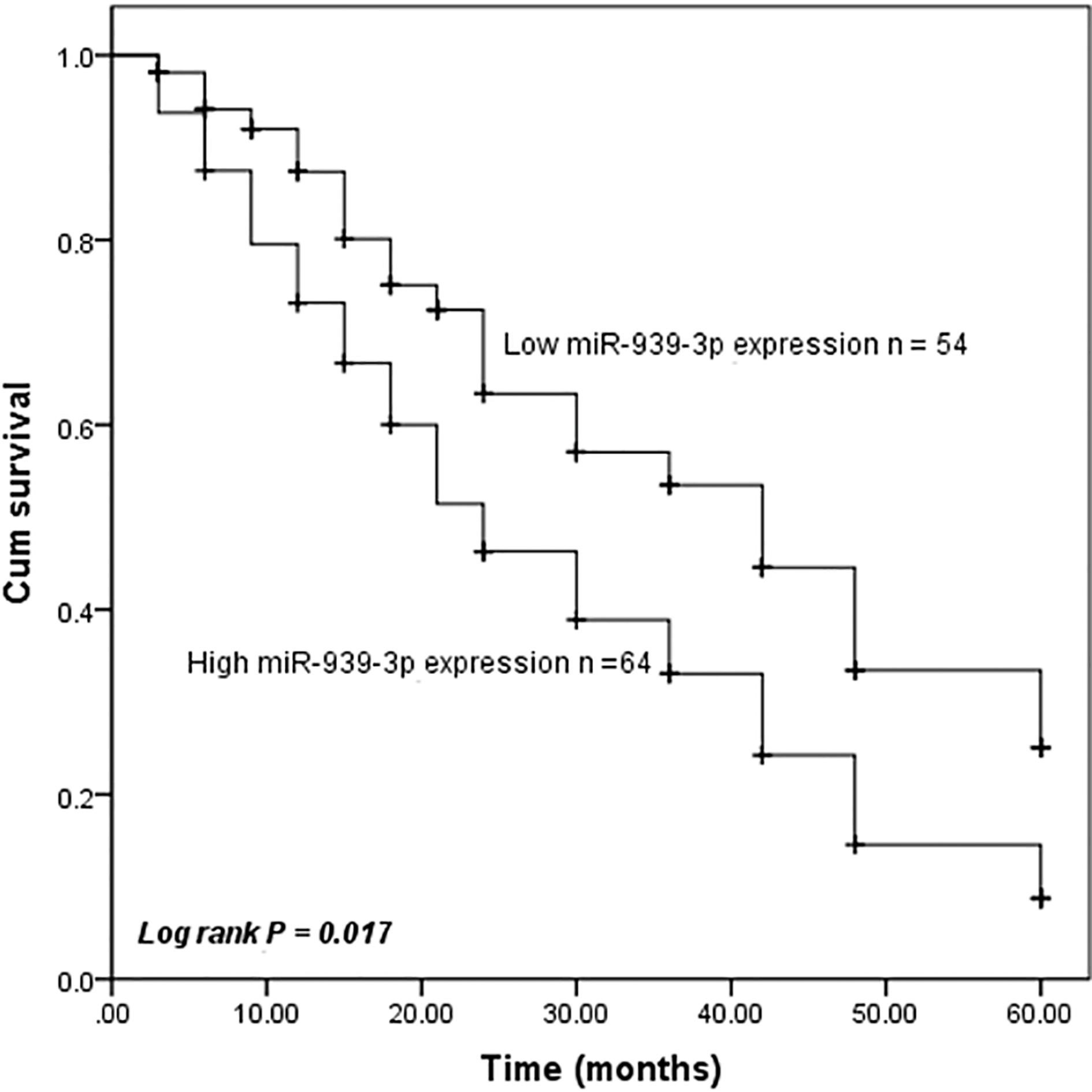
Considering miR-939-3p expression was significantly associated with lymph node metastases and TNM stage, we speculated that miR-939-3p expression might be related to the prognosis of lung cancer patients. To investigate the prognostic value of the miR-939-3p expression in lung cancer, Kaplan-Meier analysis was conducted to compare overall survival according to the miR-939-3p expression. As shown in Fig. 2, low miR-939-3p expression in tumor tissues showed a survival benefit in lung cancer patients (
Multivariate Cox analysis of clinical parameters in relation to overall survival
Multivariate Cox analysis of clinical parameters in relation to overall survival
Effects of miR-939-3p on cell proliferation in lung cancer using A549 and H1299 cells. A. The expression of miR-939-3p was measured in miR-939-3p mimic, mimic NC, miR-939-3p inhibitor, or inhibitor NC-transfected cells. B. Cell proliferation was determined using the CCK-8 assay. 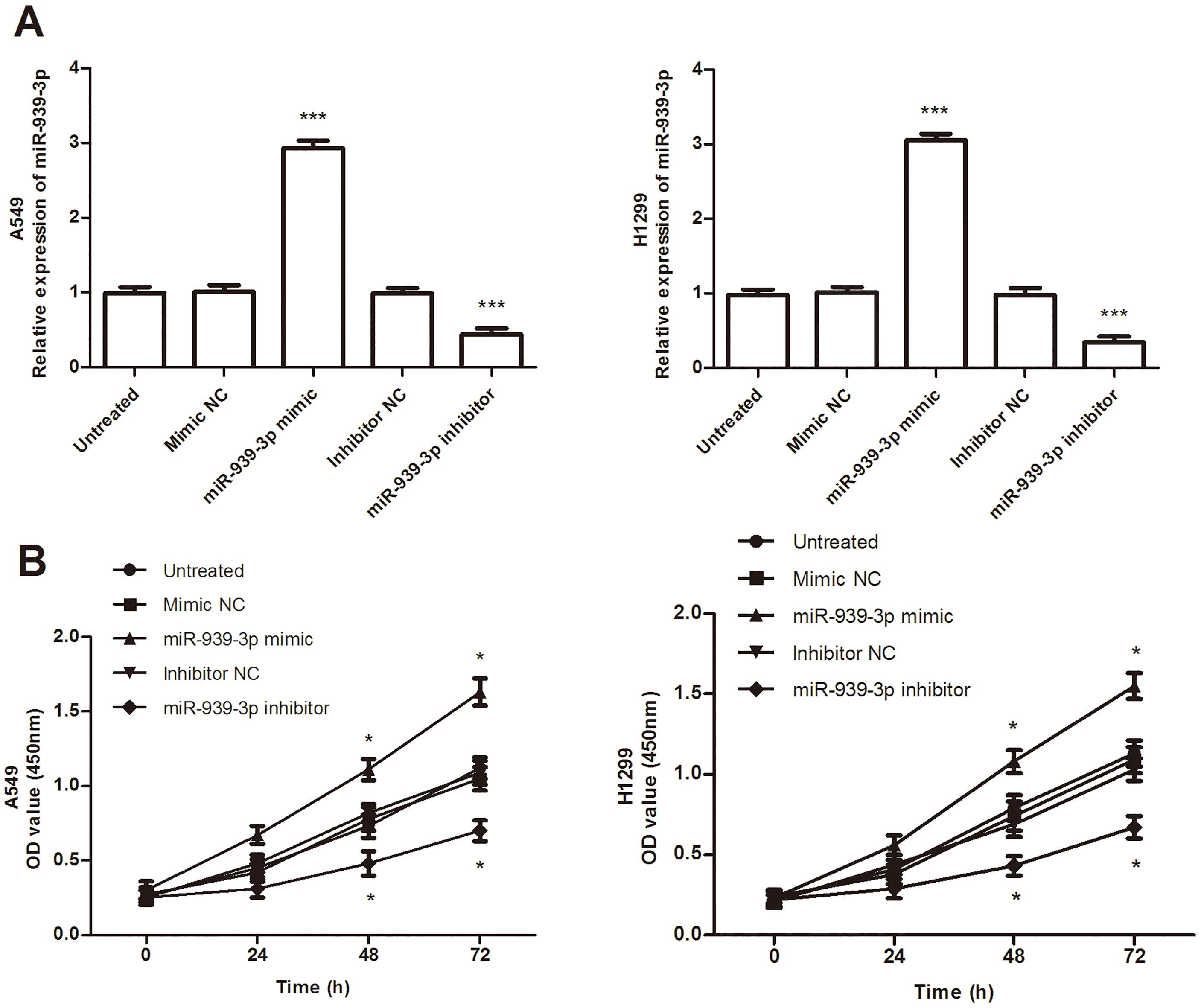
Effects of miR-939-3p on cell migration and invasion. A. Cell migration Transwell assay showed that overexpression of miR-939-3p promoted migration ability, while inhibition of miR-939-3p suppressed cell migratory ability, compared with untreated cells. B. Cell invasion Transwell assay indicated miR-939-3p promoted cell invasion. 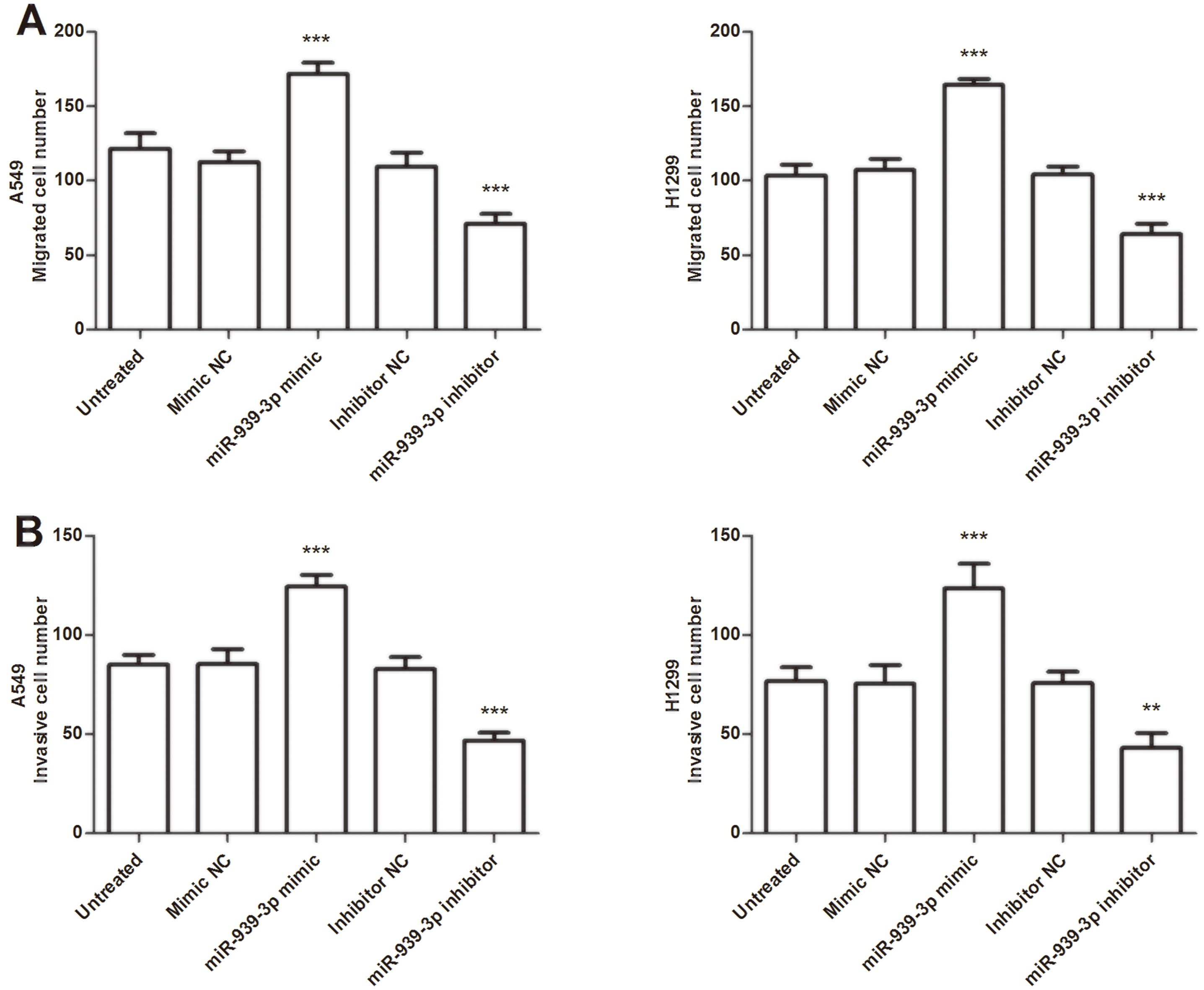
Because expression of miR-939-3p was found significantly upregulated in lung cancer tissues and cell lines, we investigated whether miR-939-3p acts as an oncogene role in cell proliferation, migration, and invasion of lung cancer. miR-939-3p mimic, miR-939-3p inhibitor, or their respective negative controls were transfected into A549 and H1299 cells, which have a relatively higher miR-939-3p expression. The qRT-PCR results showed that the expression of miR-939-3p in cells was significantly increased by miR-939-3p mimic, while decreased by the miR-939-3p inhibitor (
Discussion
Lung cancer is the main cause of cancer-related mortality, with a relatively low overall survival worldwide [20]. Identifying novel biomarkers as possible prognostic predictors or therapeutic targets is still crucial. More and more studies indicated that the diverse roles of miRNAs in the development of various malignant neoplasm [13, 21, 22]. Some miRNAs can be a diagnostic or/and prognostic biomarker for cancers. For instance, Wang et al. suggested that the high expression of miR-411 was a diagnostic and prognostic biomarker for NSCLC patients [23]. Expression of miR-206 was unregulated and the expression of miR-145 was downregulated in breast cancer, both of them might serve as an important prognostic indicator of patients with breast cancer [24]. These above studies demonstrated that abnormal expression of miRNAs has potential diagnostic or prognostic value in cancers.
In the previous studies in lung cancer, Rani et al. applied global miRNAs expression profiling approaches in serum from lung adenocarcinoma patients and identified circulating miRNAs, including miR-939, having potential as diagnostic biomarkers for lung adenocarcinoma [19]. Based on miRNA and miRNA profiling, Yang et al. explored a novel therapeutic target of NSCLC, in which miR-939-3p is found upregulated in NSCLC tissues [18]. In another study by Ma et al. also constructed a global interaction network of miRNA-miRNA in NSCLC samples and found miR-1228 and miR-939 as essential nodes in network analysis [25]. These studies showed that miR-939 is potentially important for lung cancer. However, the role of miR-939-3p has not been thoroughly investigated in lung cancer.
In the present study, we investigated the expression of miR-939-3p in lung cancer tissues and cell lines. The results showed that the expression of miR-939-3p is increased in lung cancer tissues and cell lines, which is consistent with the results in the previous study [18, 19]. Overexpression of miR-939-3p is found to be closely associated with lymph node metastasis and advanced TNM stage, which suggested miR-939-3p expression may be involved in the development of lung cancer. According to the Kaplan-Meier analysis and Cox regression analysis results, overexpression of miR-939-3p is associated with shorter overall survival of patients and is an independent prognostic factor for lung cancer.
Numerous studies also identified that some miRNAs act as an oncogene or suppressor gene and involved in the progression of various cancers [26, 27, 28]. For instance, miR-338-3p functions as a suppressor gene in bladder cancer and inhibited the proliferation, metastasis, and EMT in bladder cancer through regulating ETS1 expression [27]. miR-1260b is found upregulated in NSCLC, promotes the cell migration and invasion, and can serve as a putative target for diagnosis and treatment of NSCLC [28]. Considering the expression of miR-939-3p is increased in lung cancer tissues and cell lines, we investigated whether miR-939-3p acts as an oncogene role in cell proliferation, migration, and invasion of lung cancer. The results showed that overexpression of miR-939-3p by miR-939-3p mimic promoted cell proliferation, migration, and invasion abilities, while inhibition of miR-939-3p suppresses those abilities. The development and progression of cancers is affected by a number of genes and signaling pathways [29, 30]. In gastric cancer, miR-939 exerted its function mainly through inhibiting SLC34A2/Raf/MEK/ERK pathway [31]. In breast cancer, miR-939 was also found highly expressed and identified its extracellular protumorigenic role directly targeting VE-cadherin [32]. In ovarian cancer, miR-939 functions as a potential tumor promoter that promotes the proliferation of ovarian cancer cells by regulating the Wnt/
Taken together, miR-939-3p is upregulated in lung cancer tissues and cell lines and its overexpression promotes cell proliferation, migration, and invasion in vitro. What’s more, miR-939-3p may be an important prognostic biomarker and therapeutic target for the management of lung cancer.
Footnotes
Acknowledgments
This work was supported by Science and Technology Program of Binzhou Medical University Hospital (BY2015KJ31).
Conflict of interest
The authors declare that they have no conflicts of interest.