
Abstract
BACKGROUND:
Papillary thyroid carcinoma (PTC) is the most common thyroid malignancy which is generally accompanied by lymph node metastasis.
OBJECTIVE:
Our study evaluated whether carbon nanoparticle lymph node tracer can improve the outcomes of surgical treatment in papillary thyroid cancer (PTC).
METHODS:
Ninety-two patients were selected and underwent total thyroidectomy and lymph node resection. Our study placed 45 individuals into the treatment group (carbon nanoparticle group) and 47 cohorts in the control group (no carbon nanoparticle group).
RESULTS:
Carbon nanoparticle application remarkably improved lymph nodes detection rate in patients (4.7
CONCLUSIONS:
Our study definitively showed that carbon nanoparticles can be used to effectively treat lymphatic carcinoma. Our study presented clinical evidences for potential application of carbon nanoparticle in improving the management of PTC patients.
Keywords
Introduction
Papillary thyroid carcinoma (PTC) is the most common thyroid malignancy which is generally accompanied by lymph node metastasis [1, 2, 3, 4]. Thyroidectomy is the preferred treatment for the malignancy owing to lower recurrence rate, comparable postoperative complication rate, and parathyroid function recovery [5]. Despite this, complications such as accidental devascularization of parathyroid gland, cervical hematomas and other hemodynamic disorders could occur which present challenges to both the patients affected and the clinician treating the condition [6, 7]. Our study evaluated a technique to determine whether nanoparticles could bring better outcomes in PTC surgery.
Demographic information of subjects in the cohorts (
92) and comparison between two groups
Demographic information of subjects in the cohorts (
Carbon nanoparticles have proved to be efficacious in identifying lymph nodes in breast and thyroid cancers [8, 9, 10]. carbon nanoparticles contain particles with the average diameter of 150 nm, and their small size gives them an advantage to pass easily through the lymphatic vessels instead of the blood capillaries. Macrophages, an important part of white blood cell are able to identify and engulf these nanoparticles which make their way into the lymphatic vessels; these particles are easily identified due to their accumulation in the lymphatic system and thus, are effortlessly stained. Lymph node metastasis could be identified using this procedure and it has provided benefit to clinicians aiming to explore metastatic lymph nodes in the breast and the thyroid of affected individuals. carbon nanoparticles are characterized by the fact that they do not enter the blood circulation and pose no significant side effect to the patients. The Chinese Food and Drug Administration has approved this treatment since 2007 depicting its safe usage among cancer patients [11].
Despite the advantages of this technique, the clinical use is still limited and requires further investigation. Hence, the focus of our study was to evaluate the application of carbon nanoparticles in identifying lymph node metastasis and improving outcomes of surgical treatment in papillary thyroid cancer.
Clinical data
Our study contained 92 cases (29 males and 63 females). The study was approved by the Thyroid Surgery Department in The First Affiliated Hospital of Zhengzhou University from February 2013 to May 2015. The cohorts all displayed normal levels of PTH and serum calcium and were assigned into treatment group (45 cases) and control group (47 cases) based on the informed consent and self-selection of patients. Our study found no significant differences in gender, age, tumor pathological stage, histological grade or tumor location in the cohorts studied (Table 1). The preoperative ultrasound examination was performed on the thyroid and neck lymph nodes for all the patients by the same technician with full experiences. The ultrasound diagnosis showed 2 cases of T1aN0M0 stage, 5 cases in T1aN1aM0, 7 cases of T1bN0M0, 62 cases of T1bN1aM0, 6 cases of T2N1aM0, 2 cases of T3N0M0 and 8 cases of T3N1aM0 according to AJCC 7th Edition (2010) Thyroid Cancer TNM classification [7, 12, 13, 14]. PTCs were confirmed with both frozen pathological examination during operation and postoperative evaluation, and were shown to be well differentiated. There were 12 multifocal PTCs cases and 80 solitary cases. No subtype was further classified. There were no cases of vascular invasion, esophageal invasion, or recurrent laryngeal nerve invasion and the tumor features were summarized in Table 2. There were 12 cases of bilateral thyroid papillary carcinoma (including 3 cases of Hashimoto’s thyroiditis), 34 cases of left thyroid papillary carcinoma (including 4 cases of Hashimoto’s thyroiditis), 46 cases of right thyroid papillary carcinoma (including 6 cases of Hashimoto’s thyroiditis). In follow-up for 4 years after surgery, no death was reported for all the patients while 4 cases of cervical lymph node recurrence and metastasis were found after surgery. All patients have signed the informed consent.
Primary tumor size of subjects in the cohorts (
92)
Primary tumor size of subjects in the cohorts (
Detection of lymph node with carbon nanoparticles (CNP) suspension in total thyroidectomy. (A and B) carbon nanoparticles suspension was injected into a thyroid. (C and D) Identified black staining lymph nodes in thyroid and the resection tissues (arrow).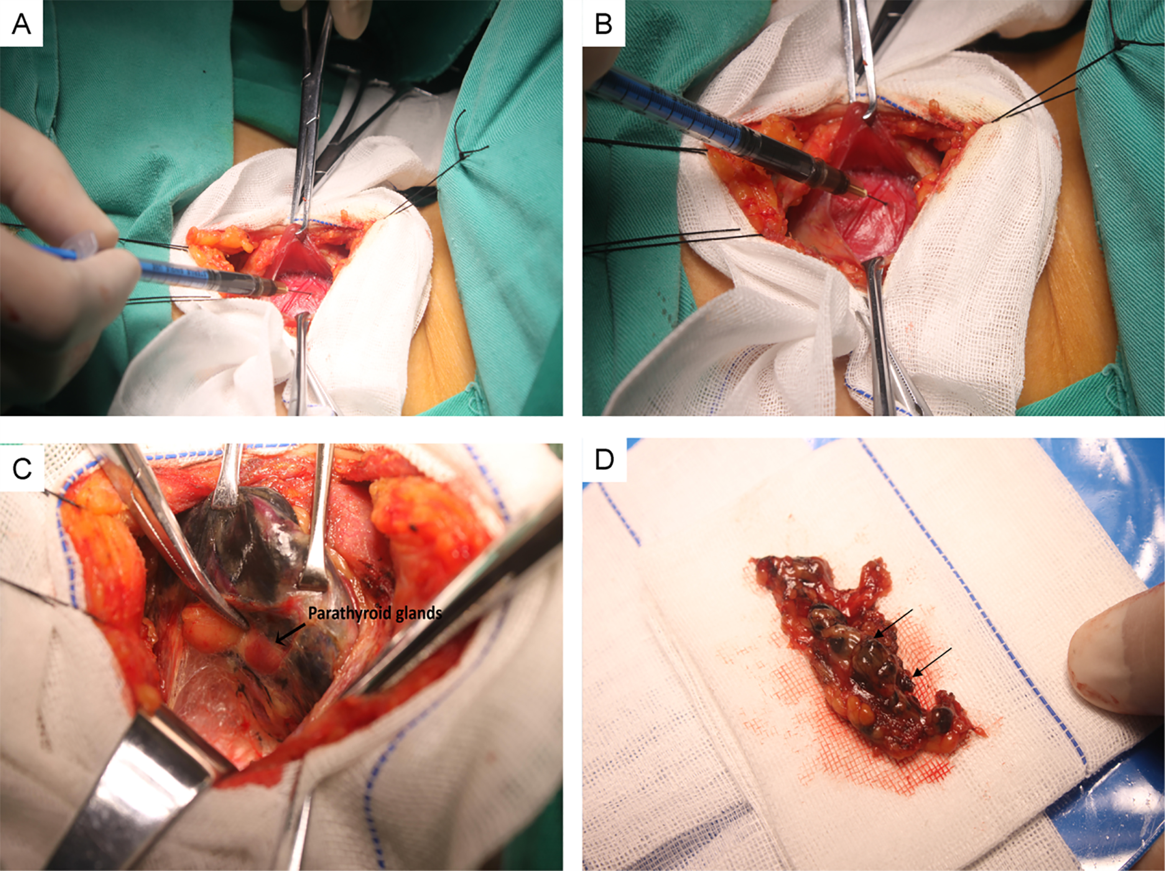
The carbon nanoparticle suspension in this study was purchased from Chongqing Lesmills Pharmaceutical Co., Ltd. National approved drug No. H20073246.
Surgical methods
Ultrasound diagnosis was implemented preoperatively by professional ultrasound technician with full experiences in the thyroid examination. Before surgeries, thyroid nodules were classified according to TI-RADS (Thyroid Imaging Reporting and Data System). Pathological diagnosis identified classic papillary thyroid tumors with no further classification of different subtypes. Level VI bilateral dissection with total/complete thyroidectomy was performed. For a brief description of surgery procedure, we first entered the lateral thyroid space, separated the upper thyroid gland and protected the superior parathyroid gland. Then we searched and protected the inferior parathyroid gland, isolated and protected the recurrent laryngeal nerve. Afterwards, we completely removed one side of the thyroid tissue and performed contralateral thyroidectomy with the same method. The removed tissue samples were fast frozen and examined. From the results, patients confirmed with papillary thyroid carcinoma were followed with the bilateral 6-area (e.g. the central area) lymph node dissection. Every patient received surgical anesthesia and had their necks hyper-extended and in a supine position. A curved incision of 2 cm was made above the sternal notch along the striate. In order to minimize damage to the lymphatic system, we only removed the front thyroid; the side and lateral thyroid were not resected. Withdrawing was performed first to prevent injecting into the blood vessels and then carbon nanoparticle suspensions (0.1–0.2 ml per injection) were gently injected into four randomly selected points in the thyroid using a skin test needle (Fig. 1A and B), After approximately 20 min, total thyroidectomy was performed (Fig. 1C and D), and then the samples were frozen and sent for further pathological confirmation before dissection of the central region of the lymph node (Fig. 2). The surgeries for both treatment group and control group were performed by the same group of physicians. Similarly, the postoperative pathological evaluations were also implemented by the same group of physicians.
Pathological images of lymph node and papillary thyroid cancer (PTC) tissues in two groups. (A and B) Lymph node and PTC images from control group (HE, x200) (C and D) Lymph node and PTC images from carbon particles group (HE, x200).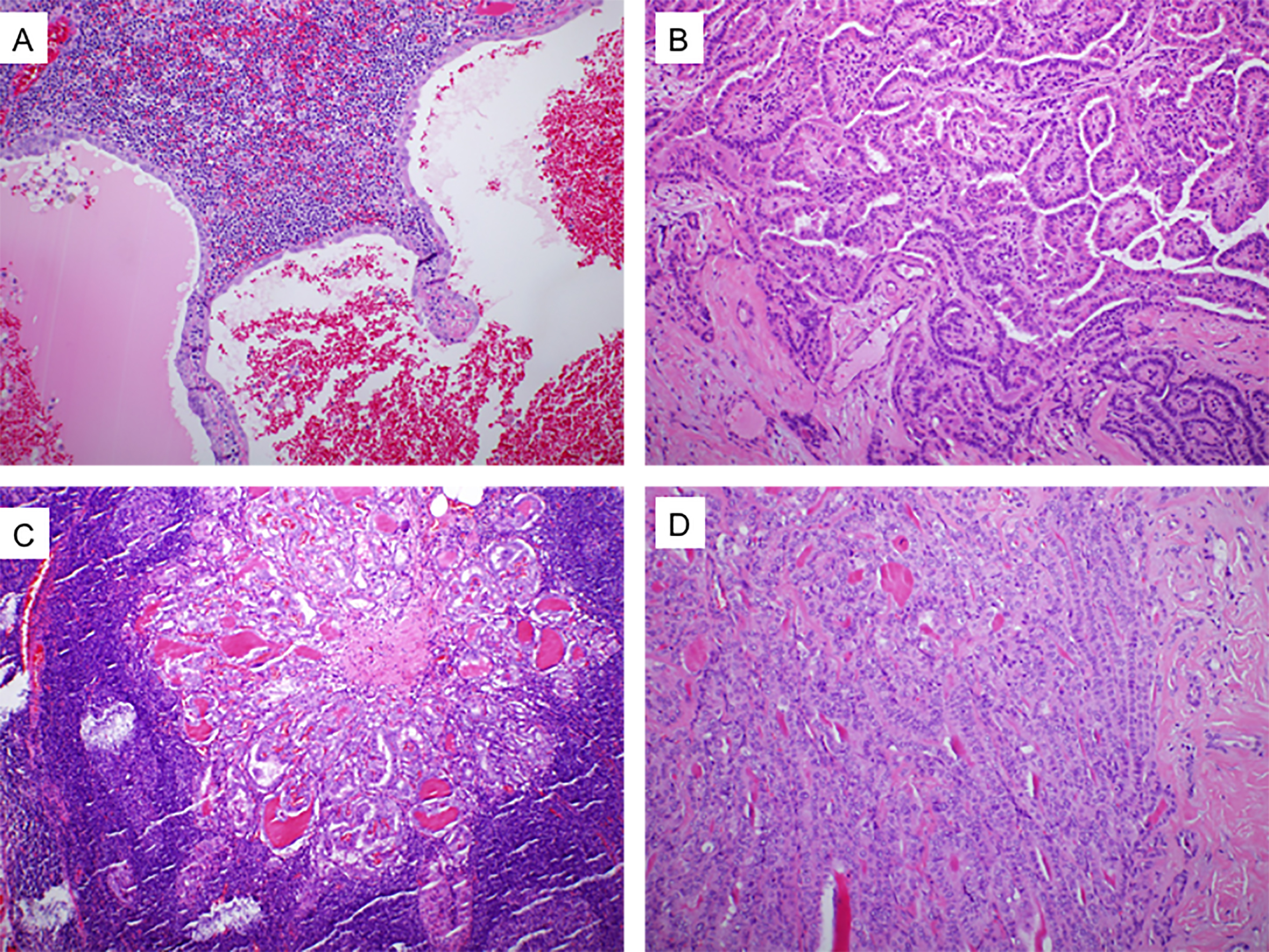
All resected metastatic lymph node specimens were examined by two pathologists independently. The number of stained lymph nodes in carbon nanoparticle group and lymph nodes in control group were recorded separately, and then compared. The presence of parathyroid was also analyzed simultaneously.
Management of postsurgical hypoparathyroidism and hypocalcemia
Normal range of Parathyroid hormone (PTH) and Calcium in serum is 15–65 ng/L and 2.0–2.7 mmol/L, respectively. PTH and calcium level were monitored 3 d after surgery. Re-examination was performed after the first and the sixth month. For patients with hypocalcemia, intravenous infusion of calcium gluconate was implemented. The dose was gradually reduced as the clinical symptoms ameliorated among patients. Specifically, we changed the intravenous infusion to oral administration of calcium and Rocaltrol (Calcitriol Soft Capsules), and withdrew the treatment after the symptoms disappeared.
Statistical analysis
Data is presented as a percentage. The statistic difference between treatment and control groups was analyzed with SPSS13.0 (SPSS Inc., Chicago, IL, USA) using Chi-square test. P value less than 0.05 was considered as significant.
Results
Number and size of the detected lymph nodes
In comparison to the control group (3.5
Lymph node detection and compliment rates in two groups
Lymph node detection and compliment rates in two groups
CNP: carbon nanoparticle.
To investigate the ability of carbon nanoparticles in identifying metastatic lymph nodes, we calculated and compared the number of detected metastatic lymph nodes between two groups. As a result, it was indicated that there is no statistical difference (
Assessment of unexpected parathyroid removal
Upon the application of carbon nanoparticles, the rate of resection for the parathyroid gland significantly decreased in the treatment group (3/45) in comparison to the control group (10/47) (
Hypoparathyroidism and hypocalcaemia
Our study reported 5 cases of detected hypoparathyroidism (11%) in the treatment group, and the control group exhibited 14 cases (29.79%) of similar cases after one month of post-surgery. Hypocalcaemia rate was 8.88% and 25.53% in two groups (Table 3). Both conditions were markedly different due to the use of carbon nanoparticles after PTC operation. All cohorts in the study experienced post-operative hypoparathyroidism and had recovered parathyroid function within six months of thyroid resection surgery. The details about hypoparathyroidism and hypocalcemia among patients were shown in Table 3.
Discussion
Our study showed that the carbon nanoparticle was effective in assisting the detection of lymph nodes. We found that the conventional lymph node evaluation and counting methods are not as accurate as the technology of carbon nanoparticles [15]. Our study used carbon nanoparticles which effectively stained lymph nodes in black color thus, it was fairly identified by surgeons [16]. The metastatic rate between the black and non-black lymph nodes was not statistically significant highlighting the inability of carbon nanoparticle in identifying metastatic lymph nodes. Our results concurred with previous studies [17].
Papillary thyroid cancer (PTC) represents 75% of all thyroid malignancies and has a longer survival period which accounts for the recurrence of this metastatic cancer [18, 19]. Although the long-term survival period is longer than other cancers, cervical lymph node metastases are frequently occurred in PTC and lead to highly recurrence of the disease, even in low-risk patients [20]. Clinicians have focused their efforts on obtaining low recurrence in the treatment of PTC [21].
Total thyroidectomy and lymph node resection are routine procedures for the treatment of PTC patients although certain medical professionals recommend prophylactic central neck lymph node resection in patients with no evident lymph node engagement [22, 23]. Post-surgical hypoparathyroidism and hyperphosphatemia are common complications among PTC patients due to inadvertent resection of parathyroid glands during thyroid resection surgeries [24, 25]. In this study, no injury was found in the recurrent laryngeal nerve among all patients who were treated with surgery. The complication of hypocalcemia observed after the operations was primarily owing to the dysfunction of parathyroid gland which suffered the destroyed blood supply after lymph node dissection. It was also attributed to the parathyroid dissection by mistake in few cases. Most patients recover their normal parathyroid function within 6 months post-thyroid surgery, and only few patients experience prolonged hypoparathyroidism [26].
Due to the variation in location, color and appearance, it can be difficult to distinguish thyroid from parathyroid gland. Together with nodal status, it would increase the risk of accidental parathyroid removal in some cases. To prevent such occurrences, surgeons must clearly distinguish the two glands appropriately. Nanotechnologies recently have been wildly used in medical study to facilitate the delivery of chemotherapeutic agent to cancer cells and accurately locate disease area [27, 28, 29]. Nanoparticles are one of the most rapidly developing areas of nanotechnologies. Previous studies have provided evidence that patients with metastatic cancer received optimal outcome following nanoparticle therapies [30]. Other studies showed that activated carbon nanoparticles (CNP) could dramatically increase lymph nodes detection sensitivity in non-small cell lung cancer and internal mammary sentinel node biopsy of breast cancer patients [31, 32, 33]. Due to the difference in permeability, particles are more likely to pass through the lymphatic vessels but not the blood capillaries. Because CNP exhibits promising bio-distribution characteristics, it can quickly adhere to lymph nodes and effectively stain the lymph nodes making it very useful for lymph node staining and thermotherapy [34, 35].
Traditional evaluation and counting of lymph nodes mainly rely on the observation of lymph nodes size and touching of lymph nodes texture by clinicians. Carbon nanoparticle is able to stain lymph nodes and assists surgeons and pathologists in dissecting, counting and evaluating lymph nodes since it is easy to detect the black-staining lymph nodes with naked eyes. In contrast, the traditional methods are likely to result in missing and mistaking dissection, as well as missing check. For instance, it is usually to mistake parathyroid gland for lymph nodes or vice versa with traditional methods. Besides, it is also prone to mistake the lymph nodes for fat and thus fail to report. According to previous studies, there is a general agreement about the extension of therapeutic lymph node dissection, while routine dissection is still controversial and may be indicated in high risk patients [36]. Actually, it was believed that routine lymph node dissection could be associated with higher morbidity rate, the uncertain clinical significance of node involvement, absence of proven benefits on survival, a consequent up staging and finally a RAI overuse with undesirable side effects, such as nausea, vomiting, ageusia, salivary gland swelling, sialoadenitis, xerostomia, pulmonary fibrosis, dental caries and second primary malignancies (0.5%) [37]. Our results confirmed significant increase in the detection of lymph nodes and decrease in the accidental resection of parathyroid gland as well as the rates of hypoparathyroidism and hypocalcaemia when compared with routine dissection.
To conclude, carbon nanoparticles can effectively stain lymph nodes which allow surgeons to correctly and efficiently identify the lymph node. We proved that application of carbon nanoparticles improved the outcomes of surgical treatment by decreasing the incidence of hypoparathyroidism in PTC patients. Therefore, carbon nanoparticles could serve as effective tracer molecules and assist the surgical treatment of PTC. However, owing to the limited samples and short-term follow-up in current study, more extensive investigation with larger group of subjects and long-term follow-up are definitely in demand, which also constitutes our future work. Besides, we also plan to investigate whether the application of carbon nanoparticles can reduce the recurrence rate, as well as the effect of on cervical lymph node dissection.
Footnotes
Acknowledgments
This work was supported by Youth Innovation funds of The First Affiliated Hospital, Zhengzhou University.
Conflict of interest
None.