
Abstract
BACKGROUND:
Recent studies have shown that Sulfatase 1 (SULF1) plays a crucial role in the genesis, development, and progression of tumors. However, there have been few studies on the role of SULF1 in pancreatic cancer.
OBJECTIVE:
The present study examined the differences in SULF1 expression levels between pancreatic cancer and normal tissues, and their correlation with the clinicopathological features and prognosis.
METHODS:
A total of 65 pancreatic cancer samples were enrolled in this study. An immunohistochemical assay were used in this study. The relationship between SULF1 expression and clinicopathological features were tested using
RESULTS:
The study showed that the SULF1 expression level was higher in pancreatic cancer tissues than in normal tissues. Analysis of the clinical and pathological data of patients revealed that high SULF1 expression was associated with later T, N, and TNM stages, higher CA19-9 levels, smaller tumor size, and poorer prognosis.
CONCLUSIONS:
These findings suggested that SULF1 could be an indicator of the clinicopathological features and prognosis of pancreatic cancer.
Introduction
Pancreatic cancer is a type of malignant tumor with very high mortality rates. In the US, it is the fourth leading cause of cancer-related deaths, and it was ranked second among deaths related to gastrointestinal cancer, after colorectal cancer [1]. The 5-year survival rate of patients suffering from this disease is only 3–6% [2]. In China, the incidence of pancreatic cancer is growing at a rate of 2.3% per year, and it is becoming more common among younger patients [3]. Surgical resection is the only possible curative treatment for pancreatic cancer, but patients are often diagnosed at later stages, and pancreatectomy is performed in only 15–20% of the patients [4]. However, even after total pancreatectomy, the lack of effective follow-up adjuvant therapy implies that the prognosis of pancreatic cancer is still very poor. Currently, the clinical diagnosis of pancreatic cancer is mainly dependent on imaging and percutaneous biopsy. Although it is possible to use the serum tumor marker CA19-9, its specificity and sensitivity are unable to meet the requirements of clinical diagnosis and treatment [5]. Therefore, in order to achieve early diagnosis, accurate recurrence prediction, and effective treatment of pancreatic cancer, the identification of sensitive and specific blood biomarkers would be crucial for improving the survival rate and quality of life of patients with pancreatic cancer [6].
Heparan sulfate proteoglycans (HSPGs) are distributed on the vast majority of animal cell surfaces, and are important components of the extracellular matrix [7]. HSPGs can bind with a variety of protein ligands, including growth factors, growth factor receptors, morphogens, cytokines, chemokines, proteases, esterases, apolipoproteins, matrix proteins, and cell adhesion factors. Hence, HSPGs are responsible for a variety of structural and signaling functions [8]. Sulfatase 1 (SULF1) is a new member of the sulfatase family that was discovered recently. It is mainly located on the cell surface. It can regulate the sulfation of surface HSPGs, and it can participate in cellular signal transduction, thereby exerting its effects on numerous physiological and pathological processes [9]. SULF1 plays a crucial role in the genesis and development of liver cancer, stomach cancer, breast cancer, gliomas, and lung cancer [10, 11, 12, 13]. It is generally believed that the effects of SULF1 on tumors can be divided into positive and negative regulation. SULF1 can inhibit cell growth via certain cytokines, such as fibroblast growth factor 2 (FGF-2), heparin-binding EGF-like growth factor (HB-EGF), and hepatocyte growth factor (HGF) [14, 15, 16]. Moreover, it can promote tumor growth by enhancing the Wnt, BMP, Hedgehog, and GDFN signaling pathways [17, 18, 19]. However, the exact regulatory and molecular interaction mechanisms of SULF1 in tumors have remained unclear until date.
Studies related to SULF1 in pancreatic cancer have found that the mRNA expression of SULF1 in pancreatic cancer tissues is 22.5 times higher than that in normal controls, and that high SULF1 expression has been detected in several pancreatic cancer cell lines [20]. However, few studies have investigated the effects of SULF1 on the genesis and development of pancreatic cancer. In addition, the relationship of SULF1 and the clinicopathological features and prognosis of pancreatic cancer are yet to be reported.
The present study analyzed the differential expression of SULF1 in pancreatic cancer tissue and cancer-adjacent normal tissue, and its relationship with clinicopathological features and prognosis. Our aim was to elucidate the role of SULF1 in the genesis, development, and prognosis of pancreatic cancer further.
Materials and methods
Expression profile data
The GSE15471 dataset was download from the the gene expression profiles in the Gene Expression Omnibus (GEO;
Samples
Sixty-five specimens of post-operative pancreatic cancer tissues and cancer-adjacent normal tissues were collected in our hospital from January 2013 to January 2017. The specimens were fixed in formalin, embedded in paraffin, and sectioned. This study was reviewed and approved by the ethics committee of the Hospital. Written consent was obtained from all participants or their guardians for the permission to use their tissue samples and medical information for scientific research. All cases received pathological confirmation of their diagnosis. Tumor-node-metastasis (TNM) staging was performed according to the 8th Edition of American Joint Committee on Cancer Staging Manual.
Immunohistochemical assay
Immunohistochemical staining was performed using the anti-SULF1 (ab133625; Abcam, 1:100, Cambridge, MA, USA) antibody. The intensity of immunohistochemical staining was classified as 0 (negative), 1 (weakly positive), 2 (moderately positive), and 3 (strongly positive). The results were finally classified as negative (0) and positive (1, 2, and 3) for subsequent analyses. If staining was observed in less than 10% of the cancer cells within a core, the staining was deemed as negative. All observations of pathological sections were double-blinded and conducted by an experienced pathologist.
Statistical analysis
Statistical analysis was performed using SPSS 19.0 (Chicago, IL, USA). To evaluate significant differences between pancreatic cancer and normal tissues, the
Relationship between SULF1 expression and clinicopathological features
Relationship between SULF1 expression and clinicopathological features
The expression of SULF1 is up-regulated in pancreatic cancer tissues (T) compared with adjacent non-tumor tissues (N) using the GSE15471 dataset. 
By analyzing chip results of 78 samples in the database GSE15471 (based on the GPL570 platform, including 39 PDAC samples and 39 normal samples), we found that SULF1 was significantly upregulated in pancreatic tissues compared normal tissues, and the difference was statistically significant (
Correlation between SULF1 expression and clinical index
Correlation between SULF1 expression and clinical index
To explore the relationship of SULF1 expression levels with the clinical and pathological features of pancreatic cancer, the 65 patients were divided into the SULF1-positive and SULF1-negative groups according to detected SULF1 expression levels. Analysis of the clinical and pathological features showed that SULF1 expression was associated with tumor T stage (
Expression of SULF1 in pancreatic cancer and normal pancreas. A, B: Immunohistochemistry was used to detect the expression of SULF1 in pancreatic cancer tissues (A) and normal pancreatic tissues (B); The positive staining was marked with arrow. SULF1 was mainly expressed in the cell membrane and cytoplasm. (C): SULF1 is expressed in both tumor tissues (T) and normal tissues (N), but the difference between the two groups was statistically significant (
Expression levels of SULF1 in pancreatic cancer tissues with different tumor sizes and TNM stage. A: Negative association of SULF1 expression with pancreatic tumor size. B: Positive association of SULF1 expression with TNM stage in pancreatic cancer.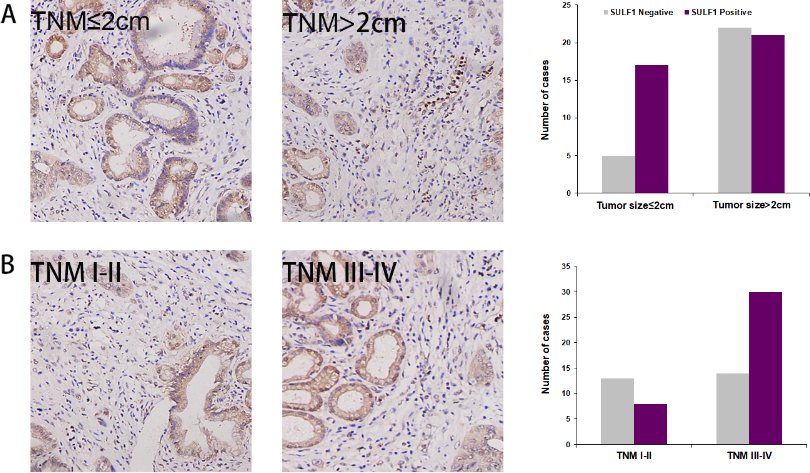
Multivariate analysis of predictive factors for survival in patients with pancreatic cancer (Cox proportional hazards model)
Correlation of SULF1 expression and overall survival in pancreatic cancer. A: Overall survival in 65 pancreatic cancer patients. B: Overall survival in the TCGA datasets.
To further assess the importance of SULF1 in for the survival time of patients with pancreatic cancer, we performed the Kaplan-Meier survival analysis, and found that among the 65 patients participating in this study, the SULF1-positive group had shorter survival time than the SULF1-negative group (
Pancreatic cancer is a highly malignant gastrointestinal tumor. In most of the patients, this disease is diagnosed at a late stage. Therefore, the rate of surgical resection is only 15–20%, and the 5-year survival rate is below 5% [21]. Statistical data from China indicated that the incidence and mortality rates of pancreatic cancer showed an increasing trend 2000 to 2011 [3]. Some reasons underlying this phenomenon are that the early diagnosis of pancreatic cancer is extremely challenging, and that many patients lose the opportunity for radical surgery before diagnosis. Another reason is that the invasiveness and metastatic tendency of pancreatic cancer often leads to very poor therapeutic efficacy and prognosis. Therefore, research related to biomarkers for early diagnosis and the prognostic factors of pancreatic cancer is extremely crucial.
SULF1 is a member of the sulfatase family, which mainly participates in signal transduction by regulating the sulfation of HSPGs on the cell surface. In the present study, we found that SULF1 expression was significantly increased in pancreatic cancer tissue. Moreover, by analyzing the relationship between SULF1 expression and clinicopathological features, we found that the tumor diameter of the SULF1-positive group was smaller than that of the SULF1-negative group. However, the T, N, and TNM stages of the SULF1-positive group occurred later than those in the SULF1-negative group. This contradiction may have been due to fact that although SULF1 can inhibit tumor cell growth, it is related to promoting interstitial proliferation of tumors and increasing vascular density, which leads to enhanced tumor invasiveness [22]. However, further investigations are required for elucidating the specific mechanisms involved. The findings of the present study suggested that the CA19-9 level was significantly higher in the SULF1-positive group than the SULF1-negative group, which indicates that SULF1 may be an important predictor of the prognosis of pancreatic cancer using CA19-9. In addition, the multivariate analysis showed that SULF1 is an independent risk factor for the prognosis of patients with pancreatic cancer. This implies that the combination of SULF1 and CA19-9 may provide better predictions for the prognosis of pancreatic cancer.
SULF1 can selectively hydrolyze the 6-O-sulfate groups of HSPGs, thereby affecting a variety of physiological and pathological processes by regulating different signaling pathways [23]. A study in 2014 found that SULF1 could inhibit the bFGF-mediated activation of the P13K/Akt and ERK pathways to induce the apoptosis of liver cancer cells. In addition, this process is related to the reduction of cyclin D1 and surviving by SULF1 expression [24]. In studies on breast cancer and cervical cancer, researchers have found that SULF1 showed tumor suppression effects by inhibiting FGF-2, HB-EGF, and amphiregulin signaling pathway [25]. Liu et al. showed that restoring SULF1 expression in cervical cancer tissues could inhibit tumor and blood vessel growth, thereby enhancing the effects of anti-tumor drugs [26]. However, SULF1 has positive regulatory effects on the signaling pathways in tumors, which may include the Wnt, BMP, Hedgehog, and GDNF signaling pathways. Research on urological cancers showed that high SULF1 expression often implies poorer prognosis [27]. The specific mechanisms of SULF1 involvement in the genesis and development of pancreatic cancer have remained unclear. Studies have shown that SULF1 can inhibit the growth of pancreatic cancer by inhibiting the FGF-2 pathway [28]. Another study showed that SULF1 could reduce the growth ability of pancreatic cancer cells by desulfurizing HSPG. However, SULF1 can promote tumor interstitial proliferation and increased vascular density, which will lead to greater tumor invasiveness [28]. Abiatari et al. [22] found that transfection of pancreatic cancer cells with the SULF1 gene can increase the invasiveness and adhesion capacity of the cells. Therefore, increased SULF1 expression can lead to later TNM stages, which implies a poorer prognosis in patients with pancreatic cancer. This result was also supported by the TCGA database. The results above indicate that SULF1 plays an important role in the progression of pancreatic cancer. However, the specific mechanisms involved are relatively complex, and require further investigation.
Conclusions
In summary, SULF1 expression was enhanced in pancreatic cancer tissue, and high SULF1 expression indicated later TNM stage, higher CA19-9 levels, and poorer prognosis in pancreatic cancer. Therefore, SULF1 could be a predictor of clinicopathological features and prognosis, and should be investigated further.
Footnotes
Conflict of interest
We declare that we have no financial and personal relationships with other people or organizations that can inappropriately influence our work. The authors declare that they have no competing interests.