
Abstract
We analyzed the expression of miR-503 in osteosarcoma tissues (OS) and discussed the clinical significance of our findings. To provide a theoretical basis for clinical applications, prognosis prediction and treatment of osteosarcoma, we studied the biological function of miR-503 and its mechanism in MG63 osteosarcoma cells. Real-time polymerase chain reaction (PCR) was used to detect the expression of miR-503 in 45 OS tissues and 20 osteochondroma tumors, analyzing the relationship between clinical pathology and follow-up data. Cox multivariate analysis revealed the clinical and pathological features of the osteosarcoma index and the influence of miR-503 expression on OS prognosis. To observe the effect on cell proliferation and invasion, MG-63 cells were transfected with miR-503. The TargetScan and PicTar bioinformatics method was used to analyze the probable target gene of miR-503 and, combined with the function of the target genes, resulted in a final validation of related pathways. miR-503 was significantly down-regulated in primary OS samples (26/45, 57.8%). The median miR-503 expression level in osteosarcoma was two-fold lower than that in osteochondroma (median expression 6.4 and 13.09, respectively,
Introduction
Osteosarcoma (OS) is the most common primary malignant bone tumor, and in most cases, the tumor cells produce osteoid or immature bone [1]. Osteosarcoma mainly occurs in teenagers, and studies have confirmed that 75% of osteosarcoma incidence occurs in 10- to 30-year-olds [2]. Invasive osteosarcoma often stems from the epiphyseal end, and the most common sites are at the long tubular bones, which contain the distal femur, the tibia and the proximal humerus [3, 4]. Fifty to seventy percent of all patients’ lesions occur around the knee joint. Since the use of preoperative chemotherapy started in the 1970s, the 5-year survival rate of OS patients has effectively improved [5]. The 5-year survival rate of metastatic osteosarcoma patients (20%) was significantly decreased than limited lesions (65%) [6]. For nearly 30 years, however, whether trying new chemotherapy drugs or combination chemotherapy, the patient survival rate has not improved. The current poor understanding of OS molecular mechanisms restrain the development of new targeted therapy [7]. Based on these problems, an accurate and effective therapeutic target that inhibits tumor cell invasion and metastasis of OS is of particular importance. We suggest that gene therapy is one method to achieve this goal.
MicroRNA (miRNA) is a non-coding RNA found in recent years. The mature miRNA is involved in gene regulation and combines with target mRNA 3’ end non-coding sequences (untranslated region, UTR) to influence the expression of the target gene [8, 9]. The first miRNA, lin4, was discovered in 1993, initiating miRNA research [10]. A growing body of evidence indicates that the pathological and physiological process of miRNAs in cancer cells play an important role in the process of cell growth, proliferation, development and apoptosis and are associated with the occurrence and development of many tumors. Many miRNAs with an abnormal expression pattern in osteosarcoma were found, including miR-143, miR-135b, miR-181a, miR-133a [11, 12, 13, 14]. These disordered microRNAs can affect biological processes, including cell growth, apoptosis, migration and invasion, and the occurrence and progress of osteosarcoma; however, how the imbalanced microRNAs are involved in the mechanism of the occurrence and development of osteosarcoma remains to be clarified.
Our recent study found that the expression of miRNA-503 was significantly decreased in osteosarcoma cells. miR-503 may play an important role in the development of OS. At present, miR-503 is one of the newly discovered microRNAs and was found to exist at a significantly lower expression level in a wide variety of tumors. studies have clearly demonstrated that miRNA-503 regulates metastasis through Rho guanine nucleotide exchanger factor 19 in hepatocellular carcinoma [15]; MicroRNA-503 inhibits tumor genesis in glioblastoma by targeting IGF-1R [16]; MiR-503 suppresses NSCLC progression by regulating PI3K p85 and IKK-beta [17]; It also regulates cisplatin resistance of SGC7901 cancer lines by targeting IGF1R and BCL2 [18]; however, miR-503 regulates different target genes in different tumors and serves varying tumor suppressor roles dependent on the molecular mechanism. The molecular mechanism of miR-503 promotion of tumorigenesis in OS is unclear.
Materials and methods
Patient tissue samples
Fresh specimens were collected from 45 OS and 20 osteochondroma cases in Liaoyang Central Hospital and Shengjing Hospital of China Medical University from 2001 to 2010, including 33 male cases and 12 female cases. Clinical classification refers to Enneking surgical staging, histological grading refers to the Price classification method, and histological type refers to the 1993 WHO classification. All 45 cases were tracked; the time of follow-up was 6 months to 3 years (average 1.5 years). Another selection of 25 osteochondroma cases admitted during the same period served as the control group, including 16 male cases and nine female cases. The average age was 22.9 (ages 15 to 44), and the median age was 24 years. The tissue samples were frozen in liquid nitrogen immediately after surgical resection. The specimens were not treated with radiotherapy and chemotherapy preoperatively, and osteosarcoma and osteochondroma were confirmed by histopathologic examination postoperatively. All of the samples’ clinical data are complete, including gender, age, tumor diameter, Enneking stage, histologic type, etc. All of the patients provided written informed consent for use of the tumor tissues for clinical research.
Cell culture and transfection
The human osteosarcoma cell line MG-63 was purchased from the Chinese Academy of Sciences in Shanghai. The MG-63 cells were cultured in Dulbecco’s modified Eagle’s medium (DMEM; Invitrogen; Thermo Fisher Scientific, Inc., Waltham, MA, USA) containing 15% fetal bovine serum (Gibco; Thermo Fisher Scientific, Inc.), 100 U/ml penicillin G (Abcam, Cambridge, MA, USA) and 100 pg/ml streptomycin (Abcam, Cambridge, MA, USA) in an incubator at 37
CCK8/Transwell method to detect cell proliferation/invasion activity
MG-63 cells were placed into 96-well plates with 2
Using the above transfection method, 1
miR-503 biological function and target gene prediction
The Landing Target Scan (
RNA extraction and real-time RT-PCR
Total RNA was extracted using the TRIzol method and was reverse transcribed into complementary DNA using a miScript Reverse Transcription kit.(Qiagen, Shanghai, China). The Qiagen miScriptPrimer assay and miScript SYBR Green PCR kit were used to amplify the miRNA-503 fragments, with RNU6A serving as the internal reference (Qiagen, Shanghai, China). The reaction conditions were as follows: 95
The relative levels of gene expression were represented as
Western blot analysis
Total MG-63 cell protein was extracted in lysis buffer (Pierce, Rockford, IL, USA) and quantified using the Bradford method. VEGFA and downstream signaling pathways’ core proteins Akt, Rictor and Erk were detected. Fifty micrograms of protein was separated using SDS-PAGE electrophoresis (12%). After the proteins were transferred to polyvinylidene fluoride (PVDF) membranes (Millipore, Billerica, MA, USA), the membranes were blocked at room temperature for 2 h; then the membranes were incubated overnight at 4
Statistical analysis
SPSS 13.0 was used for systemic analyses. The relationship between miR-503 and clinicopathological parameters was compared using a
Results
Expression of miR-503 in clinical tissue samples and OS cell lines
Expression analysis of miR-503 in tissues and cancer cell lines. A. Real-time PCR analyses of miR-503 in osteosarcoma and osteochondroma tissues. B. The expression of miR-503 in OS cancer cell lines (compared with hFOB, *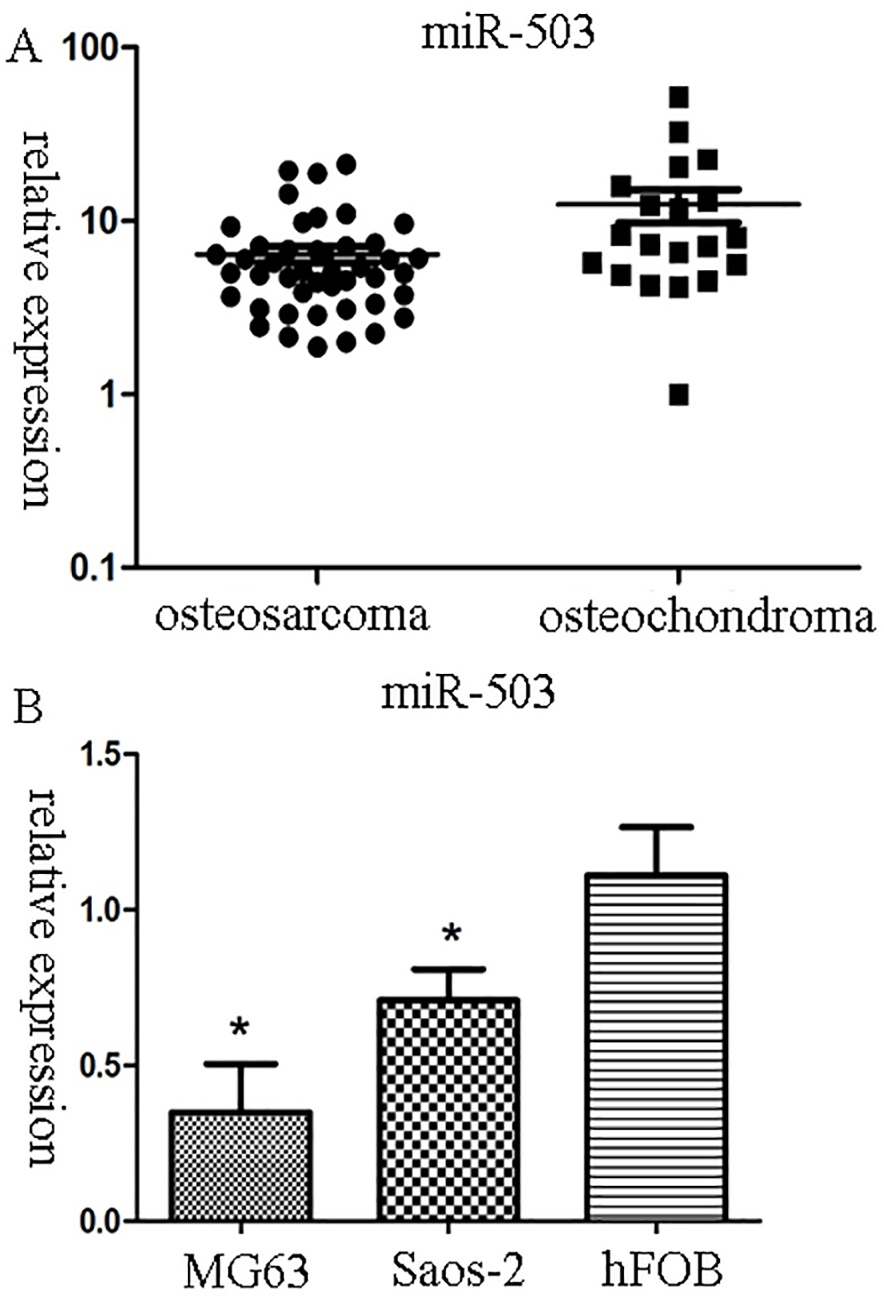
Kaplan-Meier survival analysis of OS patients (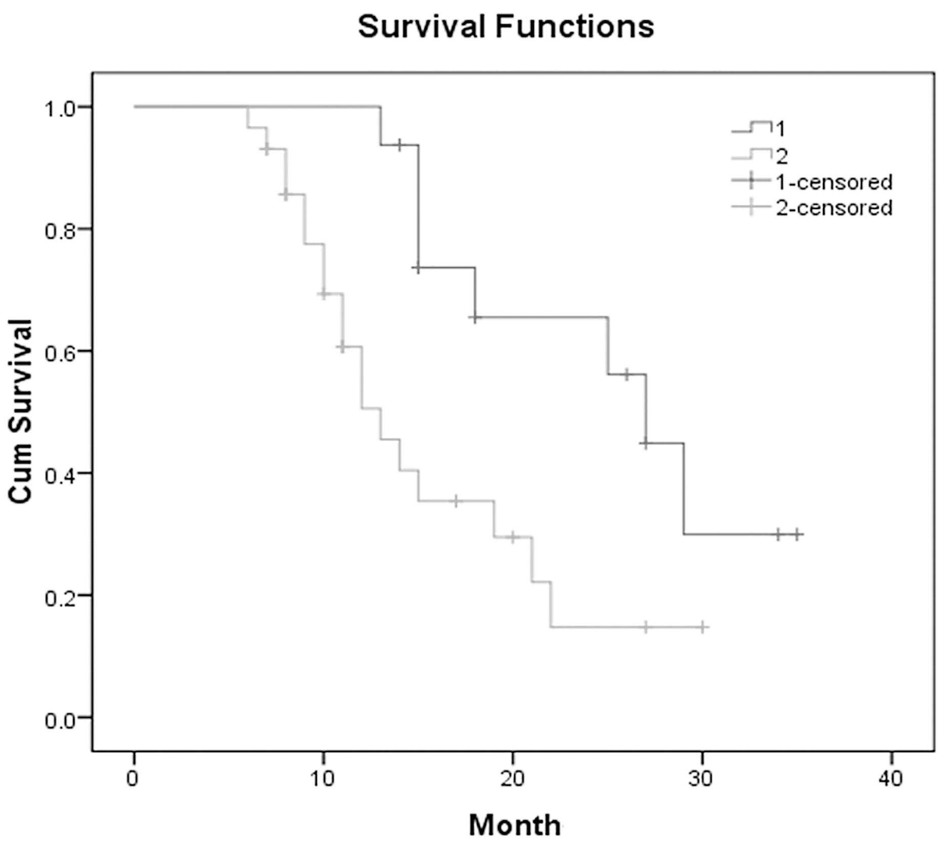
miR-503 expression was analyzed using real-time PCR in clinical samples and OS cancer cell lines (Fig. 1). Of the 65 patients (45 osteosarcoma, 20 osteochondroma), the median value of miR-503 in osteosarcoma tissues (6.4; normalized by U6 gene expression) was significantly lower than the value (13.09; normalized by U6 gene expression) in osteochondroma tissues. (
Distribution of miR-503 and clinicopathological characteristics in OS patients
Multivariate regression analysis in predicting the overall survival of osteosarcoma patients
miR-503 Function in MG63 cancer cells. A. miR-503 mimic group up-regulated miR-503 compared with the other groups. B. CCK8 method detected MG63 cell proliferation ability after miR-503 transfection. C. A transwell assay demonstrated MG63 invasion capacity among the miR-503 mimic, negative control and blank group.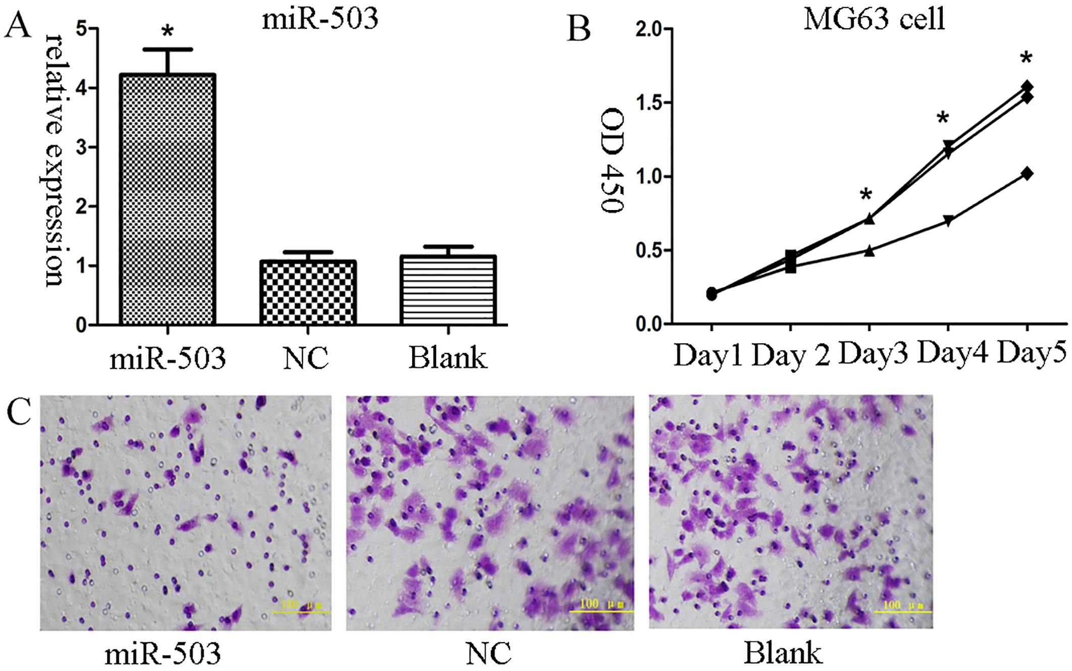
The expression of miR-503 was statistically significant compared with Enneking stage and invasion (Table 1,
miR-503 function in MG63 cancer cells after transfection
After transfecting a miR-503 mimic for 2 h, we found that the expression of the miR-503 mimic group was up-regulated 4.3-fold compared with the negative control group. The difference was statistically significant (
Additionally, we found that miR-503 may have a negative correlation with invasion. We studied the effect of miR-503 on invasion using the transwell method. The results show that compared with the blank group (78
The expression of target genes using western blot analysis and mean gray values. 1. miR-503 mimic group; 2. negative control group;3. Blank group.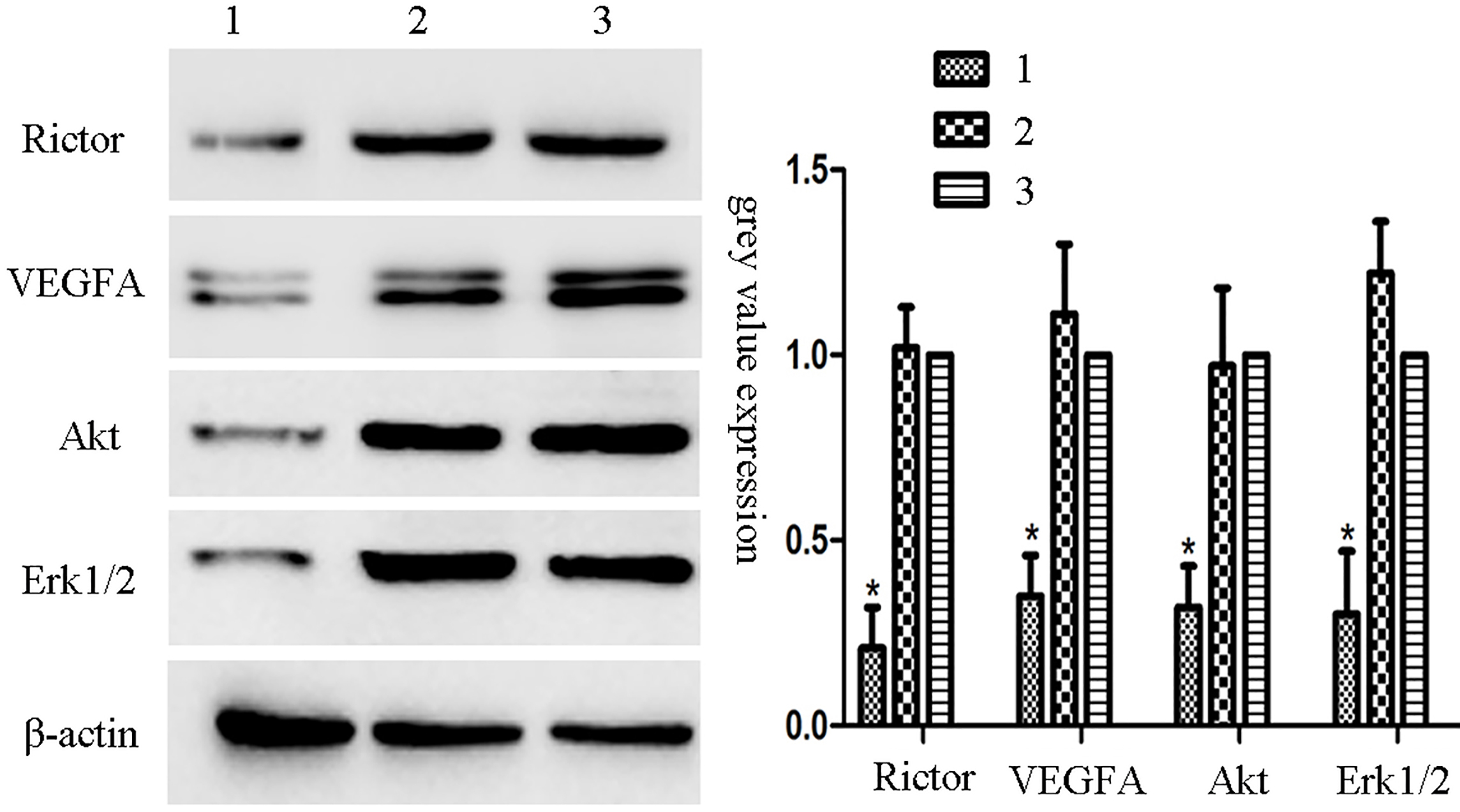
The main downstream target genes of miR-503 predicted include CCND2, VEGFA, CCND1, RICTOR, E2F3, and WNT3A (69 total predicted target genes, data not shown). GO analysis on the David database suggests that the genes regulated by miR-503 are associated with protein phosphorylation regulation, cyclin protein, etc. Pathway analysis showed that miR-503 may be related to cancer pathways, the mTOR pathway and Wnt signaling pathways. Combined with the above results, we selected target genes associated with the mTOR pathway, VEGFA, Rictor and related mTOR pathway key genes for further analysis. After transfecting the miR-503 mimic into MG63 cells, VEGFA expression was significantly lower than that of the negative control (0.35
Discussion
MiRNAs are endogenous, 22-nt-long noncoding RNA gene products. Through complementary combination with the 3’ end of target mRNA at the translation section (3’ UTR) or direct combination with the mRNA translation region causing mRNA splicing, miRNAs negatively regulate target genes [19]. We studied the expression of miR-503 in osteosarcoma and explored the clinical significance of miR-503 down-regulation expression in osteosarcoma tissues. We found that the expression of miR-503 in osteosarcoma tissues and cancer cells is significantly lower compared to that of the controls. Statistical analysis showed that miR-503 expression levels, invasion, and Enneking stage are closely related, but no correlation exists with gender, age, histologic type, tumor location or tumor size. A lower expression of miR-503 and a shorter overall survival in patients with osteosarcoma are correlated. Through the COX proportional hazards regression analysis, the expression levels of miR-503 were identified as a significant independent risk factor for survival in patients with osteosarcoma. Prior studies have proven that the difference in microRNA expression is associated with patients’ overall survival rate, including miR-181 [20], miR-140 [21], miR-15b [22] and miR-34 [23], etc. We aim to continue to expand the sample numbers in future work, to provide data for miR-503 in order to more accurately predict the prognosis of patients with osteosarcoma, and, together with the above validated microRNAs, to provide a comprehensive evaluation and clinical prognosis of patients.
Additionally, the role and mechanism of miR-503 in osteosarcoma are discussed in this research. Upon transfection of osteosarcoma MG63 cells with miRNA-503, we found that the expression of miRNA-503 and the proliferation of MG63 cells are negatively correlated using the CCK8 cell proliferation assay. The cell proliferation rate in MG63 cells with up-regulated miRNA-503 expression clearly declined; therefore, in osteosarcoma, miRNA-503 inhibits tumor growth and proliferation. Using a transwell assay, the invasion of transfected miRNA-503 cells decreased significantly. Through bioinformatics analysis, we found that miR-503 could combine with the VEGFA 3’ UTR translation region and inhibit VEGFA translation. Therefore, VEGFA may be negatively regulated by miR-503.
Osteosarcomas are highly vascular tumors, and prior research has confirmed that VEGF is the most powerful factor promoting tumor angiogenesis [24]. VEGFA is the most important member of the VEGF family, which regulates angiogenesis, A high level of serum VEGF is related to osteosarcoma metastasis, recurrence of a poor tumor chemotherapy effect and shorter survival time [25]. Thus, blocking angiogenesis may be useful for the treatment of osteosarcoma. Claesson-Welsh showed that regulated tumor angiogenesis through the MAPK/ERK, MAPK/P38, PI3K/AKT and FAK signaling pathways [26]. In this study, MG63 cells were induced by the high level expression of miR-503, VEGFA and corresponding downstream target proteins’ expression decreased. Conversely, inhibiting miR-503 expression in MG63 cells, VEGFA and its downstream target proteins’ expression increased. Therefore, we speculate that the down-regulated expression of miR-503 in MG63 cells attenuated the inhibition effect of VEGF; VEGFA expression increased, and its downstream signaling pathways PI3K/Akt and the core target protein expression in the MAPK/Erk also increased. Conversely, when miR-503 expression was up-regulated, the inhibitory effect of VEGFA expression was enhanced and the VEGFA expression was reduced; concomitantly, the expression levels of core target proteins in the PI3K/Akt and MAPK/Erk pathways were reduced.
Another target gene regulated by miR-503 is Rictor, a 200 kd protein, which was found to be involved in cytoskeleton regulation signaling pathways by Sarbassov et al. [27]. Rictor is the core scaffolding protein of mTORC2. Recent studies have shown that the Rictor-mTORC2 compound has an important effect on cancer [28, 29]; by improving the VEGF expression level, the compound can promote endothelial cell survival and migration to promote angiogenesis, suggesting that the compound is also an important regulatory factor, which plays an important role in the process of tumor angiogenesis, tumor invasion and EMT [30]. Therefore, we speculate that the low expression of miR-503 may lead to Rictor over-expression. The Rictor-mTORC2 complex transduces signals to the VEGFA signaling pathways, causing MG63 cell growth, invasion and metastasis. Furthermore, VEGFA over-expression promotes tumor angiogenesis. Both interactions eventually lead to a poor prognosis of osteosarcoma.
The mechanism of miR-503 down-regulated expression in osteosarcoma is still unclear. Previous research has shown that the changes in miRNA expression may be caused by genetic or epigenetic changes, including changes in DNA methylation and histone acetylation, chromosome segment lack and repeat, abnormal transcription factors and abnormal processing of mature microRNAs [31]. Therefore, we speculate that these may underlie the molecular mechanism of miR-503 down-expression, and future work will address this hypothesis.
In conclusion, the mTOR/VEGF signaling pathway plays an important role in the development of osteosarcoma due to the negative regulation of VEGFA expression by miR-503. VEGFA may be involved in the inhibition of osteosarcoma blood vessel growth, indicating that miR-503 can be used as a potential inhibitor of osteosarcoma. This work provides an essential theoretical basis for the targeted therapy of osteosarcoma.
Footnotes
Conflict of interest
The authors have no conflicts of interest.