
Abstract
BACKGROUND:
LncRNAs are involved in the metastasis and recurrence of human tumors, including colorectal cancer (CRC). We previously reported that lncRNA AB073614 promotes tumor proliferation and metastasis and predicted a poor clinical outcome of CRC patients. Herein, we investigated the underlying mechanism of lncRNA AB073614-related metastasis in CRC.
MATERIAL AND METHODS:
The expression of lncRNA AB073614 in CRC tissues were evaluated by quantitative real-time PCR (qRT-PCR). Transwell assay was performed to detect the effects of lncRNA AB073614 on cell migration and invasion. Epithelial-mesenchymal transition (EMT) molecular markers and Janus kinase/signal transducer and activator of transcription (JAK/STAT3) pathway proteins expression levels were detected by Western blot and Immunofluorescence.
RESULTS:
We confirmed that lncRNA AB073614 was highly expressed in the colorectal cancer tissues. LncRNA AB073614 knockdown in SW480 and HCT116 cells significantly promoted the protein expression levels of E-cadherin and Occludin, and decreased the expressions of N-cadherin and Vimentin, then further decreased the cell migration and invasion ability. Interestingly, the expression of phosphorylated STAT3 was also down-regulated. Furthermore, SW480 and HCT116 cells were transfected with lncRNA AB073614 vector and treated with a JAK inhibitor, AT9283. The results showed that lncRNA AB073614 regulated EMT through JAK-STAT3 signaling pathway.
CONCLUSION:
All these results indicate that lncRNA AB073614 can induce the expression of EMT cell markers and regulate the process of EMT of CRC cells through regulating the JAK/STAT3 pathway activation.
Background
As a main public health issue, malignant tumor seriously threatens the life and health of human and also is one of the main mortality factors [1]. Colorectal cancer (CRC) is one of the most common malignant tumors [2]. The statistical result of the worldwide morbidity and mortality of all malignant tumors shows that the morbidity of CRC ranks the third among males with malignant tumors; and that of females with malignant tumors ranks the second; while the mortality of CRC among males with malignant tumors ranks the fourth; that of females ranks the third [2]. Currently, the main treatment to cure CRC is radical operation of surgical excision [3]. The main reason for the failure of surgical operations lies in the metastasis and recurrence of tumors, which is also the main reason for the mortality of patients [4]. Recent studies showed epithelial-mesenchymal transition (EMT) is the main mechanism for tumor metastasis [5, 6]. The EMT of tumor cells refers to the biological process of the transformation of tumor cells from epithelial cells to mesenchymal cells [5, 6]. EMT is mainly manifested by the disappearance of epithelial phenotype, such as the down-regulated expression or gradual vanishing of epithelial cell markers like E-cadherin and
The signal pathway of Janus kinase/signal transducer and activator of transcription (JAK/STAT) mediates the transcription pathway of various cytokines, participates widely in the regulation of biological processes like cell proliferation, differentiation, apoptosis, and immune responses [8]. The activation of JAK/ STAT participates in the occurrence and development of various diseases including solid tumor, lymphoma, leukemia, and chronic inflammation [8, 9, 10, 11]. The basic transfer process of signal pathway is that the association of cytokines and their receptors causes the dimerization of receptors, making receptor-coupling JAKs approach each other and activated through the tyrosine phosphorylation; the activated JAKs further catalyze the phosphorylation of the tyrosine on the receptors and form corresponding STATs docking sites, making STATs associated with receptors via structural domain SH2 and hence realizing their phosphorylation under the activation of JAKs; and then STATs form homodimer/heterodimer and enter into cell nucleus to bind to the promoter of target genes, so as to activate the transcription and expression of corresponding genes [8, 10, 11]. As an important member of signal pathway of inflammation, STAT3 plays a significant role in the occurrence, development, and invasion of tumors [12]. After activation, JAK/STAT pathway can inhibit cell apoptosis and promote the biological effect of the proliferation and invasion of tumor cells [11, 12]. Activated STAT3 can destroy to varying degree extracellular matrix and lead to the degradation and destruction of basement membranes, providing suitable environment for the early metastasis of tumor cells [12, 13]. In addition, STAT3 can also promote EMT process and propel the transformation of chronic inflammation to cancer [13, 14].
Difference in the lncRNA AB073614 expression in CRC patients grouped by clinicopathological characteristics
Difference in the lncRNA AB073614 expression in CRC patients grouped by clinicopathological characteristics
*The relative expression of lncRNA AB073614 was calculated using 2
Epigenetic regulation plays an important role in every stage of the occurrence and development of life, while RNA molecule can be seen in various epigenetic regulation processes [15]. Studies in recent years showed that RNA is not confined to central dogma as the intermediate of the transmission of genetic information from DNA to proteins, but widely participates in various biological functions [16]. During the analysis and determination on 1% human genome functional elements by the Encyclopedia of DNA Elements Project, it was discovered that the entire genome was widely activated and transcribed, generating a large amount of non-coding RNA (ncRNA) [16]. Among the genome of mammals, only 1% to 2% proteins belong to coding protein, while 70%–90% transcripts fall into ncRNA [16, 17]. Among these transcripts, except microRNA (miRNA), tRNA, and rRNA which are well-known to people, those ncRNA with less than 200 nucleotides are referred as small non-coding RNAs, and those with 20
Clinical samples
This study was approved by the Medical Ethics Committee of the Fifth Affiliated Hospital, Sun Yat-sen University. Written informed consent was signed by all the 43 participants. CRC specimens and corresponding adjacent normal tissues were collected from 43 cancer patients undergoing surgery for colorectal cancer at the Fifth Affiliated Hospital of Sun Yat-sen University from 2014 to 2016. All samples were collected from patients prior to receiving any preoperative chemotherapy or radiotherapy, and the tumor samples were pathologically confirmed and evaluated for tumor content by a pathologist (median tumor content in the samples was 80–90%). The tissues were stored in liquid nitrogen until use. The characteristics of 43 patients are summarized in Table 1.
Cell culture
The human CRC cell line SW480 and HCT116 were purchased from the American Type Culture Collection (USA). The cells were cultured in Leibovitz’s L-15 medium (Gibco, USA) containing 10% fetal bovine serum and 1% streptomycin/penicillin at 37
Plasmid and cell treatments
Overexpression of lncRNA AB073614 in cells was realized through pcDNA-AB073614 transfection. The AB073614 sequence was synthesized and subcloned into the pcDNA3.1 vector (Invitrogen, USA). The empty pcDNA3.1 vector was used as a control. Plasmid vectors (pcDNA-AB073614 and pcDNA3.1) were prepared using DNA Midiprep kits (Qiagen, UAS), and were transfected into SW480 or HCT116 cells. LncRNA AB073614 siRNA and negative control (NC) siRNA were obtained from GenePharma (Shanghai, China). siRNA oligonucleotides (10 nmol/L) were transfected into SW480 or HCT116 cells using Lipo- fectamine™ 3000 (Life Technologies, USA). As a JAK inhibitor, AT9283 (0.1
Quantitative real-time PCR (RT-PCR)
Total RNA was extracted from SW480 and HCT116 cells and each sample of CRC and paired adjacent normal tissues using TRIzol reagent (Invitrogen, USA) according to the manufacturer’s manual. cDNA synthesis was performed using a PrimeScript RT Reagent Kit (Takara, China). The PCR amplification was performed with the conditions of 95
Expression of lncRNA AB073614 in CRC and corresponding adjacent tissues examined by RT-PCR, 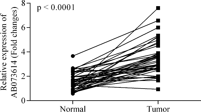
Cell migration and invasion ability assessments were performed using Transwell polycarbonate membrane inserts (Millipore, Germany). 48 h post-transfec- tion, the cells (1
Western blot
SW480 and HCT116 cells were harvested and lysed using RIPA buffer with proteinase inhibitor cocktail (Sigma-Aldrich, USA). The protein concentration was measured using BCA reagent (Thermo Fisher Scientific, USA). Equal quantities (20
Immunofluorescence
Cells were fixed with 3% paraformaldehyde for 30 min and then permeabilized with 0.1% Triton X-100 for 30 min on ice. Antibodies (Anti-E-cadheren, Occludin, N-cadheren, and Vimentin; 1:100) were added and incubated overnight at 4
Statistical analysis
Data are presented as mean
Knockdown of lncRNA AB073614 decreases the aggressiveness of CRC cells in vitro. (A, B) Knock down of lncRNA AB073614 by si-AB073614 transfection in SW480 and HCT116 cells. (C, D) Representative transwell images of migrated and invaded SW480 and HCT116 cells underwent different transfections. (E, F) Quantitative analysis of migrated and invaded cells. Numbers are presented as mean 
LncRNA AB073614 is significantly up-regulated in CRC tissues
In order to evaluate the changes of lncRNAAB073614 expression in colorectal cancer, 43 cancer patients undergoing surgery for colorectal cancer were recruited in this study. As shown in Fig. 1, the expression level of lncRNA AB073614 was significantly higher in the colorectal cancer tissues compared with the corresponding adjacent normal tissues (
Knockdown of lncRNA AB073614 decreases the aggressiveness of CRC cells in vitro
To examine whether lncRNA AB073614 was indeed involved in the regulation of CRC pathological courses, we first knocked down lncRNA AB073614 expression in CRC cell line SW480 and HCT116 by siRNA transfection. The efficacy of knockdown was confirmed by RT-PCR analysis (
LncRNA AB073614 knockdown inhibits EMT in CRC cells. (A, B) RT-PCR analysis of EMT-related gene expressions in si-AB073614 transfected SW480 and HCT116 cells. (C, D) Western blot analysis of EMT-related and JAK/STAT3 signal pathway-related protein expressions in si-AB073614 transfected SW480 and HCT116 cells. (E, F) Immunostaining of SW480 cells; E-cadherin (red), N-cadherin (red), Occludin (green), Vimentin (green), DAPI (blue). Data are presented as mean 
Overexpression of lncRNA AB073614 increases the aggressiveness of CRC cells in vitro. (A, B) Overexpression of lncRNA AB073614 by pcDNA-AB073614 transfection in SW480 and HCT116 cells. (C, D) Representative transwell images of migrated and invaded SW480 and HCT116 cells underwent different transfections and AT9283 treatment. (E, F) Quantitative analysis of migrated and invaded cells. Numbers are presented as mean 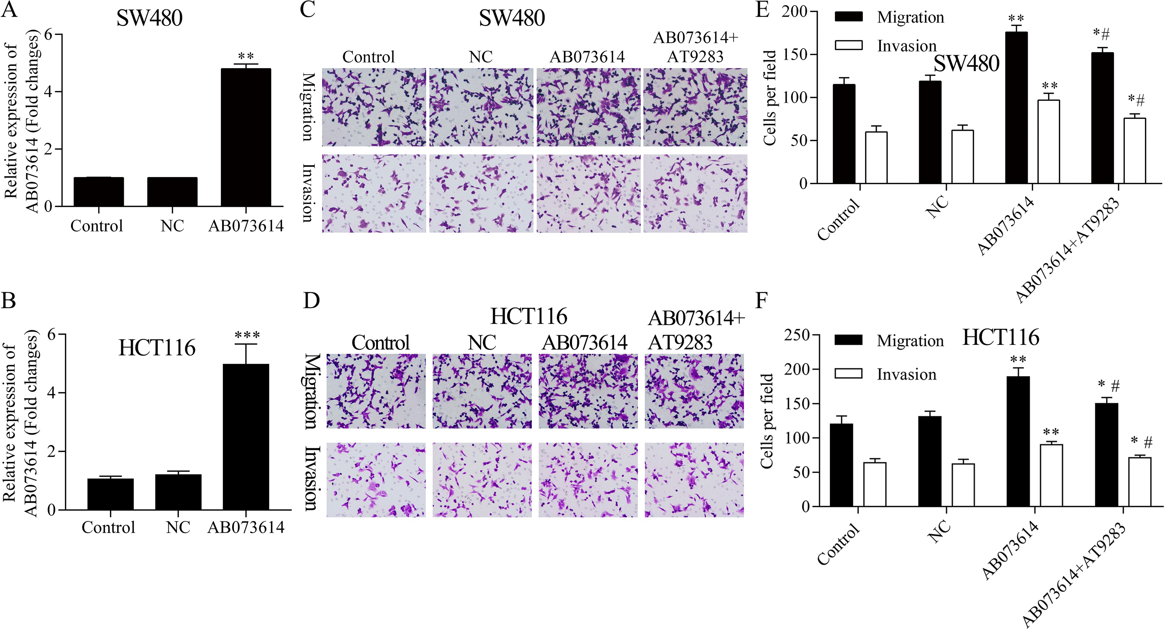
AT9283 treatment can reverse the EMT of CRC cells induced by AB073614 overexpression. (A, B) RT-PCR analysis of EMT-related gene expressions in pcDNA-AB073614 transfected and (or) AT9283 treated SW480 and HCT116 cells. (C, D) Western blot analysis of EMT-related and JAK/STAT3 signal pathway-related protein expressions in pcDNA-AB073614 transfected and (or) AT9283 treated SW480 and HCT116 cells. (E, F) Immunostaining of SW480 and HCT116 cells; E-cadherin (red), N-cadherin (red), Occludin (green), Vimentin (green), DAPI (blue). Data are presented as mean 
EMT plays a critical role in cell invasion. Therefore, we further studied whether EMT can be regulated by lncRNA AB073614 was further studied. As shown in Fig. 3A–D, the protein expression levels of E-cadherin and Occludin were significantly promoted after lncRNA AB073614 knockdown when compared with the NC group (
Overexpression of lncRNA AB073614 increases the aggressiveness of CRC cells in vitro
A lncRNA AB073614 overexpressed SW480 and HCT116 cell line were established by pcDNA-AB073614 transfection in order to evaluate the meta- static ability of CRC cells. The RT-PCR result showed that the expression of lncRNA AB073614 was significantly up-regulated compared with the NC group (
AT9283 treatment can reverse the EMT of CRC cells induced by AB073614 overexpression
RT-PCR and Western blotting results showed that, after overexpression of lncRNA AB073614, the protein expression levels of E-cadherin and Occludin were significantly decreased when compared with the NC group (
Discussion
Our previous study had demonstrated that overexpressed lncRNA AB073614 predicted a poor clinical outcome of CRC patients. Knockdown of AB073614 expression significantly inhibited the proliferation and metastasis of CRC cells which was associated with the PI3K/AKT signaling pathway. However, we here emphasized on the underlying mechanism of metastasis-induced by lncRNA AB073614, especially the critical role of epithelial-mesenchymal transitions (EMT) and the key STAT3 signals in lncRNA AB073614-induced EMT in CRC. EMT refers to the transformation process that epithelial cells lose the original epithelial phenotype but instead obtain mesenchymal one [6]. Recent studies hold that it not only exits during the embryonic development of multicellular organism, but also plays a vital role in the metastasis and invasion of tumor cells [5, 6]. The main molecular feature of EMT is the deficiency of the expression and function of epithelial markers like E-cadherin and Occludin, and meanwhile the excessive expression of mesenchymal cell markers like N-cadherin and vimentin, thus changing the morphology of cells [5, 6]. At the same time, the interaction between cells and cell matrix makes the extracellular matrix degraded, thus making cells more aggressive [6]. There have been studies proved that EMT participates in the metastasis and invasion of multiple epithelial cancers, such as breast cancer, ovarian cancer, prostatic cancer, and gastric cancer [27, 28, 29, 30], and meanwhile plays an extremely important role in the molecular mechanism of the invasion and metastasis of CRC [6, 31].
Eepithelial biomarkers mainly include adhesive proteins, such as E-cadherin,
Invasion and metastasis is one of the features of malignant tumors and is also the main cause for the mortality of tumor patients [34]. JAK/STAT is an extremely quick signal pathway from the extracellular to cell nucleus [9]. In recent years, it has been discovered that the activation of JAK/STAT, especially STAT3, exerts important effects on the occurrence and development of tumors [12, 13]. The expression of STAT in many human malignant tumor tissues and cell lines is high, while in normal tissues, STAT is seldom, even does not be activated [12]. The formation of tumors is the result of the overall dysfunction of cell signal regulation pathway and its constructing network, in which the excessive activation of STAT plays a role in controlling the abnormal proliferation, invasion and metastasis, angiogenesis, and immune evasion of tumor cells, giving play to a vital function in the occurrence, development and evolution of tumors [12]. Currently STAT3 is considered as a likely cancer gene, and the abnormal signal pathway participated by it may play a significant role in the invasion and metastasis of tumors [13, 14]. In this study, we found that the phosphorylated STAT3 (pSTAT3) expression was decreased when the expression of lncRNA AB073614 was knocked down. Furthermore, the JAK inhibitor, AT9283, treatment was able to block the STAT3 phosphorylation induced by lncRNA AB073614 overexpression, and reverse the up-regulated migration and invasion abilities of SW480 and HCT116 cells brought by the lncRNA AB073614 overexpression.
Conclusion
Together, all these results indicate that lncRNA AB073614 can induce EMT of CRC cells through the regulation of the JAK/STAT3 pathway activation. In recent years, the in-depth studies on lncRNA have changed people’s original concept that lncRNA is the by-product of gene transcription and made breakthroughs in the occurrence and development mechanism of cancers [17, 20]. LncRNA activates the expression of related transcripts via signal pathway, thus affecting the expression of epithelial and mesenchymal cell markers and regulating the process of EMT [22]. However, many problems remain unsolved. For example, there are many signal pathways in the EMT process of lncRNA-mediated tumor cells, but it remains unclear whether the pathways can interact with each other and what their specific molecular mechanism is. In addition, the correlation between most lncRNAs in tumors and EMT as well as their molecular mechanism remains to be further studied. Meanwhile, with the studies on STAT going deeper, tumor treatment with STAT3 as molecular target becomes possible. Deeper studies on STAT3 and lncRNA can provide new clues and target spots for the development of specific therapies of CRC and other malignant tumors.
Footnotes
Acknowledgments
This work was funded by the Science and Technology Foundation of Zhuhai (No. ZHW2015-200).
Conflict of interest
None.