
Abstract
OBJECTIVE:
To investigate the expression and role of long non-coding RNA (lncRNA) small nucleolar RNA host gene 12 (SNHG12) in papillary thyroid carcinoma (PTC).
METHODS:
The relative expression levels of lncRNA SNHG12 (hereinafter referred to as SNHG12) in 42 pairs of PTC tissues and para-carcinoma tissues were detected via quantitative reverse transcription polymerase chain reaction (qRT-PCR). SNHG12 specific interference sequences were designed and synthesized. The relative expression level and transfection efficiency of SNHG12 in PTC cells were detected via qRT-PCR. After the interference in SNHG12 expression, the change in cell proliferation capacity was detected via methyl thiazolyl tetrazolium (MTT) assay, the change in cell cycle distribution was detected via flow cytometry, the changes in cell migration and invasion capacities were detected via Transwell assay and wound healing assay, and the changes in expressions of molecular markers of Wnt/
RESULTS:
The results of qRT-PCR showed that the SNHG12 expression was up-regulated in 30 pairs of PTC tissues and cells. The results of MTT assay showed that the cell proliferation capacity was inhibited after the interference in SNHG12. The results of flow cytometry showed that the cell cycle progression was blocked in G1-G0 phase after the knockdown of SNHG12 expression. The results of Transwell assay and Western blotting showed that the interference in SNHG12 could inhibit the invasion and metastasis capacities of tumor cells through influencing the Wnt/
CONCLUSION:
The SNHG12 expression is relatively high in PTC tissues and cells. In-vivo/in-vitro experiments prove that SNHG12 can promote the proliferation and metastasis of PTC cells through influencing the Wnt/
Keywords
Introduction
Papillary thyroid carcinoma (PTC) is the most common type of thyroid cancer, accounting for about 90% of all pathological types [1]. PTC is a kind of low-grade malignant tumor, clinically manifested as thyroid masses and accompanied by multi-focal and regional lymph node metastasis [2]. After the effective and reasonable treatment, the 5-year survival rate of PTC is about 90% [3]. But PTC has a high degree of invasion with the development trend of dedifferentiation, finally developing into poorly-differentiated thyroid cancer or undifferentiated cancer and leading to the decline in the survival rate and life quality of patients. Therefore, it is significant to study the infiltration and metastasis processes of PTC cells, investigate the molecular mechanism of cancer cell metastasis, search the differential expression of PTC and predict the biomarkers of metastasis and molecules in intervention treatment for further improving the cure rate and survival rate of patients.
Long non-coding RNA (lncRNA) is a class of non-coding RNA with the length of more than 200 nucleotides, which is transcribed from RNA polymerase II. Its sub-cells are located in the nucleus or cytoplasm without the function of protein coding [4, 5, 6]. In the field of non-coding RNA, lncRNA has become a hotspot after microRNA. More and more studies have confirmed that lncRNA is involved in the tumor invasion, metastasis, autophagy, differentiation and other biological processes and plays an important role in the development and progression of human diseases, especially the tumors [7, 8]. It is reported that the high expression of lncRNA HOXA11-AS in gastric cancer can promote the tumor cell invasion and metastasis, and its potential molecular mechanism is the epigenetic regulation of downstream target genes
LncRNA small nucleolar RNA host gene 12 (SNHG12) is located in chromosome 1p35.3 region with a total length of 963 bp. In triple-negative breast cancer, C-MYC can induce the up-regulation of SNHG12 expression, promote tumor invasion and metastasis, and inhibit the apoptosis [11]. In human osteosarcoma and colon cancer, the high expression of SNHG12 plays a similar role to “oncogene” in promoting the development of tumors [12, 13]. So far, the expression level and biological effects of SNHG12 in PTC have not been reported. In this study, it was found for the first time that SNHG12 was highly expressed in PTC tissue and cells, and promoted tumor cell proliferation and metastasis through the regulation of Wnt/
Materials and methods
Tissue samples and cell culture
Forty-two cases of PTC samples and para-carcinoma tumor-free thyroid tissue samples resected in The Second Affiliated Hospital of Xi’an Jiaotong University from January 2015 to June 2016 were collected. All cases were confirmed via pathological diagnosis. The patients did not receive the chemotherapy, radiotherapy or other treatments of thyroid cancer before operation. PTC cell lines, K1, BCPAP and TPC-1, and one normal thyroid cell line Nthy-ori3-1 were purchased from Institute of Biochemistry and Cell Biology of Chinese Academy of Sciences. Cells were cultured in the 1640 medium (GIBCO-BRL) containing 10% fetal bovine serum (10% FBS), double-antibody 100 U/mL penicillin and 100 mg/mL streptomycin (Invitrogen) at 37
SNHG12 interference sequences and quantitative reverse transcription polymerase chain reaction (qRT-PCR) primers
SNHG12 interference sequences: siRNA-1#, 5’-TG ACATGCAAGGAGACTTC-3’, 2# 5’-CTACCATGC CTGAACCTTA-3’, 3# 5’-CGTACTCGCCGGTGTG ACT-3’, SNHG12 and GAPDH primers: SNHG12 (forward), 5’-TCTGGTGATCGAGGACTTCC-3’, and (reverse) 5’-ACCTCCTCAGTATCACACACT-3’; G-APDH, (forward) 5’-ACACCCACTCCTCCACCTTT-3’ and (reverse) 5’-TTACTCCTTGGAGGCCATGT-3’. The above sequences were synthesized by Invitrogen (Shanghai).
Detection of the SNHG12 expression via qRT-PCR
The total RNA was extracted from the PTC tissues, corresponding para-carcinoma tissues and PTC cells using the Trizol kit, and the RNA concentration was measured using the ultraviolet spectrophotometer. cDNA was synthesized according to the operation steps of the PrimeScript TM RT Master Mix (Perfect Real Time) kit. qRT-PCR reaction system (20
Detection of the PTC cell proliferation capacity via MTT assay
After the digestive passage of PTC cell lines in the logarithmic growth phase, the SNHG12 gene silencing group and the control group were inoculated onto the 96-well plate (3
Detection of the changes in cell cycle and apoptosis via flow cytometry
The concentration of PTC cell lines in the logarithmic growth phase was adjusted to be 3
Detection of the changes in migration and invasion capacities via Transwell assay
The interference sequences and control sequences were transiently transfected into PTC cells. After the cell fusion reached 80%–90%, the cells were digested using trypsin and re-suspended using the serum-free medium; the single-cell suspension was prepared and the density was adjusted to be 3
Wound-healing assay
At
Detection of the changes in Wnt/
-catenin signaling pathway expression via Western blotting
The cells in experimental group and control group were collected and added with the cell lysis buffer. The total protein was extracted and quantified using the Bradford method, followed by sodium dodecyl sulfate polyacrylamide gel electrophoresis (SDS-PAGE). The separated proteins were transferred onto the polyvinylidene fluoride (PVDF) membrane, sealed using 5% skim milk, and added with 1:1000 rabbit anti-human antibody
SNHG12 expression level in PTC tissues and cells. A: Detection of the relative expression levels of SNHG12 in 42 pairs of PTC tissues and para-carcinoma tissues via qRT-PCR. There are 30 cases of up-regulation and 12 cases of downregulation. B: Detection of the relative expression levels in PTC cells and normal thyroid cells via qRT-PCR. C&D: Design and synthesis of SNHG12 specific interference sequences, transient transfection of PTC cells, and detection of transfection efficiency via qRT-PCR (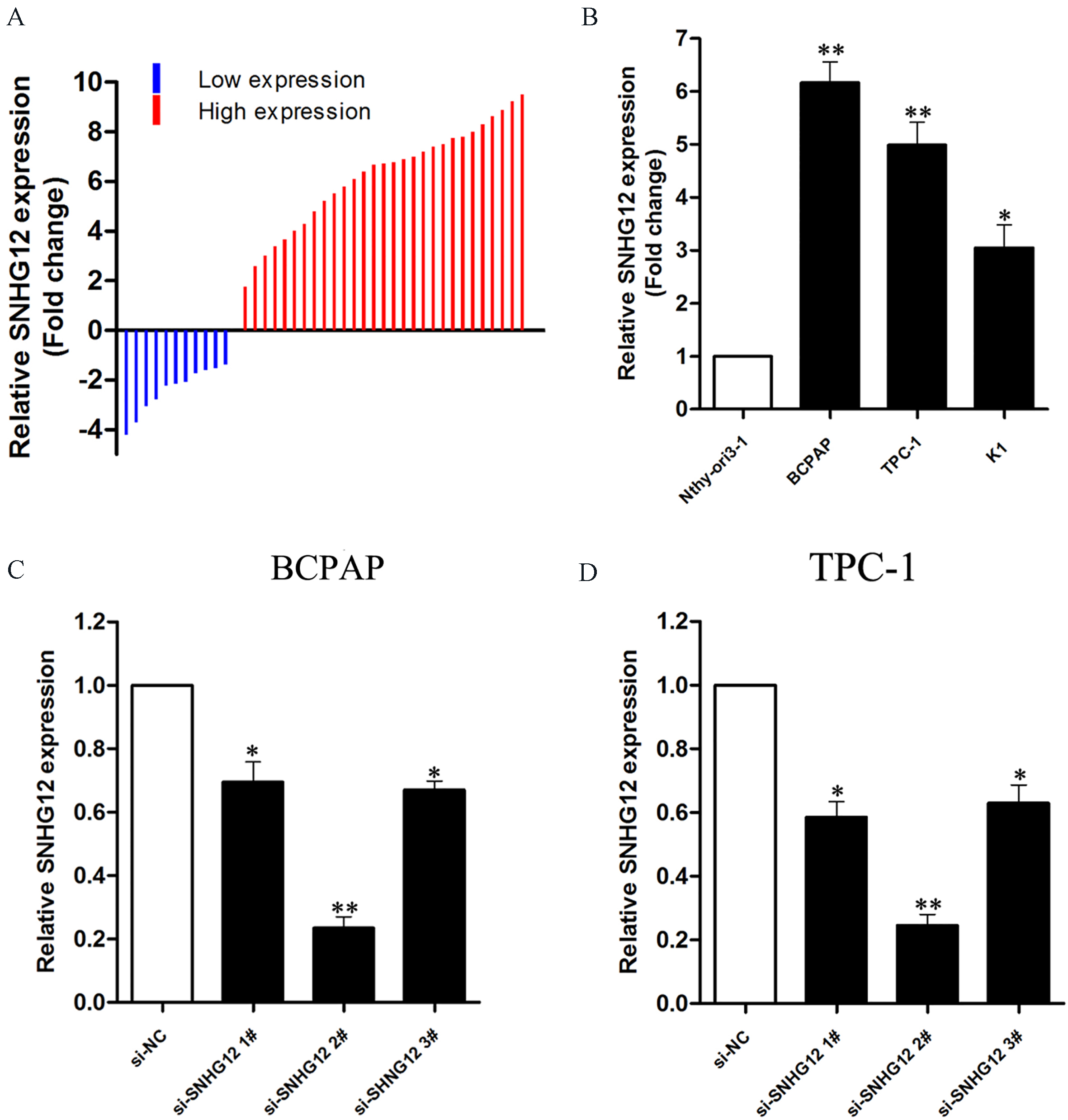
Effect of SNHG12 on proliferation of PTC cells. A&B: The results of MTT assay show that the cell proliferation is inhibited after knockdown of SNHG12 expression. C&D: Flow cytometry shows that the cell cycle progression is blocked in G1-G0 phase after the interference in the SNHG12 expression in PTC cells (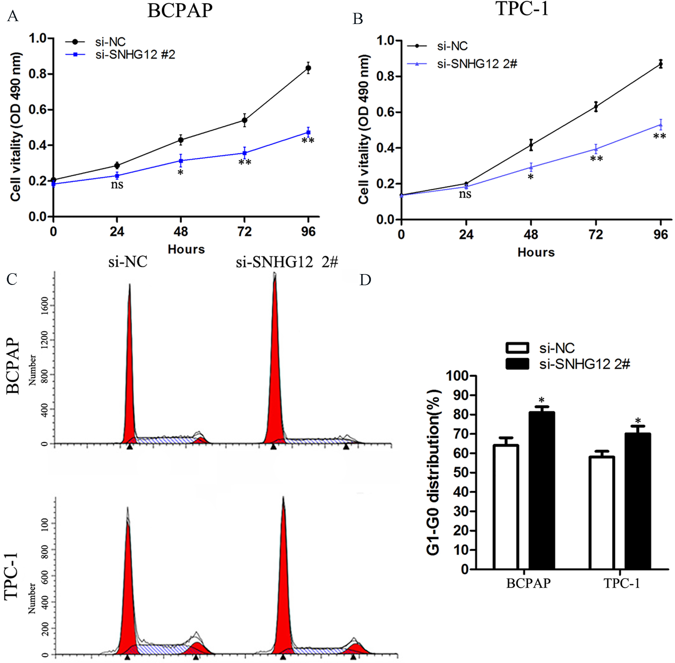
Effect of SNHG12 on apoptosis and metastasis of PTC cells. A&B: The results of flow cytometry show that the apoptotic rate is significantly increased after the interference in the SNHG12 expression in PTC cells. C&D: In PTC cells, after the interference in the lncRNA00673 expression, the tumor cell migration and invasion capacities are decreased in Transwell assay. E&F Representative images of scratch wound-healing migration assay in thyroid cancer cell lines transfected with si-NC or si-SNHG12 (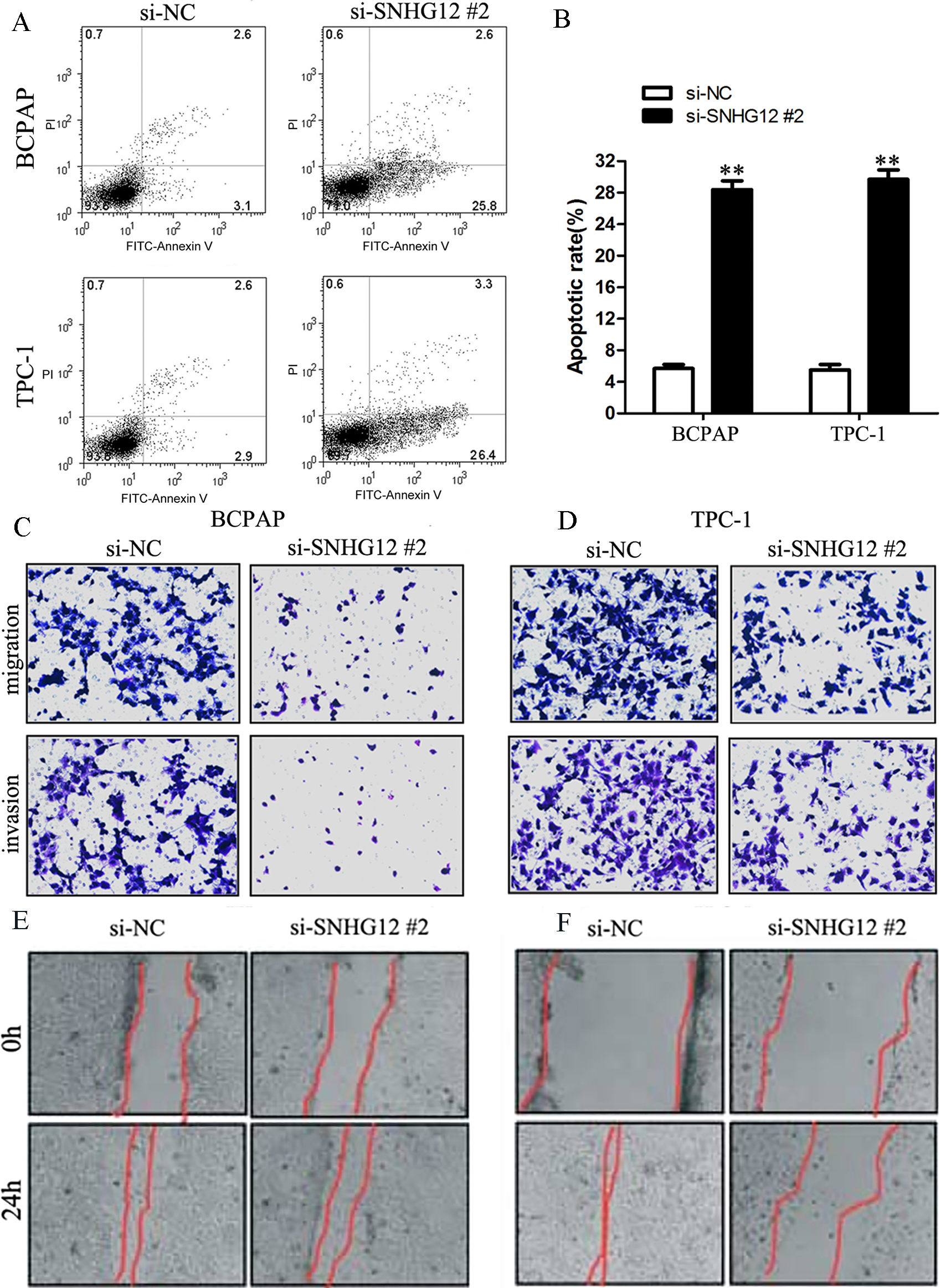
Detection of effect of SNHG12 on tumor cell metastasis via in-vivo experiment. A: The empty vector and sh-SNHG12 are transfected into BCPAP cells to construct the stable cell lines. The treated cells are injected into the nude mice through the caudal vein, the nude mice are executed after 21 d and the lung tissues are removed for photograph. B: Section and HE staining of lung tissues of nude mice. C: Detection of the relative expression level of SNHG12 in metastatic tumors via qRT-PCR. D: After the interference in SNHG12 in PTC cells, the changes in the molecular marker expression of Wnt/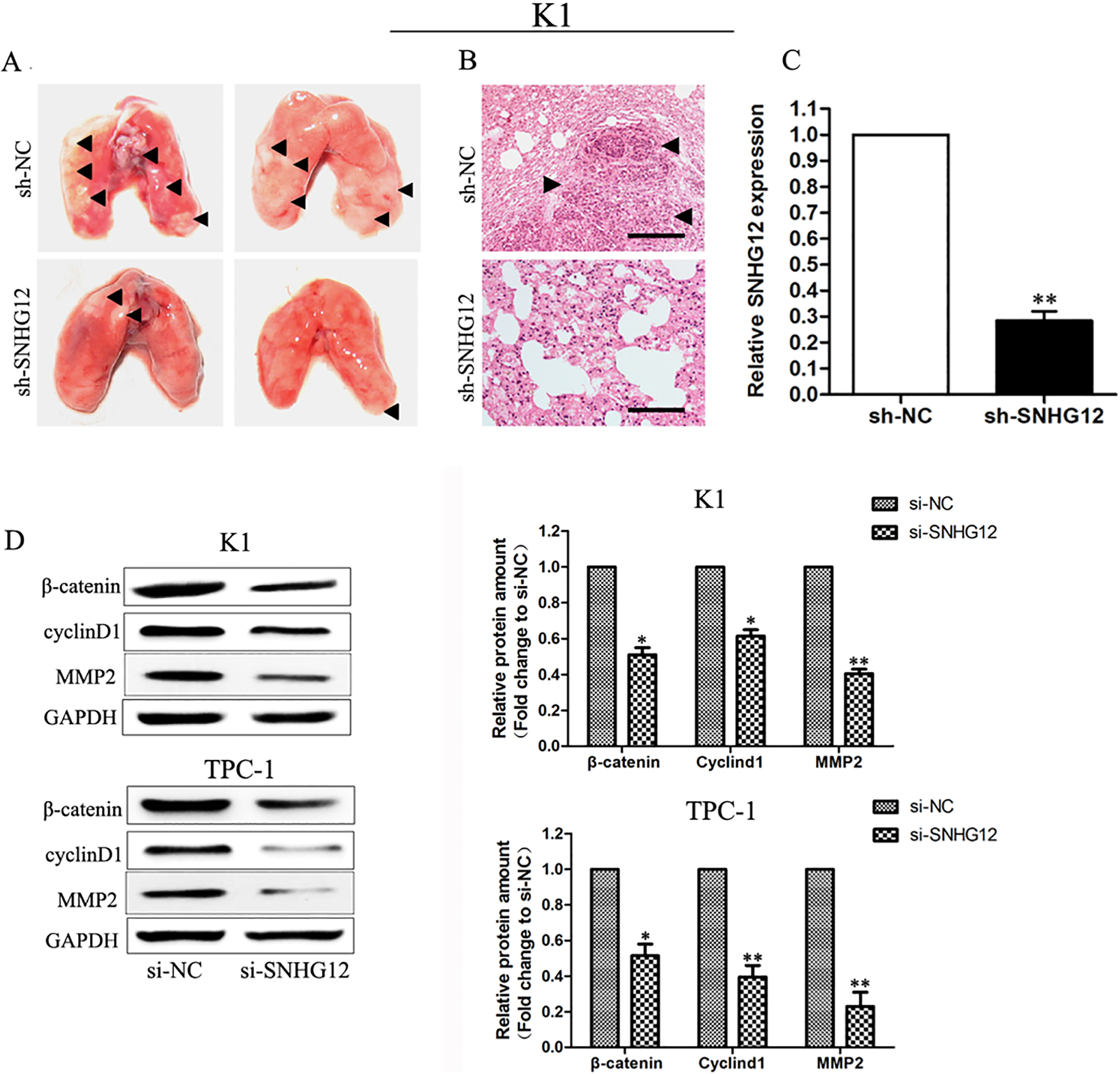
BALB/c male nude mice aged 4–5 weeks were selected as the objects of study. Sh-SNHG12 and control sequences were transfected into PTC cells to establish the stable cell lines. Then, the cells in experimental group and control group were injected into the nude mice via the caudal vein. Nude mice were fed in the specific pathogen-free (SPF) experimental animal room and observed every 3 days. The status of nude mice was observed and they were weighted; nude mice were executed after 28 d and the lung tissues and liver tissues were taken for HE staining. The number and size of metastasis nodes were observed.
Statistical analysis
All experiments were repeated for 3 times and were analyzed by using Statistical Product and Service Solutions (SPSS) 15.0. Data were expressed as mean
Results
Relative expression level of SNHG12 in PTC tissues and cells
Forty-two cases of PTC tissues and para-carcinoma tissues were collected. The relative expression level of SNHG12 in PTC tissues was detected via qRT-PCR. The results showed that the SNHG12 expression was up-regulated in 30 cases of PTC tissues compared with that in para-carcinoma tissues (Fold Change
Research on the effect of SNHG12 on PTC cell function via in-vitro experiment
First, MTT assay was used to detect the effect of SNHG12 on the proliferation capacity of PTC cells. The results showed that there was no significant difference in the number of viable cells between the two groups within 24 h after transfection (
Research on the effects of SNHG12 on the PTC cell invasion and metastasis via in-vivo experiment
The metastatic tumor model of nude mice was established, and the effects of knockdown of SNHG12 expression on the invasion and metastasis capacities of PTC cells were studied via the in-vivo experiment. First, the cell line BCPAP with the highest transfection efficiency was screened as the model cells. The cells in the experimental group and the control group were injected into the nude mice via the caudal vein and observed every 3 days. The status of nude mice was observed and they were weighted. Nude mice were executed after 21 d and the lung was taken and photographed. The number of metastatic tumor on the lungs of nude mice in the experimental group was significantly decreased compared with that in the control group. Then, the metastatic tumor was fixed and sliced, followed by immunohistochemical assay. HE staining proved that the metastatic tumor of nude mice was successfully constructed (Fig. 4B). The metastatic tumor on the lungs of nude mice was taken and the total RNA of tumor tissues was extracted. The relative expression level of SNHG12 in metastatic tumor tissues was detected via qRT-PCR, and the results showed that compared with that in the control group, the expression level of SNHG12 in the metastatic tumor was significantly decreased in the experimental group (Fig. 4C).
SNHG12 affected the PTC cell function through regulating Wnt/
-catenin signaling pathway
Wnt/
Discussion
Thyroid cancer is common in middle-aged women, and the incidence rate of PTC is the highest in malignant thyroid tumors, showing an increasing trend year by year. Surgical treatment is the preferred means for PTC, and other methods include iodine-131 radioactive therapy and thyroid hormone suppressive therapy [17, 18, 19]. In the early stage, PTC has no obvious symptoms with strong invasion capacity, so lymph node metastasis or invasion to surrounding organs and tissues has often occurred when diagnosed [20]. According to reports, the 5-year survival rate after PTC infiltration and distant metastasis is only 35% [21]. Therefore, how to improve the survival rate of PTC and life quality of patients, deeply study the effective gene targets of PTC and search more effective gene therapy urgently need to be addressed.
With the rapid development of high-throughput sequencing technology, the researchers have found a class of non-coding RNA with the length of more than 200nt, which is abnormally expressed in a variety of tumors and closely related to the changes in tumor biological behaviors [22, 23]. In PTC, lncRNA also plays a similar role to “oncogene” or “cancer suppressor gene” and involved in its development. The expression of lncRNA BANCR is relatively low in PTC tissues and cells. Overexpression of BANCR can inhibit the proliferation and promote apoptosis of PTC cells, and its potential molecular mechanism is partly through the regulation of ERK1/2 and p38 expression [24]. Zhu et al. [25] found that lncRNA HOTAIR is highly expressed in PTC tissues and cells and promotes the proliferation of PTC cells. And our research results found for the first time that SNHG12 was highly expressed in PTC tissues and cells, and the inhibition of SNHG12 expression could promote the apoptosis and inhibit the proliferation and metastasis.
It is reported that Wnt signal pathway can be involved in the physiological processes, such as embryonic development, cell immunity and maintenance of homeostasis, through regulating the DV1 protein, Axin protein, APC protein and
It was found for the first time in this study that SNHG12 is highly expressed in PTC tissue and cells, and the knockdown of SNHG12 expression can inhibit the proliferation, invasion, metastasis and apoptosis of PTC cells partially through Wnt/