
Abstract
BACKGROUND:
This research was aimed to study the expression of Serine/arginine rich splicing factor 2 (SRSF2) in tissues of hepatocellular carcinoma, and explore the relationship between the expression and the clinic pathological and prognosis of human hepatocellular carcinoma (HCC).
METHODS:
One hundred and fifty-three pairs HCC tissues and adjacent normal tissue were collected from January 2010 to March 2013. The expression of SRSF2 gene was detected by immunohistochemistry, western blotting and real-time quantitative polymerase chain reaction (PCR), and the relationship between the expression and the clinic pathological and prognosis of HCC being analyzed.
RESULTS:
In 153 cases of hepatocellular carcinoma, SRSF2 was highly expressed in 93 cases, low expression of 60 cases, immunohistochemistry score (6.50
CONCLUSION:
SRSF2 is highly expressed in hepatocellular carcinoma and its expression increases with the degree of tumor differentiation and TNM staging. It is related to lymph node metastasis and metastasis of tumor cells, and is positively related to serum alpha fetoprotein content, and affects the postoperative survival time of HCC patients.
The base clinical data of the HCC patients
The base clinical data of the HCC patients
Alternative splicing (AS) refers to the process in which different mRNA splicing isoforms are produced through alternative splicing of the same mRNA precursor (different combinations of alternative splicing sites), and it plays a significance role in proteome diversity [1]. Most splicing factors are RNA binding proteins, which affect protein expressions by selection of different splicing sites, thus leading to alterations in protein expression levels and their functions [2]. In recent years, many studies [3, 4, 5] have pointed out that abnormal AS exists widely in many types of cancer cells, which significantly impacts cell cycle regulation, cell migration/invasion and other biological characteristics of cancer cells. More importantly, the specific splicing factors that are usually up-regulated in cancer cells not only promote cancer cell survival and disease progression, but also may be of predictive value on the prognosis of the patients [6, 7]. Lack of safe, efficient and easily controllable vehicles may be a key barrier to the development of gene therapy. Non-viral systems such as p53 gene delivery using non-viral methods attract more and more attention, especially chemical approaches [8]. Acid-labile amidized cationic polymers could make the polymers inert in the bloodstream but activate to recover such abilities as cellular ad- sorptive and endo/lysosome-lysing ability in the target sites by responding to the pH gradients in the tumoral interstitium or endo/lysosomes [9, 10]. Serine/arginine rich splicing factor 2 (SRSF2) protein is an important member of the Serine/arginine rich (SR) protein family. It plays an important role in transcriptional activation, RNA stability and mRNA translation through binding with enhancer sequences and acting as a splicing activator. Studies [11] have shown that the abnormal expression of SRSF2 is related to the genesis, development and adverse prognosis of myelodysplastic syndromes. In addition, animal experiments [12] have shown that SRSF2 plays a key role in liver development, and that the abnormal expression of SRSF2 leads to the death of the mice due to acute liver failure. However, there have been few reports on the expression of SRSF2 in human hepatocellular carcinoma (HCC) tissues and the correlation of its expression with clinical pathology or prognosis. In this study, we examined the expression level of SRSF2 protein in HCC tissues and the normal adjacent tissues through immunohistochemistry and Western blots, and analyzed the correlation between SRSF2 expression and hepatoma cell differentiation, histological grading as well as other clinic pathological features, thus providing certain theoretical basis for further studies of HCC pathogenesis and the development of new targeted drugs.
Materials and methods
Clinical specimens
Specimens of HCC tissues and the adjacent normal tissues were collected from 153 patients with hepatocellular carcinoma between January 2010 and May 2012 at Qilu Hospital Affiliated to Shandong University. None of the patients were treated with chemoradiotherapy or biological therapy before the resection, and all of them were diagnosed as HCC by pathological examination. The clinical data of the patients were shown in Table 1.
After the 153 specimens of HCC tissues and the adjacent normal tissues were removed by surgery, an appropriate proportion of each specimen was sent to the pathology department to be made into paraffin sections, while the remaining tissues with the size of a soybean were directly frozen in liquid nitrogen. This study was approved by the ethics Association of Qilu Hospital Affiliated to Shandong University. All the patients have signed informed consents and volunteered to join the study.
Reagents and instruments
Immunohistochemistry kit (BOSTER, Wuhan, China); Tissue total protein extraction kit (Sangon Biotech, Shanghai, China); Tissue total RNA extraction kit (QIGEN, Dusseldorf, Germany); Reverse-transcription kit (Takara, Tokyo, Japan); SRSF2 and GAPDH rabbit monoclonal antibodies, goat anti-rabbit secondary antibody (Abcam, Cambridge, USA).
Immunohistochemistry
The paraffin sections were subjected for immunohistochemical detection of SRSF2 expressions according to the instructions in the commercial kit. PBS was used as the negative control antibody. At least 5 visual fields were selected for assessment in each paraffin section, and the proportion of positive cells and the dyeing conditions were recorded (SRSF2 was expressed only in the nucleus). Immunohistochemical scoring criteria: the proportion of positive cells
Western blot assay
The specimens frozen in liquid nitrogen were subjected for tissue total protein extraction by the commercial kit. Proteins were loaded for SDS-PAGE and transferred to a wet NC membrane (400 mA, 1.5 h). The membrane was blocked with 5% no-fat milk (1 h at room temperature), incubated with primary antibodies SRSF2 and GAPDH (ab204916 and ab9484, 4
The expression of SRSF2 in HCC tissues and the adjacent normal tissues. A–E: immunohistochemistry detection of the strongly positive expression (A), positive expression (B) and weakly positive expression (C) of SRSF2 in HCC tissues, as well as the weakly positive expression (D) and negative expression (E) of SRSF2 in the adjacent normal tissues; (F) Van chart statistics of the immunohistochemical score of SRSF2 in different tissues; 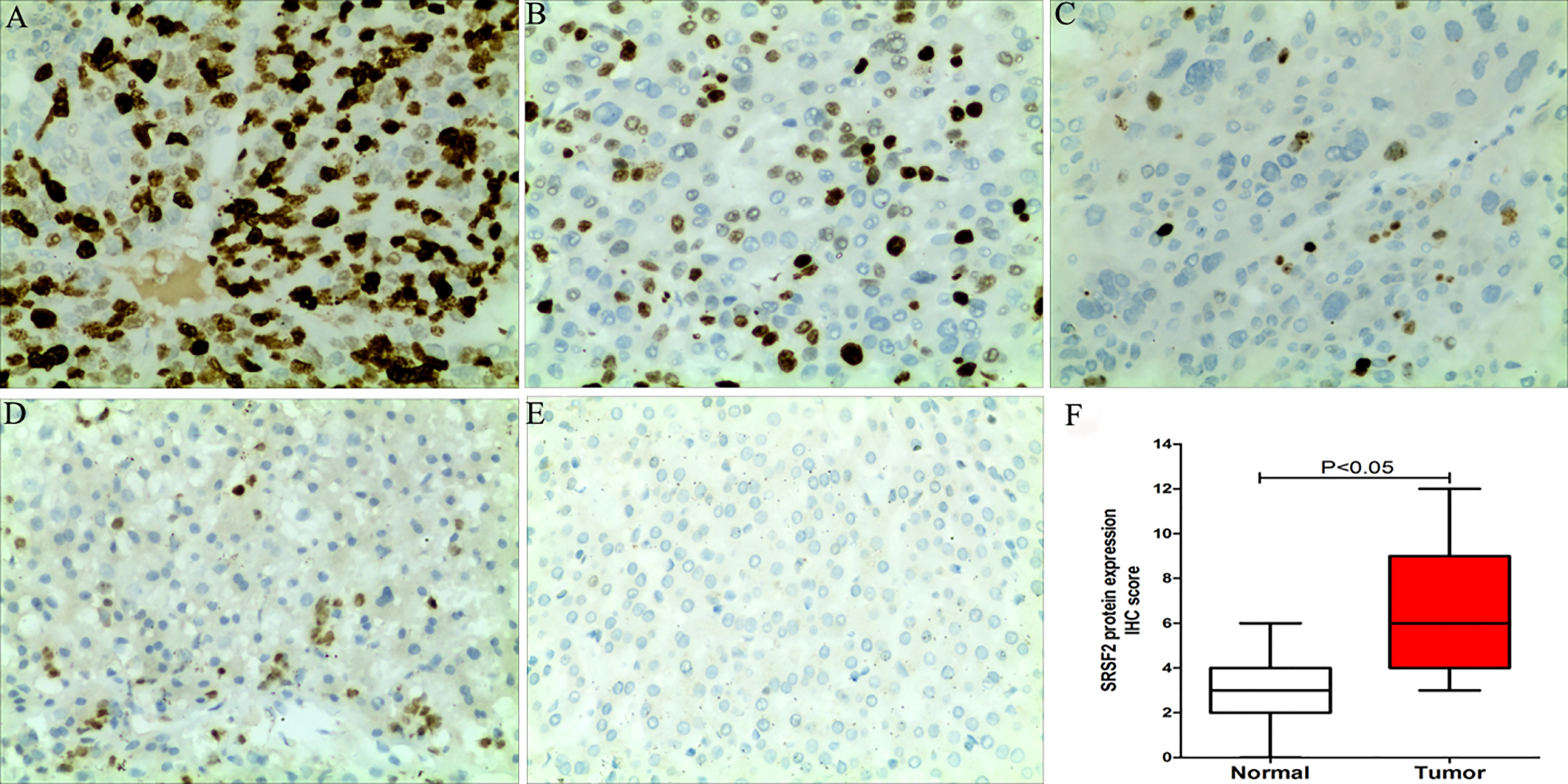
The relationship between SRSF2 expression and clinicopathological features in HCC tissues. Legends: A: 4 cases of well-differentiated (WD) HCC tissues, moderately differentiated (MD) HCC tissues or poorly differentiated (PD) HCC tissues, respectively; B: 4 cases of HCC tissues with non-lymph node metastasis (NLNM) and lymph node metastasis (LNM), respectively; C: 4 cases of HCC tissues with no distant metastasis (NDM) or distant metastasis (DM), respectively; D: 4 cases of HCC tissues at TNM stage I, TNM stage II, TNM stage III or TNM stage IV, respectively. E: 4 cases of the negative expression of SRSF2 in paracancerous tissue (PT).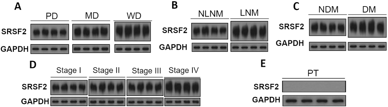
Statistical analysis was performed using the SPSS 20.0 statistical program.
Results
Clinical data of the 153 HCC patients
Among the 153 HCC patients, 113 were male and 40 were female; their age ranged from 34 to 75 years, with the mean age of (50.6
Immunohistochemistry detection of SRSF2 protein expressions
The expressions of SRSF2 protein in the 153 cases of HCC tissues and the adjacent normal tissues were detected and scored by immunohistochemistry. The results showed that all the HCC tissues contained SRSF2 positive cells without any negative expression; the adjacent normal tissues showed no strongly positive or positive expressions of SRSF2, and all of them showed weakly positive or negative expressions of SRSF2; the immunohistochemical score of SRSF2 in HCC tissues was (6.50
The relationship between SRSF2 expression and the clinic pathological features of HCC tissues
The expressions of SRSF2 protein in the 153 cases of HCC tissues were detected and scored according to the procedures described in 1.3, and those with the immunohistochemical score
Immunohistochemistry detection of the relationship between SRSF2 protein expression and clinical pathology
Immunohistochemistry detection of the relationship between SRSF2 protein expression and clinical pathology
Correlation between the expression of SRSF2 protein in HCC tissues and the serum alpha-fetoprotein (AFP) level.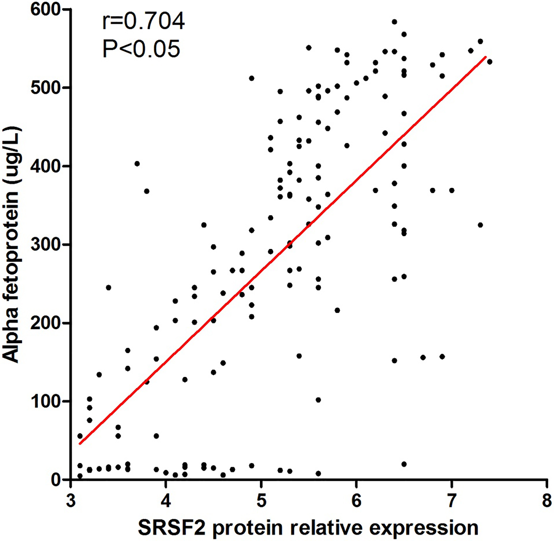
Relationship of SRSF2 expression and the postoperative survival rates of HCC patients.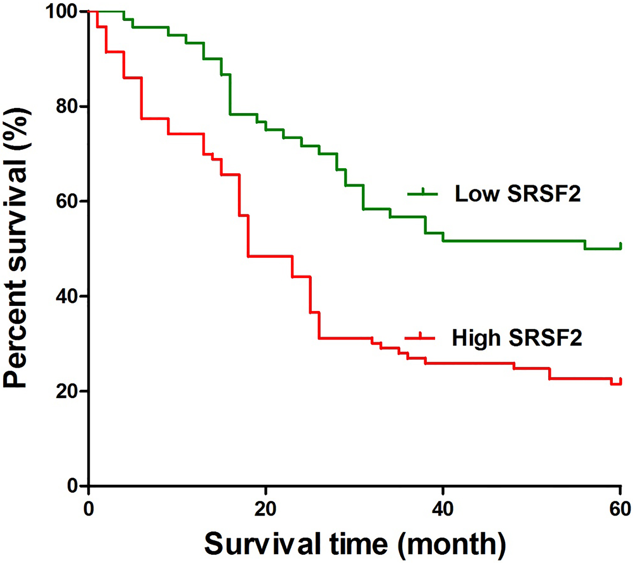
Western blot was performed to determine the relative expressions of SRSF2 protein in the 153 cases of HCC tissues, and SPSS20.0 was used to analysis the correlation between SRSF2 expression in HCC tissues and the level of serum AFP in the patients by Pearson methods. The results showed that the relative expression of SRSF2 in HCC tissues was positively correlated with the level of serum AFP (
Relationship between SRSF2 expression and prognosis in HCC patients
The 153 HCC patients were followed up for 5 years after operation, and their postoperative survival time was recorded. The results showed that the 1-year, 2-year, 3-year, 4-year and 5-year survival rates of the patients with SRSF2 overexpression were 74.19%, 44.09%, 26.88%, 24.73% and 21.51%, respectively, while the 1-year, 2-year, 3-year, 4-year and 5-year survival rates of the patients with SRSF2 low expression were 93.33%, 71.67%, 56.67%, 51.67% and 50.00%, respectively; suggesting that the HCC patients with SRSF2 low expression were correlated with better survival rates than those with SRSF2 overexpression, as shown in Fig. 4.
Discussions
Hepatocellular carcinoma is the fifth most common malignant cancer ranking after lung cancer, gastric cancer, esophageal cancer and breast cancer. There are a large number of HBV infected people in China, making it with high incidence of hepatocellular carcinoma. New HCC cases account for about 55% in the world every year [13]. According to the statistics, although the 1-year survival rate after HCC radical resection has increased from 39.3% to 87%, the 5-year postoperative survival rate is still only 15%–40%, and metastasis of cancer cells is one of the major causes of adverse prognosis. A number of studies on the molecular mechanism of cancer cell invasion and migration have indicated that the aberrant overexpression of splicing factors plays an important role in promoting the invasion/migration ability of cancer cells [3, 4, 5]. SR protein family is the most well-studied splicing factors: Li et al. [14] point out that SRSF10 regulates the production of lipin1
We further analyzed the relationship between SRSF2 expression and the clinicopathological features of HCC patients, and found that the expression of SRSF2 in HCC tissues was not correlated to sex, age, tumor size or T staging. However, the expression of SRSF2 in HCC tissues increased with the status of tumor differentiation as well as TNM staging, and was significantly increased in the HCC tissues with lymph node metastasis, distant metastasis or portal vein invasion, suggesting that overexpression of SRSF2 may be involved in the progression of HCC. In addition, Pearson correlation analysis showed that the relative expression of SRSF2 protein in HCC tissues was positively correlated with serum alpha fetoprotein (
Previous studies have pointed out that the abnormal expression of splicing factors is not only related to the genesis and development of multiple diseases, but also has certain predictive value for the prognosis of patients [6, 7]. In this study, we followed up the 153 HCC patients for 5 years, and found that the survival rates of the 93 HCC patients with high SRSF2 expressions were significantly lower than those of the 60 patients with low SRSF2 expressions 1–5 years post operation, suggesting that the expression of SRSF2 in HCC tissues could affect the postoperative survival of HCC patients.
To sum up, SRSF2 was overexpressed in HCC tissues. Its expression increased with tumor differentiation and TNM staging, and was related with lymph node metastasis as well as distant metastasis of tumor cells. Moreover, the expression of SRSF2 was positively correlated with serum content of alpha fetoprotein, and could affect the length of postoperative survival in HCC patients.
Footnotes
Acknowledgments
This research was supported by the Fund of the Key Research and Development Program of Shandong Province (2016GSF201180).
Conflict of interest
None declared.