
Abstract
BACKGROUND:
Abnormal expression of miR-192 has been observed in a variety of human cancers, but the expression pattern of miR-192 and its prognostic value in pediatric acute myeloid leukemia (AML) is poorly known.
OBJECTIVE:
This study was to explore the expression status of miR-192 and its clinical significance in pediatric patients with AML.
METHODS:
Quantitative RT-PCR was carried out to detect miR-192 expression level in the serum from 97 AML cases and 50 healthy controls.
RESULTS:
The results showed that downregulation of serum miR-192 was observed in pediatric AML patients and strongly correlated with aggressive clinical features. Increased serum miR-192 expression occurred more frequently in the AML subjects with favorable risk cytogenetics. Moreover, serum miR-192 expression showed good performance to screen pediatric AML subjects from normal controls. Furthermore, serum miR-192 was identified as a independent prognostic indicator for both overall survival and event free survival. In addition, low serum miR-192 expression significantly contributed to poor prognosis in the whole cohort of AML patients or the AML patients with intermediate-risk cytogenetics.
CONCLUSIONS:
Collectively, serum miR-192 potentially can be a reliable biomarker for the diagnosis and prognosis in pediatric AML.
Introduction
Pediatric acute myeloid leukemia (AML), which accounts for 15–20% of pediatric leukemia, is a cytogenetically, and molecularly heterogeneous malignancy [1, 2]. Due to improvements in chemotherapy-based regimen and hematopoietic stem-cell transplantation, the survival rate of AML patients has been greatly improved in the past several decades [3, 4]. However, relapse remains the major cause of treatment failure, and the prognosis of AML is still difficult to be assessed [5]. Thus, it is necessary to identify effective biomarkers for the diagnosis and the prediction of treatment response of AML patients.
Association between serum miR-192 expression and different clinical features in pediatric AML patients
Association between serum miR-192 expression and different clinical features in pediatric AML patients
BM, bone marrow; PLT, platelet; WBC, white blood cells; FAB, French-American-British classification.
MicroRNAs (miRNAs) are a class of small non-coding RNAs that function by directly binding to the 3’ untranslated region of target messenger RNA (mRNA), resulting in degradation of mRNA and/or translational inhibition [6, 7]. Deregulation of miRNAs has been found to be involved with multiple cellular events including cell proliferation, differentiation and apoptosis [8]. Increasing evidence has shown that miRNAs can exert either oncogenic or anti-oncogenic properties in the initiation and progression of AML. For instance, miR-335 overexpression was found in AML patients and its upregulation significantly contributed to poor prognosis [9]. In contrast, miR-29a was greatly decreased in AML, and low miR-29a expression was closely associated with shorter survival [10]. Additionally, mature miRNAs have been demonstrated to be remarkably stable in plasma and serum, which led to a rapidly growing interest in using miRNAs in circulation system as diagnostic and prognostic biomarkers [11].
MiR-192, located on chromosome 11 [12], has been proven to play a tumor suppressive role in leukemia [13, 14, 15]. In addition, the aberrant expression of miR-192 has been found in osteosarcoma [16], prostate cancer [17], hepatocellular carcinoma [18], colorectal cancer [19], lung cancer [20], pancreatic ductal adenocarcinoma [21], and esophageal squamous cell carcinoma [22]. However, the status of miR-192 expression and its prognostic significance in AML remained uncovered. To address this problem, in the current study we aimed to explore the diagnostic and prognostic value of serum miR-192 in the pediatric patients with AML.
Patients and blood collection
This prospective cohort study was approved by the Ethics Committee of Affiliated Hospital of Binzhou Medical University. Written informed consent was obtained from all the participants or their guardians prior to serum collection. All specimens were handled and made anonymous according to the ethical and legal standards.
A total of 147 serum samples were obtained from 50 normal healthy children and 97 de novo pediatric AML patients before receiving any therapy. All the participants were aged between 3 to 14 years old. The AML patients were classified according to the French-American-British (FAB) and World Health Organization criteria. All AML cases received regular follow-up. Overall survival (OS) was defined as the time from diagnosis to the date of death. Event-free survival (EFS) was defined as the time from diagnosis to the date of induction failure or relapse or death. Detailed clinical features of 97 pediatric AML patients were provided in Table 1. The follow-up time was 60 months.
A. Serum miR-192 levels in pediatric AML patients were significantly lower when compared to healthy controls. B. Serum miR-192 levels in pediatric AML patients with favorable risk cytogenetics were greatly higher than those with intermediate/unfavorable risk cytogenetics.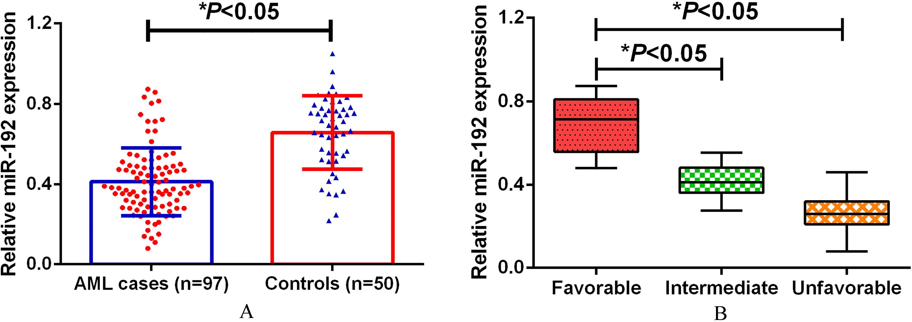
Up to 5 ml blood was withdrawn from each participant. All blood samples were centrifuged at 3000 g for 10 min and 12000 g for 5 min at 4
The QIAamp RNA Blood kit (Qiagen, Hilden, Germany) was used to extract the total RNA in serum samples following the manufacturer’s protocol. Reverse transcription was performed with the Prime-Script RT reagent kit (TaKaRa, Dalian, China). Quantitative RT-PCR was processed on ABI 7500 fast real-time PCR system (Applied Biosystems, Foster City, USA) using the Maxima SYBR Green qPCR Kit (Thermo Scientific, CA, USA). The relative miR-192 expression was determined by the comparative 2
Statistical analysis
As the data were not subjected to normal distribution, the relative serum miR-192 expression between groups were performed using the Mann-Whitney U test or Kruskal-Wallis test. The median value of miR-192 expression was used as the cut-off value to divide the AML patients into high miR-192 group and low miR-192 group. The Pearson Chi-square test was employed to evaluate intergroup differences. Receiver operating characteristic (ROC) curve and the area under the curve (AUC) was used to assess the diagnosis value of serum miR-192 expression. Multivariate logistic analysis for the correlation with the risk of survival and recurrence to AML was carried out. Survival curves were constructed by Kaplan-Meier survival analysis, and differences were compared using the log-rank test. The statistical analyses were processed with MedCalc 12.1.4.0 (MedCalc, Mariakerke, Belgium) and GraphPad Prism 6 (GraphPad Software Inc., La Jolla, CA, USA) software. Results were considered statistically significant when
Results
Down-regulation of miR-192 in pediatric AML patients
Quantitative RT-PCR results revealed that miR-192 expression in pediatric AML patients was significantly lower than that in normal controls (
More importantly, ROC curve analysis showed that serum miR-192 was a potential indicator for distinguishing pediatric AML patients from normal controls with AUC of 0.829, and the sensitivity and specificity were 79.4% and 82.0%, respectively (Fig. 2).
Multivariate analyses of the impact of parameters on overall survival and event free survival in 97 pediatric AML patients
Multivariate analyses of the impact of parameters on overall survival and event free survival in 97 pediatric AML patients
ROC analysis using miR-192 for screening AML cases from normal controls.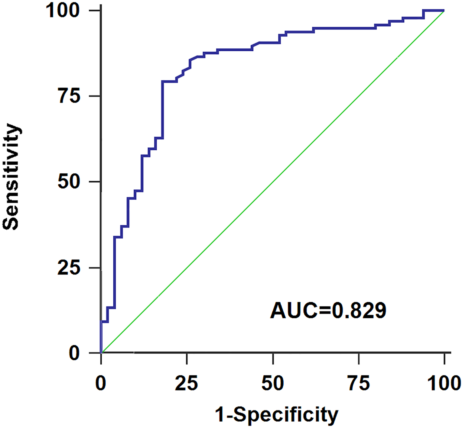
To analyze the correlation between serum miR-192 level and the clinical characteristics of AML patients, 97 pediatric AML subjects were assigned to miR-192 high (
Association between serum miR-192 expression with treatment response
Of 97 pediatric AML subjects, 58 cases achieved a CR. The expression levels of serum miR-192 were compared in 58 AML patients before or after achieving CR, and the analysis demonstrated that serum miR-192 levels were markedly increased in AML patients after treatment, suggesting that serum miR-192 expression was closely associated with treatment response (Fig. 3).
Serum miR-192 expression levels in 58 AML subjects before and after complete remission.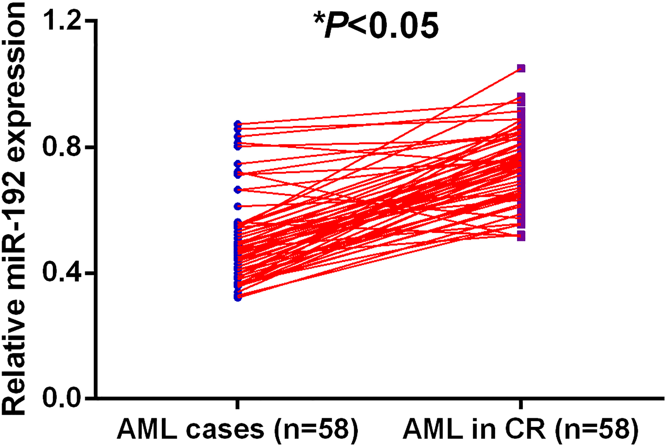
In the multivariant analysis model, cytogenetics (OS: HR
The Kaplan-Meier analysis revealed that pediatric AML patients with lower serum miR-192 level had significantly worse OS (
A. AML patients with low serum miR-192 expression had poorer OS. B. AML patients with low serum miR-192 expression had poorer EFS.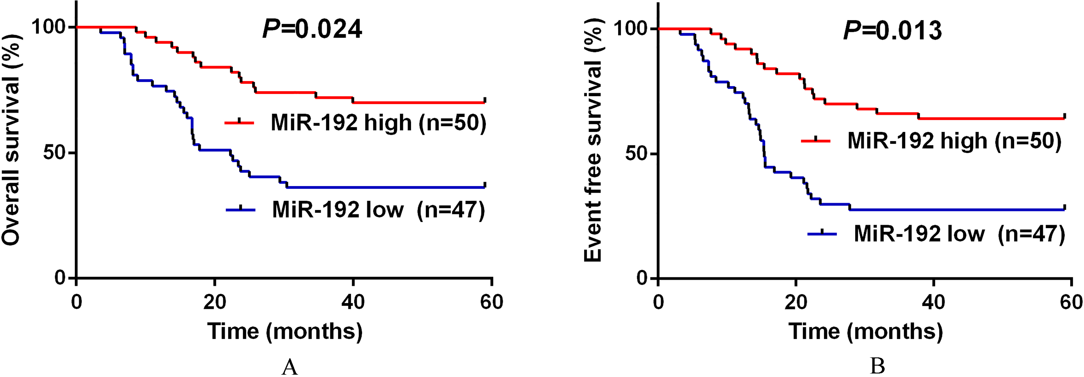
A. For the AML patients in the intermediate risk cytogenetic subgroup, low serum miR-192 expression was closely associated with poorer OS. B. For the AML patients in the intermediate risk cytogenetic subgroup, low serum miR-192 expression was closely associated with poorer EFS.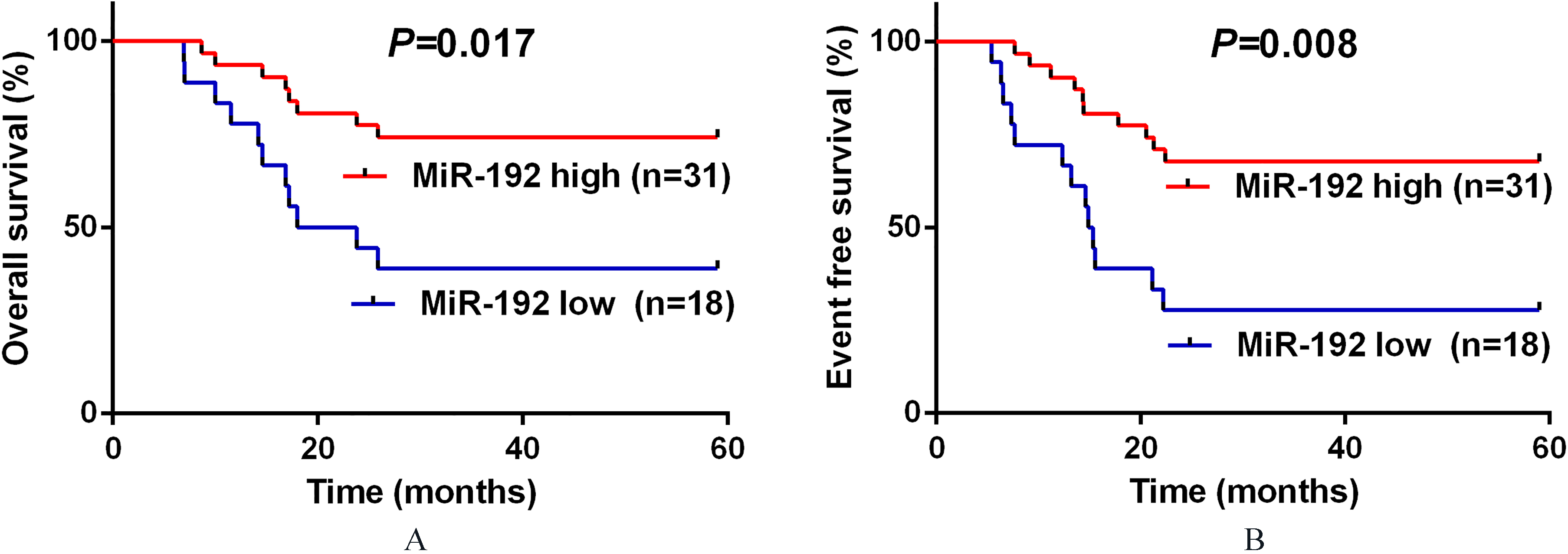
AML is one of the most common childhood cancers with high mortality. In the current study, we showed that serum miR-192 expression in pediatric AML patients was greatly down-regulated when compared to normal controls. Moreover, serum miR-192 levels were significantly increased in the AML subjects with favorable risk cytogenetics. ROC analysis showed that serum miR-192 expression could effectively identify pediatric AML cases from healthy controls. Furthermore, increased serum miR-192 expression was observed in patients achieving a complete response, and negatively associated with the aggressive clinical variables. Then, low serum miR-192 level was frequently correlated with shorter OS and EFS of the whole cohort of AML patients, as well as the AML patients with intermediate-risk cytogenetics. Also, serum miR-192 expression can serve as an independent prognostic indicator for pediatric AML patients. To the best of our knowledge, this study is the first report on the clinical significance of serum miR-192 in pediatric AML patients. The possible reasons accounting for missing miR-192 in the circulation system in pediatric patients with osteosarcoma might be as follows: First, the tumor cells per se directly secreted less miR-192 in the circulation system. In addition, the missing miR-192 in AML might be exosomic. It is possible that less miR-192 is packaged into the exosomes in AML cells, led to decreased miR-192 in the serum indirectly. We have added these information in the introduction and discussion respectively.
These results were consistent with other studies regarding leukemia. Ke and colleagues showed that miR-192 expression was greatly reduced in AML specimens. Moreover, loss of miR-192 stimulated cell proliferation and suppressed G0/G1 cell cycle arrest, cell apoptosis in vitro through targeting CCNT2 [13]. In acute lymphoblastic leukemia (ALL) , Sayadi et al found that miR-192 upregulation induced ALL cell proliferation arrest, and the cells transduced with miR-192-overexpressing virus dramatically promoted cell apoptosis [14]. In chronic lymphocytic leukemia (CLL), miR-192 expression in CLL patients was markedly lower than that in healthy controls [15].
MiR-192 was proposed as a tumor suppressor in several types of cancers. In osteosarcoma, miR-192 expression was significantly decreased both in cancer tissues and cell lines. Up-regulated miR-192 expression strongly restrained tumorigenicity in vitro by regulating TCF7 [16]. In prostate cancer, miR-192 expression was reduced in cancer cells. Moreover, miR-192 inhibition significantly stimulated cancer cell proliferation, colonyforming ability, and migration via targeting nin one binding protein [17]. In hepatocellular carcinoma, in vitro and in vivo studies revealed that miR-192 overexpression remarkably repressed cancer cell metastasis. In addition, low miR-192 expression was associated with poor outcome in patients [18]. Chiang et al. showed that miR-192 expression was underexpressed in colorectal cancer tissues and cell lines. Furthermore, loss of miR-192 was correlated with worse clinical variables [19]. In lung cancer, miR-192 expression was markedly downregulated in cancer tissues. Additionally, ectopic miR-192 expression suppressed cancer cell proliferation and inhibited carcinogenesis in vivo by directly silencing retinoblastoma 1 expression [20].
More interestingly, miR-192 might play a completely different role in some cancer types. Zhao and colleagues demonstrated that miR-192 levels were increased in pancreatic ductal adenocarcinoma patients. Moreover, enforced miR-192 expression induced cancer cell proliferation, migratory capacity, cell cycle progression, and inhibited cell apoptosis [21]. In esophageal squamous cell carcinoma, Li et al found that miR-192 was significantly upregulated both in cancer tissues and cells. In addition, repression of miR-192 promoted cell apoptosis and decreased cell proliferation by inversely regulating Bim [22]. Hence, the precise mechanism on the contradictory roles of miR-192 in various types of cancer was still limited and further exploration was required.
Taken together, our data offer the convincing evidence that serum miR-192 expression was markedly decreased in pediatric AML patients and closely associated with poor clinical outcome. Moreover, pediatric AML patients with low serum miR-192 expression had worse prognosis. Therefore, serum miR-192 might serve as a promising diagnostic and prognostic biomarker of this disease.
Footnotes
Conflict of interest
We deny any conflict of interest.