
Abstract
Thyroid carcinoma is one of the most frequent malignant tumors of the endocrine system, which accounts for nearly 1% population in newly diagnosed carcinoma worldwide and the incidence has an increasing tendency in recent years. To explore whether miR-135a-5p could affect the proliferation, invasion and migration of thyroid carcinoma cells by targeting VCAN. The expression levels of miR-135a-5p and VCAN were detected in human thyroid carcinoma tissues and cells, para-carcinoma tissues, as well as human normal thyroid cells using RT-qPCR and Western blot. In addition, dual-luciferase reporter gene assay, MTT assay, colony formation assay, wound healing assay, Transwell assay, flow cytometry analysis and in vivo tumorigenesis assay were also conducted. The results demonstrated that miR-135a-5p was down-regulated while VCAN was up-regulated both in thyroid carcinoma tissues and cells. Furthermore, the up-regulation of miR-135a-5p inhibited cell proliferation, invasion and migration of thyroid carcinoma. Dual-luciferase reporter gene assay provided evidence indicating that miR-135a-5p targeted VCAN in thyroid carcinoma. MiR-135a-5p could inhibit cell proliferation, invasion and migration of thyroid carcinoma by targeting VCAN. MiR-135a-5p and VCAN might emerge as a target for the treatment of thyroid carcinoma.
Keywords
Introduction
Thyroid carcinoma is one of the most frequent malignant tumors of the endocrine system, which accounts for nearly 1% population in newly diagnosed carcinoma worldwide and the incidence has an increasing tendency in recent years [1]. Thyroid carcinoma can be mainly classified as papillary thyroid carcinoma (PTC), follicular thyroid carcinoma (FTC), Hürthle cell carcinoma (HCC), anaplastic thyroid carcinoma (ATC), and medullary thyroid carcinoma (MTC) [2]. Among the subtypes of thyroid carcinoma, PTC accounts for about 80% of the total incidence, while FTC, HCC and poorly differentiated thyroid carcinoma (PDTC) have a high risk in metastasizing to lung or bone [3]. Nevertheless, the mortality remains high as many patients have no response to these conventional thyroid carcinoma therapies including radioiodine, radiotherapy, surgery, and chemotherapy [4]. Thus, new treatments like gene therapy are needed urgently. Although significant progress has been made these years in the understanding of molecular and gene basis of the formation of thyroid carcinoma, there is still a great need of understanding the undiscovered mechanisms in tumorigenesis and metastasis to prevent, diagnose, and treat thyroid carcinoma.
MicroRNAs (miRNAs, miR) are non-coding single-stranded RNAs (around 21 nucleotides) that are involved in regulating the gene expression by binding to the 3
Versican (also known as CSPG2 or VCAN), a chondroitin sulfate proteoglycan as one of the main component of the extracellular matrix (ECM), presents a significantly higher level in many diseases, interacting with hyaluronan (HA), than that in normal tissues [14]. The N-terminal globular (G1) domain and C-terminal globular (G3) domain of VCAN play a role in regulating migration, adhesion and proliferation in normal tissues, as well as an interactive role in varieties of intracellular and extracellular molecules [15]. Its overexpression associated with the development and relapse of tumor has been reported in many types of carcinoma cells, such as colorectal carcinoma, breast carcinoma, gastric carcinoma etc. [16, 17, 18]. It is suggested that VCAN is involved in promoting proliferation, motility, and invasion of malignant tumor cells, therefore the inhibition of its synthesis or process may be a potential therapeutic target in carcinoma [19]. But up to now, the expression and regulation of VCAN gene in thyroid carcinoma cells remain unclear.
Several miRNAs, including miR-144, miR-133a and miR-136, have been reported as potential regulator of structural ECM proteins, collagen and VCAN in the gathering, adhesion, and organization of the ECM [19]. Previous studies also proved that the level of VCAN in carcinoma cells can be controlled by the miRNA-mRNA regulation network. For example, a study indicated that the migration of malignant melanoma cell could be inhibited by targeting VCAN gene with a miRNA, which revealed the possibility of study on the targeting relationship between miRNA and VCAN [20]. In the present study, we aimed to verify the expression and function of both miRNA-135a-5p and VCAN in thyroid carcinoma cells and find the certain relationship between them.
Materials and methods
Clinical specimens
Fifty-three pairs of human thyroid carcinoma and para-carcinoma tissues (distance of 3 cm from the lesion) were collected from patients (19 males and 34 females) diagnosed with thyroid carcinoma who had undergone tumor resection at People’s Hospital of Weifang during Sep 2014 to Sep 2016. Tissues were snap-frozen in liquid nitrogen after surgery and reserved at
Cell culture
The human thyroid carcinoma cell line FTC-133, human PTC cell lines TPC1 and K1, human squamous thyroid carcinoma (STC) cell line SW579, and human normal thyroid cell line HT-ori3 (ATCC, Maryland, USA) were used in this research. FTC-133 cells were cultured in dulbecco’s modified eagle medium (DMEM) with 10% fetal bovine serum (FBS) and Ham’s F12 (1:1). K1 and TPC1 cells were cultured in 1640-RPMI medium with 10% FBS. SW579 cells were also cultured in DMEM with 10% FBS. HT-ori3 cells were cultured in Ham’s F12 with 5% FBS, supplemented with 1 mU/mL thyrotrophic hormone, 10 ng/mL hydrocortisone, 5
Cell transfection
Cells (5
RT-qPCR primer sequences
RT-qPCR primer sequences
F: Forward primer; R: Reverse primer.
All cellular RNA was extracted from tissues and cells using QIAzol (Qiagen, Hilden, Germany). Then they were transcribed into cDNAs, which were amplified following the manufacturer’s specification of PrimeSeript TM Reverse Transeriptase kit (TaKaRa, Japan) and the ABI 7900 real time PCR instrument (ABI, California, USA). U6 and GAPDH were used as standard for calculating relative miRNA and mRNA levels respectively by the 2
Western blot
Total proteins were extracted from cells after 24 h transfection. The concentration of protein was determined using Bradford method. The relative proteins were separated by SDS-PAGE under 220 v and transferred to the PVDF membranes. The membranes were incubated overnight with primary antibodies of VCAN and
MTT assay
K1 cells were plated onto 96-well plates at a density of 1
Colony formation assay
K1 cells of 1
Wound healing assay
The cells were inoculated into 6-well plates. When the cell confluence achieved about 90%, scratches were created by scraping the cell layer across each well. Detached cells were rinsed out using serum-free medium. The remaining cells were cultured for another 12 or 24 hours in 5% CO
Transwell assay
The invasion of transfected cells was determined using Transwell assay. The membrane of upper chambers was coated with 50 mg/L Matrigel (1:8). A total of 100
Dual-luciferase reporter gene assay
The amplification of VCAN 3
The expressions of miR-135a-5p and VCAN in thyroid tissues and cells. (A) RT-qPCR was used to detected miR-135-3p expression in thyroid tissues (
MiR-135a-5p directly targets VCAN. (A) The predicted miR-135a-5p binding site on wild-type VCAN 3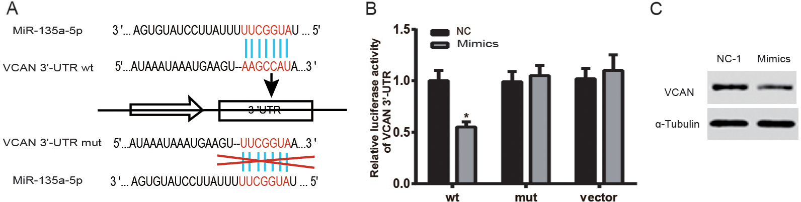
MiR-135a-5p inhibits cell proliferation, migration and invasion of thyroid carcinoma by targeting VCAN. (A) Cell viability was determined by MTT assay on K1 cells from different groups (Control, cells without transfection; NC-1, cells transfected with miR-135a-5p Negative control; NC-2, cells transfected with Pgv208-GFP vectors; Mimics, cells transfected with miR-135a-5p mimics; cDNA VCAN, cells transfected with VCAN cDNA; Mix, cells transfected with miR-135a-5p mimics and VCAN cDNA); (B) colony formation assay was used to detect the proliferation of cells in different groups (control, NC-1, NC-2, mimics, cDNA VCAN, Mix), the results were shown as the percentage colony number of control group; (C) cell migration was detected by wound healing assay, the relative wound closure was quantified as the ratio of the migration distance to control distance after 12 h and 24 h; (D) cell invasion was detected by Transwell assay, invaded cells were stained and counted in three randomly selected visual fields. *
MiR-135a-5p affects cell cycle and apoptosis of thyroid carcinoma by targeting VCAN. (A) The cell cycle of K1 cells from different groups (Control, cells without transfection; NC-1, cells transfected with miR-135a-5p Negative control; NC-2, cells transfected with Pgv208-GFP vectors; Mimics, cells transfected with miR-135a-5p mimics; cDNA VCAN, cells transfected with VCAN cDNA; Mix, cells transfected with miR-135a-5p mimics and VCAN cDNA) was detected by flow cytometry with PI staining; (B) the cell apoptosis of cell from different groups (Control, NC-1, NC-2, mimics, cDNA VCAN, Mix) was analysed by flow cytometry with Annexin V-FITC/PI staining. *
MiR-135a-5p suppresses tumor growth of thyroid carcinoma in vivo. (A) The tumors from the nude mice injected with K1 cells from different groups (Control, cells without transfection; NC-1, cells transfected with miR-135a-5p Negative control; NC-2, cells transfected with Pgv208-GFP vectors; Mimics, cells transfected with miR-135a-5p mimics; cDNA VCAN, cells transfected with VCAN cDNA; Mix, cells transfected with miR-135a-5p mimics and VCAN cDNA) were separated five weeks after the injection; (B) The volume of tumor from different groups (Control, NC-1, NC-2, mimics, cDNA VCAN, Mix) was counted each 5 days and tumor growth curve was calculated at 30 days after injection, *
Approximately 2
As for apoptosis, the cells (2
In vivo tumorigenesis assay
0.2 mL K1 single-cell suspension (2
Statistical analysis
SPSS 20.0 (Chicago, Illinois, America) was employed for statistical analysis and GraphPad Prism 6.0 (GraphPad Software, Inc., CA, USA) was used for plotting. The data were presented as mean
Results
MiR-135a-5p and VCAN expression in thyroid tissues and cells
RT-qPCR was applied to detect the expression levels of miR-135a-5p in 53 pairs of thyroid carcinoma and para-carcinoma tissues. As presented in Fig. 1A, miR-135a-5p expression was dramatically down-regulated in thyroid carcinoma tissues compared with para-carcinoma tissues (
MiR-135a-5p directly targets VCAN
To confirm that whether miR-135a-5p could target VCAN, plasmids carrying miR-135-5p or VCAN 3
MiR-135a-5p suppresses cell proliferation and invasion of thyroid carcinoma through targeting VCAN 3
-UTR
Cell proliferation
To investigate the effects of miR-135a-5p on K1 cell proliferation, we carried out MTT and colony formation assays. As shown in the MTT assay results (Fig. 3A), there was no obvious difference in cell numbers among groups 48 h after transfection, whereas, 72 h after transfection, substantial decreased cell number was observed in Mimics group and significant increased cell number was seen in cDNA VCAN group (
Cell migration and invasivion
To define the role of miR-135a-5p in cell migration and invasion, we conducted wound healing and Transwell assays. As expected, migration rate of cells in the Mimics group markedly decreased, while a significantly risen migration rate was observed in cells transfected with cDNA VCAN (
We also conducted Transwell assay to explore the effect of miR-135a-5p on cell invasion. Similarly, the number of invaded K1 cells was significantly smaller in Mimics group (
MiR-135a-5p affects cell cycle and apoptosis of thyroid carcinoma by targeting VCAN
Cell cycle
Flow cytometry was applied to analyze cell cycle, results were presented in Fig. 4A. Apparently, significant cell number reduction in S and G2/M, and accumulation in G0/G1 were observed in Mimics group (
Cell apoptosis
As shown in Fig. 4B, comparing with NC groups, apparent increase in cell apoptosis rate was noticed in the Mimics group while significant reduction was observed in the cDNA VCAN group (
MiR-135a-5p suppresses tumor growth of thyroid carcinoma in vivo
To investigate whether miR-135a-5p overexpression repressed tumor growth in vivo, we conducted the nude mice tumorigenesis assay. Photographs of tumors obtained from mice 5 weeks after the injection were presented in Fig. 5A, the tumor volume was remarkably smaller in the Mimics group, in contrast with the cDNA VCAN group. Obviously, lower tumor growth rate and apparent reduction in tumor volume were noticed in the Mimics group, while higher tumor growth rate and tumor volume increase were seen in cDNA VCAN group (
Discussion
As one of the most frequent malignant carcinomas of the endocrine system with an ever-increasing incidence, thyroid carcinoma has been studied a lot on its pathogenesis and therapies. However, the molecular and gene basis of its development is still unclear, which is proved to be a difficulty to surmount through. In recent years, researchers began focusing on the regulating role of miRNAs in tumors, and several miRNAs have been proved to function by overexpressing or suppressing the carcinoma progress. For instance, Qiu et al. found that the overexpression of miRNA-613 down-regulated cell proliferation, migration, and invasive ability in PTC by targeting SphK2 [21]. Minna et al. identified that miR-451a, targeting AKT/mTOR pathway, was suppressed in thyroid carcinoma cells and inhibited the neoplasia progress [22]. Similarly, miR-191 was also reported by Colamaio et al. as a suppressor in FTC and PTC, playing a negative role in pathogenesis by targeting CDK6 [23]. In the present study, we demonstrated that miR-135a-5p was remarkably suppressed in human thyroid carcinoma tissues. The expression of miR135a-5p was inversely correlated with the expression of VCAN gene. Moreover, functional assays proved that the overexpression of miR-135a-5p inhibited cell proliferation, migration and invasion of thyroid carcinoma cells, and slowed the growth of thyroid carcinoma in vivo by targeting VCAN 3
Recently, miR-135a-5p has been identified as a biomarker in tumor tissues as its ectopic expression. Golubovskaya et al. confirmed that the overexpression of miR-135 inhibited the invasion of cancer cells and increased the sensitivity to chemotherapy by targeting focal adhesion kinase, which could be a key for tumor therapy [10]. MiR-135a-5p has also been proved as a tumor suppressor in gastric carcinoma and gallbladder carcinoma by targeting very low density lipoprotein receptor (VLDLR) and AP-2
Several target genes of miR-135a-5p have been discovered in previous studies. For further understanding the mechanism of miR-135a-5p in thyroid carcinoma cells, we identified versican (VCAN) as a potential target based on bioinformatics analysis. As a main component of ECM, the overexpression of VCAN was indicated to be associated with improvement of cell proliferation, migration and invasion in malignant tumor tissue [19]. We observed in the present study that the expression level of VCAN in thyroid carcinoma cells was remarkably higher than other tissues, accelerating cell proliferation and migration as well as increasing the invasion of thyroid carcinoma cells, which supported that VCAN could be a potential therapeutic target in thyroid carcinoma cells.
Up to now, although the mechanism that miR-135a-5p could target VCAN is agreed by bioinformatics analysis, there is no experimental evidence published yet. Our experiment of dual-luciferase reporter gene assay proved that VCAN 3
For the first time, our study investigated the functional mechanism of miR-135a-5p as a suppressor in thyroid carcinoma cells as well as targeting and regulating VCAN, which would provide evidence for further studies on the miR135a-5p/VCAN pathway and its functional roles in thyroid carcinoma cells. However, there are still several limitations in the study. Firstly, since thyroid carcinoma is classified as several types with different characteristics, the result of this study may not be appropriate for all types of thyroid carcinoma. Secondly, we cannot exclude that other genes may be targeted by miR-135a-5p, which can result in the regulation of thyroid carcinoma cells in this experiment. Lastly, VCAN may be regulated by other miRNAs besides miR-135a-5p, thus further study on the functional mechanism of VCAN is also needed.
In summary, our data put forward a new idea that miR-135a-5p can suppress cell progress including cell proliferation, migration, and invasion in thyroid carcinoma by targeting VCAN 3
Conflict of interest
None.