
Abstract
The aim of this study was to demonstrate the potential of a wireless pixelated β+-sensitive intracerebral probe (PIXSIC) for in vivo positron emission tomographic (PET) radiopharmacology in awake and freely moving rodents. The binding of [11C]raclopride to D2 dopamine receptors was measured in anesthetized and awake rats following injection of the radiotracer. Competitive binding was assessed with a cold raclopride injection 20 minutes later. The device can accurately monitor binding of PET ligands in freely moving rodents with a high spatiotemporal resolution. Reproducible time-activity curves were obtained for pixels throughout the striatum and cerebellum. A significantly lower [11C]raclopride tracer-specific binding was observed in awake animals. These first results pave the way for PET tracer pharmacokinetics measurements in freely moving rodents.
IN HUMANS, neuronal activation patterns correlated to behavior are recorded easily using positron emission tomography (PET) or functional magnetic resonance imaging. In animals, the combination of PET tracer pharmacokinetics measurements with behavioral studies would constitute a breakthrough for the understanding of functional processes in the brain. 1 However, PET requires anesthesia or severe animal restraints, which prevent simultaneous behavioral activities. For the past 10 years, several instruments dedicated to behavioral neuroimaging have been proposed. 2 In the simplest approach, behavioral experiments are carried out after injection of the tracer, the animals being left free in an open field. 3 After 30 minutes, animals are anesthetized and brain images are acquired with microPET. Although attractive, this method obviously restricts the combination of PET and behavioral studies to specific protocols and prevents simultaneous recordings. Another approach, the RatCAP, relies on a wearable PET device allowing for imaging the entire brain. 4 In a third approach, a motion tracking system is used within a PET scanner. 5 Nevertheless, it still requires constraining the animal in a specific holder. 5 In all approaches, either the bulkiness of the system or the global constraints strongly limit animal motion.
We propose a strategy for in vivo recording of PET tracer time-activity curves (TACs) in freely moving rats using a radiosensitive pixelated intracerebral probe and its backpack supporting the microdriver and the radiofrequency transmitter: PIXSIC. We have previously shown its ability to measure TACs in anesthetized rats. 6 Here we demonstrate that the probe can provide kinetics measurements of [11C]raclopride, a well-known dopaminergic D2 receptor tracer, in freely moving rats using chronically implanted wireless probes.
Materials and Methods
PIXSIC Design
PIXSIC relies on a silicon probe 200 μm thick, 690 μm wide, and 17 mm long hosting 10 pixels of 200 × 500 μm implanted in the brain region of interest (Figure 1). The pixel sensor is based on a reverse-biased, high-resistivity silicon diode. PIXSIC enables high-sensitivity time-resolved measurements in a volume defined by the range of beta particles within living tissues.7,8 The pixelated detection scheme adds “imaging” features by recording several TACs along the probe position. The probe design is light, compact, energetically autonomous, and based on a wireless radiofrequency data exchange that empowers free behavioral activity during acquisition. 6

PIXSIC system. The head socket (A) bears the silicon probe, whose sensitive part is implanted into the brain. This probe is wire bonded to an application-specific integrated circuit (ASIC) that is protected by a hull. The backpack is carried by the rat and includes radio transmission components and a power supply that enables the autonomous and wireless operation of the system (B), a connection cable with the head socket (C), and a harness (D). The animal is equipped with a jugular catheter for radiotracer injections (E).
Autoradiography
Probe implantations in high-density D2 regions were optimized with autoradiography. Animals were anesthetized (single intraperitoneal injection of urethane 1.7 g/kg). The tail vein was catheterized for radiotracer injection. Twenty minutes after injection of 74 MBq of [11C]raclopride, the rat was sacrificed; its brain was removed and frozen in isopentane at −30° C. Coronal sections of 20 μm were obtained with a cryostat at −20° C, thaw-mounted on glass microscope slides, and juxtaposed to a phosphor imager for 60 minutes. After development, optical densities were measured for the cortex, the striatum, and the cerebellum using MultiGauge software (Fujifilm, Tokyo, Japan).
Animal Procedure
Adult male Sprague Dawley rats (350–400 g) were housed under standard conditions of temperature and humidity and under artificial light from 8 am to 8 pm. All experiments were conducted in accordance with European guidelines for the care of laboratory animals (2010/63/EU), and all experiments were approved by the animal use ethics committee of the University of Lyon.
Surgery
All animals (n = 6) were anesthetized with isoflurane (induction 3.5% in air and maintenance 2% in air) after a subcutaneous injection of buprenorphine for analgesia (30 ng/kg of rat; Buprecare, Axience SAS, Pantin, France). Chronic intravenous silicone catheters were surgically implanted into the right jugular vein situated dorsally between the scapulae (Vascular Access Button, Instech Solomon, Plymouth Meeting, PA). Next, as described elsewhere,6,9 two probes were stereotaxically implanted for each animal, in the striatum (a region highly expressing D2 receptors; anteroposterior +0.6, lateral +3.0 from bregma, and dorsoventral −7.0 below dura) and the cerebellum (a reference region containing negligible densities in D2 receptors; anteroposterior −12.0, lateral −0.5 from bregma, and dorsoventral −6.0 below dura). Both probes were cemented on the skull (Heraeus, Hanau, Germany). Rats were left in their cages to recover from surgery for at least 6 days before experimentation.
[11C]Raclopride Synthesis
[11C]Raclopride was synthesized (radiochemical yield of 25% decay corrected) in an automated synthesizer (CERMEP Biomedical Cyclotron, Lyon, France). Chemical and radiochemical purity were determined by high-performance liquid chromatography (> 98%). Specific activity of the injected radiotracer ranged from 50 to 285 GBq/μmol (1.35 to 7.70 Ci/μmol), and the corresponding injected raclopride mass ranged from 0.65 to 3.7 nmol/kg.
PIXSIC Scanning Procedure
Over 45 seconds, 74 MBq (2 mCi) of [11C]raclopride in 0.5 mL saline was injected into the dorsal vein. The TACs were recorded for 60 minutes. The following three protocols were applied:
Protocol 1 (anesthetized rat recordings): three rats were anesthetized with isoflurane. Then PIXSIC was set in place (probes and electronics backpack) prior to the [11C]raclopride injection.
Protocol 2 (awake rat recordings): three rats were equipped with PIXSIC without any anesthesia. Then each rat was placed in a box (30 × 30 times 35 cm), allowing for free movement. [11C]Raclopride was injected after 20 minutes' habituation.
Protocol 3 (displacement): protocol 2 + 30 minutes after the injection of [11C]raclopride, unlabeled raclopride was injected (2 mg/kg in 1 mL/kg saline) in the vein catheter.
Data Analysis
PIXSIC data were averaged every minute. The corresponding TACs were normalized for radioactive decay, and the detection sensitivity of each pixel was measured as described elsewhere. The specific binding ratio (B/F) was calculated as described elsewhere 10 with the following equation:

B represents the specific binding signal, defined as the difference between total regional radioactivity of the striatum and that of the cerebellum. The cerebellum was used as a reference tissue to evaluate free radioligand concentration and nonspecific binding in the brain (F). This equation integrates the time curves for B (B(t)) and F (F(t)) from 30 to 50 minutes to make it possible to compare the values obtained in awake and anesthetized animals. Statistical analyses were conducted for repeated measurements by comparing the means obtained from the awake and anesthetized animals for each time point by a two-way analysis of variance (ANOVA), followed by post hoc Bonferonni tests. A p value less than .05 was considered significant.
Results
The aim of the study was to demonstrate the ability of PIXSIC to record [11C]raclopride TACs in freely moving rats and to compare results obtained in awake and anesthetized rats. Figure 2 details the location of the two probes in the brain by superimposing schematic pixelated probes with two [11C]raclopride autoradiograms at the striatal and the Probe positions were determined using surgical stereotaxic coordinates confirmed by histologic controls. Figure 3 shows the corresponding TACs recorded along the probe in awake and anesthetized rats and the corresponding specific bindings. TACs were acquired at least 6 days after the surgical implantation of the probes to exclude postimplantation inflammatory reactions. TACs were derived from the average of pixels from specific areas (4 pixels in the cortex, 6 in the striatum, and 10 in the cerebellum). The recorded TACs present a good consistency with previous [11C]raclopride recordings with PET scanners.4,11 Moreover, two major differences appear between anesthetized and awake recordings: (1) the tracer concentration level at tracer equilibrium is much higher in anesthetized animals, and (2) the specific binding in the striatum is much higher in the anesthetized animals, whereas it is similar in the cortex for both cases.
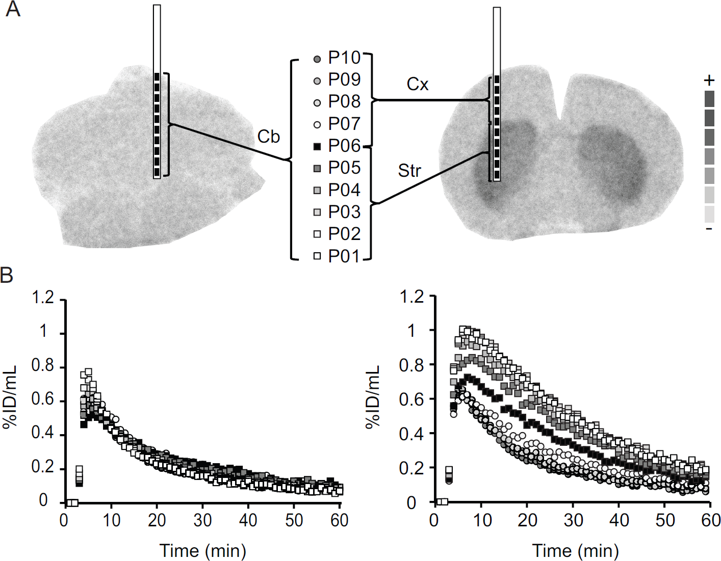
Localization of one PIXSIC probe in the rat cerebellum (left) and the second probe in the cortex (Cx) and striatum (Str) region (right) in [11C]raclopride autoradiography (A). Silicon probe sizes were 200 μm thick, 690 μm wide, and 17 mm long. The radiosensitive pixels (200 μm x 500 μm) are numbered from P01 to P10 (P01–P06 are located in the striatum and P07–P10 are located in the cortex). Corresponding time-activity curves (B) after bolus administration of [11C]raclopride (74 MBq; 2 mCi; IV, n =1 rat). ID = injected dose. cerebellar levels.

Cortical, striatal, and cerebellar time-activity curves after intravenous bolus administration of [11C]raclopride in awake freely moving (n = 3) (A) and anesthetized (n = 3) rats (B). Corresponding specific binding in the striatum and cortex of awake and anesthetized rats (C). Specific binding was obtained by subtracting the cerebellum radioactivity (as a region without D2 receptors) from the radioactivity found in the cortex or striatum. ID = injected dose.
A calculation of the B/F ratio in the striatum gives values significantly higher in anesthetized than in awake rats (3.23 ± 0.5 and 1.79 ± 0.2, respectively). In contrast, values are similar in the cortex: 0.3 ± 0.1 and 0.4 ± 0.2 in anesthetized and awake rats, respectively. Both results provide evidence of the fact that animal anaesthesia and/or motion interfere with the tracer availability, uptake, and binding. Finally, competitive binding experiments were performed to ascertain the specificity of the [11C]raclopride uptake in the striatum for both cases. As expected, tracer binding rapidly decreases in competition conditions (Figure 4).

Alteration of specific binding of [11C]raclopride in the striatum and the cortex after intravenous injection of 2 mg/kg raclopride (arrow) in awake freely moving rats (n = 3). ID = injected dose.
Discussion
In this study, striatal, cortical, and cerebellar TACs were obtained in anesthetized and freely moving rats with PIXSIC after intrajugular injections of [11C]raclopride. The procedure allowed for wireless recording of the radioactivity kinetics along the probes implanted in freely moving rats. Clear correlations were obtained between the pixel locations and the distribution of the radioligand. More precisely, the radioactivity counts were very low for all the pixels of the probe placed within the cerebellum and for pixels of the second probe partially located in the cortex. In contrast, considering each pixel, there was a slight increase in the radioactivity counts through the dorsoventral axis of the striatal probe, confirming D2 concentration heterogeneity and its progressive density increase. 12
In agreement with other results,3,4,13 and despite the different methodologies (clinical PET, microPET, RatCAP system), specific binding of [11C]raclopride is much higher in anesthetized than in freely moving rats. As suggested previously, potential causes of this effect are the enhancement of peripheral metabolism in the awake case, changes in peripheral metabolism and blood flow, and interference of the anesthetic with the dopaminergic transmission system (neurotransmitter concentration modifications). This may include changes in the rate of dopamine release, which can compete with [11C] raclopride and modify its binding to D2 receptors, autoreceptor effects, or the influence on tracer recapture through blood-brain barrier transporters.4,13,14 These results further highlight the potential bias induced by anesthetics on quantitative measurements and confirm the interest in developing preclinical approaches in awake animals to facilitate comparison with PET human data.
The TACs recorded with probes chronically implanted in the brain did not show significant changes compared to TACs recorded in acute experiments. Although some tissue structures are slightly damaged due to probe implantation, these results demonstrate that invasiveness does not affect the pharmacokinetics recordings.
Due to the small weight and dimensions of PIXSIC components (head socket and backpack), they do not interfere with the movements of the rodent. Another advantage of the system is that it is not sensitive to ambient light. 15 Yet some limitations remain due to the fragility of the probes and their connections with the backpack that need to be solved for the next PIXSIC generation. Another crucial limitation to be reduced is linked to the relatively low sensitivity of the probe. We determined that PIXSIC's single-pixel sensitivity is in the range of 30% of the sensitivity of the beta-microprobe and a last-generation microPET camera. 6 This leads to higher doses of radiotracer to be injected for a PIXSIC acquisition than for a classic microPET acquisition (74 MBq in our conditions versus 18 MBq) and to an averaging of data every minute. Since specific activities of the brain PET radiotracers are classically obtained between 37 and 74 GBq/μmol and cannot systematically be guaranteed at higher values, particular attention will have to be given to the injected amounts of radiotracer to stay within the range of tracer doses. Further studies in instrumentation would be desirable to improve the sensitivity of the current probes.
Conclusion
PIXSIC is a reliable approach for studying the dynamic neurobiological processes underlying behavior in rodents. PIXSIC provides accurate pharmacologic data in awake and freely moving animals, which can be used to assess receptor occupancy, availability of transporters and enzymes, and glucose metabolism in particular brain regions. This technological approach constitutes a breakthrough toward recordings of radiolabeled molecules kinetics in freely moving rodents and their comparison with the anesthetized model. The next step will consist of determining the most appropriate kinetic model with arterial input function measurements. Another challenge will be the correlation of molecular data with behavioral measurements through specific and relevant paradigms chosen to stress the potential of such a combination.
Footnotes
Acknowledgments
Financial disclosure of authors: This work was supported by the French Research National Agency (ANR) under grant no. ANR-09-blan-0102-01. This work was performed within the framework of the LABEX PRIMES (ANR-11-LABX-0063) of Université de Lyon, within the program “Investissements d'Avenir” (ANR-11-IDEX- 0007) operated by the French National Research Agency (ANR).
Financial disclosure of reviewers: None reported.