
Abstract
Using adenovirus (Ad)-based vectors is a promising strategy for novel cancer treatments; however, current tracking approaches in vivo are limited. The C-terminus of the Ad minor capsid protein IX (pIX) can incorporate heterologous reporters to monitor biodistribution. We incorporated metallothionein (MT), a low-molecular-weight metal-binding protein, as a fusion to pIX. We previously demonstrated 99mTc binding in vitro to a pIX-MT fusion on the Ad capsid. We investigated different fusions of MT within pIX to optimize functional display. We identified a dimeric MT construct fused to pIX that showed significantly increased radiolabeling capacity. After Ad radiolabeling, we characterized metal binding in vitro. We explored biodistribution in vivo in control mice, mice pretreated with warfarin, mice preimmunized with wild-type Ad, and mice that received both warfarin pretreatment and Ad preimmunization. Localization of activity to liver and bladder was seen, with activity detected in spleen, intestine, and kidneys. Afterwards, the mice were euthanized and selected organs were dissected for further analysis. Similar to the imaging results, most of the radioactivity was found in the liver, spleen, kidneys, and bladder, with significant differences between the groups observed in the liver. These results demonstrate this platform application for following Ad dissemination in vivo.
Barriers to Gene Therapy Approaches for Cancer
As the limits of existing treatments for cancer are recognized, it has become clear that novel therapies must be considered for successful treatment; cancer therapy using adenovirus (Ad) vectors is a promising strategy. 1 The existing approaches to Ad-based cancer therapy include (a) mutation compensation, (b) molecular chemotherapy, (c) genetic immunopotentiation, (d) genetic modulation of resistance or sensitivity, and (e) oncolytic therapy or virotherapy. Despite promising results, major limitations have been defined in the application of serotype 5 Ads based on clinical and preclinical studies such as a high prevalence of Ad-neutralizing antibodies, 2 downregulation of the coxsackie-adenovirus receptor (CAR), 3 and Ad sequestration by the liver. 4 In addition, current clinical biodistribution tracking of Ad vectors in vivo is limited to invasive procedures such as biopsies, which are sampling error prone, cannot be repetitively performed, and do not give a full representation of the pharmacokinetics involved.
Current Detection Methods Are Inadequate for Ad Vector Systems
Several imaging approaches have attempted to track Ad biodistribution, particularly the use of reporter genes, such as green fluorescent protein (GFP), 5 somatostatin receptor type 2 (SSTR-2),6–8 sodium iodide symporter (NIS),9,10 firefly luciferase,11,12 and herpes simplex virus thymidine kinase (HSV-TK),13,14 as well as soluble marker peptides such as human carcinoembryonic antigen (hCEA) 15 and β- human chorionic gonadotropin (β-hCG). 16 Despite their utility for assessing gene delivery and expression, these reporters and markers by themselves are not suitable for monitoring physical biodistribution. The major drawback to approaches that tag viruses with reporter genes is that these systems require initial viral infection and subsequent cellular expression of a reporter gene to allow noninvasive imaging.
Thus, detection is restricted to infected cells expressing the reporter gene, which does not represent the physical distribution of the virus itself. Likewise, the use of reporter systems cannot reflect the pharmacodynamics of viral vector particles and the kinetics of vector clearance.
Capsid Protein IX is a Potential Imaging Location for Labeling Ads
We and other groups defined the C-terminus of protein IX (pIX) as a locus presenting incorporated heterologous ligands17,18 as well as for incorporating reporter genes, such as HSV-TK, 19 luciferase, 20 and fluorescent proteins.21–23 To extend this concept, we devised a novel approach to incorporate the human metallothionein (MT) protein as a fusion to the Ad minor capsid protein pIX. 24 MT is a ubiquitous, low-molecular-weight, metal-binding protein that participates in heavy metal metabolism and detoxification. 25 Mammalian forms of MT bind seven metal ions in tetrahedral metal-thiolate clusters, including 99mTc,26,27 a commonly used medical isotope useful for radioimaging by single-photon emission computed tomography (SPECT). Our previous study demonstrated the feasibility of 99mTc binding in vitro to the pIX-MT fusion on the capsid of Ad virions using a simple transchelation reaction. 24 We showed the capability for in vivo SPECT imaging of a mouse after administration of a 99mTc-radiolabeled Ad. However, this study was limited by low incorporation of 99mTc into the MT construct.
Incorporating Human MT into pIX for Analysis of Biodistribution
In our current study, we sought to investigate different fusions of MT within pIX to optimize 99mTc radiolabeling for functional display of this gene product. We identified a dimeric MT construct that shows a significant increase in 99mTc-radiolabeling activity and characterized metal binding. Furthermore, we explored the ability to noninvasively observe Ad biodistribution and the kinetics of uptake in vivo on a whole-body level. These results demonstrate the possibility of detecting Ad dissemination for monitoring safety in vivo.
Materials and Methods
Cell Culture
The human embryonic kidney epithelial cell line (HEK293) and the human lung adenocarcinoma epithelial cell line (A549) were obtained from the American Type Culture Collection (ATCC, Manassas, VA). The cell lines were grown in high-glucose Dulbecco's Modified Eagle Medium (DMEM, Invitrogen, Carlsbad, CA) containing 100 IU/mL penicillin, 100 μg/mL streptomycin, and 2 mM L-glutamine (Invitrogen) and supplemented with 10% fetal bovine serum (Atlanta Biologicals, Lawrenceville, GA). The cells were maintained at 37°C and 5% CO2 under humidified conditions and subcultured using a 0.25% (w/v) trypsin- 0.53 mM ethylenediaminetetraacetic acid (EDTA) solution (Invitrogen).
Animals
Female C57BL6 mice at 4 to 6 weeks of age were obtained from Charles River Laboratories (Wilmington, MA). All animals received humane care based on guidelines set by the American Veterinary Association. The experimental protocols involving live animals were reviewed and approved by the Institutional Animal Care and Use Committee of LSU Health Sciences Center in Shreveport.
Recombinant Ad Construction
An initial pIX-MT intermediate sequence in pUC57 was constructed by synthesizing a 932 bp DNA fragment (GenScript, Piscataway, NJ) corresponding to amino acids 106 to 140 of the Ad serotype 5 pIX protein-coding sequence, followed by a 15-amino acid linker (GGGGSGGGGSGGGGS), amino acids 1 to 61 of the human MT 1A sequence, a 16 bp DNA linker sequence (TGAGCTAGCGACGTCA), and the 583 bp DNA sequence immediately upstream of the pIX gene (bp 4032–4614, GenBank accession AY339865). Additional DNA fragments were synthesized containing spacer sequences consisting of a 30 A spacer of 25 amino acids corresponding to an alpha helical linker 18 ; a 45 A spacer of 38 amino acids from ApoE4 cDNA (bp 373-472, GenBank accession XM_008844); an ABD spacer of 46 amino acids from the albumin-binding domain 3 (ABD) of streptococcal protein G (bp 880-925, Genbank accession X06173); a 75 Å spacer of 63-amino acid fusion consisting of the 30 Å and 45 Å spacers; and a red fluorescent protein (RFP) spacer equivalent to 238 amino acids from the monomeric RFP gene (bp 1-711, Genbank accession AY678264.1). These synthetic DNA fragments were subcloned directly into the MfeI – BstXI sites of the AdenoVatorCMV5 shuttle vector (QBiogene, Carlsbad, CA). A turboGFP (tGFP) complementary DNA sequence (Axxora, San Diego, CA) was also subcloned into the CMV5 expression polylinker site of pAdenoVatorCMV5 for use as a reporter gene, and the CMV5 promoter was replaced with the human survivin promoter gene sequence from −230 to +30 bp. The resulting pAdenoVator-Survivin-tGFP-pIX-MT shuttle vectors were used to construct recombinant Ad plasmids by homologous recombination with pAdEasy1 (containing the E1 and E3 deleted Ad5 backbone) in Escherichia coli using methods previously described. 28 The resultant recombinant plasmids were linearized with Pac I and transfected into HEK293 cells to generate and rescue Ad virions. The wild-type Ad and Ad-tGFP-pIX-MT vectors used as controls have been described previously. 24
Virus Propagation and Purification
Viruses were propagated in HEK293 cells, which do not express wild-type pIX. Viruses were purified by double CsCl ultracentrifugation and dialyzed against Dulbecco's phosphate-buffered saline (PBS) containing 10% glycerol. Final aliquots of virus were analyzed for viral particle (vp) titer using absorbance at 260 nm and a conversion factor of 1.1 × 10¹². Multiplicity of infection (MOI) was determined using an Adeno-X Rapid Titer Kit (Clontech, Mountain View, CA) and represents the number of infectious units (ifu) of virus. Viruses were stored at − 80°C until use.
Ad Replication Assay
The HEK293 cell line was infected with each Ad construct at 10:1 MOI. The infected cells were incubated for up to 3 days at 37°C. After each time point, replicate cells were collected and resuspended in 0.2 mL PBS. Total genomic DNA was extracted using a Qiagen DNA-mini kit (Valencia, CA), and Ad E4 copy number was determined by quantitative polymerase chain reaction (PCR). For this assay, the following sets of primers and probes were designed and used: sense (5′-GGGTCGCCACTTAATCTACCT-3′), antisense (5′-GCAAGGCGCTGTATCCAA-3′), and probe (5′FAM-CGCTTGTGGTATGATGGCCACGTTAMRA-3′).
Western Blot Analysis
Purified Ad virions were lysed in RIPA buffer, resolved on precast 4 to 20% gradient sodium dodecyl sulfate-polyacrylamide gel electrophoresis (SDS-PAGE) gels (Thermo Scientific Pierce, Rockford, IL), and transferred to nitrocellulose membranes. Staining was performed using a rabbit antiadenovirus pIX polyclonal antibody 29 or a monoclonal anti-Ad hexon monoclonal antibody (clone 65H6, Thermo Fisher Scientific, Rockford, IL), followed by a secondary horseradish peroxidase (HRP)-conjugated goat antirabbit mouse IgG antibody (Santa Cruz Biotechnology, Santa Cruz, CA) or HRP-conjugated goat antimouse IgG antibody (Santa Cruz Biotechnology). Specific protein bands were detected by chemiluminescence using Amersham ECL Plus reagents (GE Healthcare, Piscataway, NJ).
Radiolabeling of Virus and Gel Chromatography
For radiolabeling Ad virions, 10 to 40 mCi of 99mTc-pertechnetate in 0.5 mL saline was reduced in the presence of glucoheptonate 30 using a modified kit (10 mg sodium glucoheptonate and 25 μg SnCl2) for 15 minutes at room temperature. The 99mTc-glucoheptonate (225 μL) was then added directly to aliquots of Ad (0.1-10 × 1011 vp in 75 μL containing 3.3 mg/mL bovine serum albumin). Incubation with Ad aliquots proceeded for 15 to 120 minutes at 37°C. Purification of radiolabeled Ad virions from free 99mTc and 99mTc-glucoheptonate was performed by size-exclusion chromatography using Sephacryl S-100 HR (GE Healthcare). Viral particle titer of each fraction was determined by measuring the absorbance at 260 nm in a spectrophotometer, using a conversion factor of 1.1 × 10¹² vp per absorbance unit. 31 The radioactivity of each fraction was measured with a CRC-25R dose calibrator (Capintec, Ramsey, NJ).
Metal Competition Assay
Aliquots of the radiolabeled Ads were incubated in the absence or presence of nonradioactive competitor metal ions. After addition of competitor metal using 100 X stock solutions, the aliquots were incubated at room temperature or at 37°C for 15 minutes. The Ad-bound and free 99mTc were separated using P-30 micro spin columns (Bio-Rad, Hercules, CA) and quantified using a dose calibrator (Capintec).
SPECT/CT Scanning of Ad-Infected Mice
Groups of mice were preimmunized with wild-type Ad by intraperitoneal injection with 1 × 10¹⁰ vp at 3 weeks and 1 week prior to SPECT imaging. To deplete vitamin K- dependent coagulation factors, groups of mice were injected subcutaneously with 133 μg of warfarin in 100 μL of peanut oil at 3 and 1 days prior to SPECT imaging. Mice were initially injected via the tail vein with approximately 750 μCi of the radiolabeled Ad, and image acquisition was started at 60 minutes afterwards. The animals were anesthetized using isoflurane and fixed in a prone position on the bed and center of rotation relative to the gantry of a dedicated small-animal trimodality positron emission tomography (PET)/SPECT/computed tomography (CT) system (TriFoil Imaging, Northridge, CA). Three-dimensional (3D) SPECT imaging was performed in a step-and-shoot manner using the following acquisition parameters: 64 projections, 30 seconds/projection (35-minute image acquisition), with a 140 keV photopeak 6 10% window. SPECT 3D reconstructions were performed using an ordered subset expectation maximization (OSEM) iterative reconstruction algorithm with five iterations of four subsets (TriFoil Imaging). Immediately after each SPECT imaging was performed, a CT image was acquired using the same coordinates with 256 projections and 1024 × 1024 projection matrix size and a voltage of 60 kV; CT reconstructions were performed using filtered backprojections. The SPECT and CT images were visualized and fused using Amira 3D analysis software (FEI Visualization Sciences Group, Burlington, MA).
Ex Vivo Analysis
At the completion of imaging, each mouse was euthanized with a lethal dose of isoflurane followed by cervical dislocation. Tissues and organs, including liver, spleen, stomach, kidneys, heart, lungs, pancreas, small intestine, large intestine, peritoneal fat pad, bladder, and ovaries and uterus, and blood were removed and weighed, and the radioactivity was measured using a NaI (Tl) well counter and MAESTRO multichannel analyzer software (Advanced Measurement Technology, Oak Ridge, TN). Tissue and organ uptake was calculated as percentage of injected dose per gram of tissue (%ID/g), corrected for decay.
Statistical Analysis
All data are expressed as means ± SEM. The in vitro experiments were performed in triplicate. Statistical analysis was carried out with the Student t-test using GraphPad Prism version 5.0 software (GraphPad, La Jolla, CA). Statistical significance was set at p < .05.
Results
Constructing Ad Vectors Containing pIX-MT Fusions
As shown in Figure 1, our initial pIX-MT fusion was separated by a 15–amino acid flexible linker (GGGGS) X 3. In this study, we hypothesized that by increasing the distance between the MT and the pIX C-terminus using peptide linkers, a better display and accessibility of each individual MT moiety of the pIX C-terminus tetramer cluster would be obtained for radiolabeling. To test whether insertions of alpha-helical spacers between the pIX C-terminus and the MT sequence could improve radiometal binding, we made fusion constructs with additional spacers of varied lengths joining the pIX C-terminus to MT (see Figure 1B). These constructs included three alpha-helical spacers of 26 amino acids (30 A), 33 amino acids (45 A), and 78 amino acids (75 A), as previously described. 18 A spacer corresponding to the ABD from streptococcal protein G (46 amino acids) was also included. 32 Finally, a longer spacer consisting of 251 amino acids encoding RFP was included. Additional pIX fusions were made using an alternative isoform of MT, MT3, as well as an MT1A-MT1B dimer for comparison with MT1A.
We used the E. coli recombination system 28 to generate recombinant replication–deficient Ads containing an E1A expression cassette with tGFP sequence under control of the human survivin promoter. 33 After construction, the propagated Ad viruses were purified by double CsCl ultracentrifugation. After purification, the recombinant pIX-MT fusion proteins were examined by Western blot analysis and were clearly detected using an anti-MT antibody (Figure 2A). These results indicate correct introduction of the pIX-MT protein constructs into the viral capsid and demonstrate that the spacer sequences do not impair the incorporation of pIX into Ad capsids. For the Ad construct containing a pIX-RFP-MT fusion (Ad5- pIX-RFP-MT), two bands were identified. These likely represent an intact pIX-RFP-MT fusion protein and a pIX cleavage product due to an Ad protease cleavage site within the RFP sequence. 34 As a control, all Ad constructs showed equivalent 130 kDa bands by Western blot analysis using an anti-Ad hexon protein (Figure 2B).

Construction of adenoviruses containing pIX-MT variants. A, Schematic representation of pIX localization within the Ad5 capsid. B, Schematic representation of pIX-MT variants.
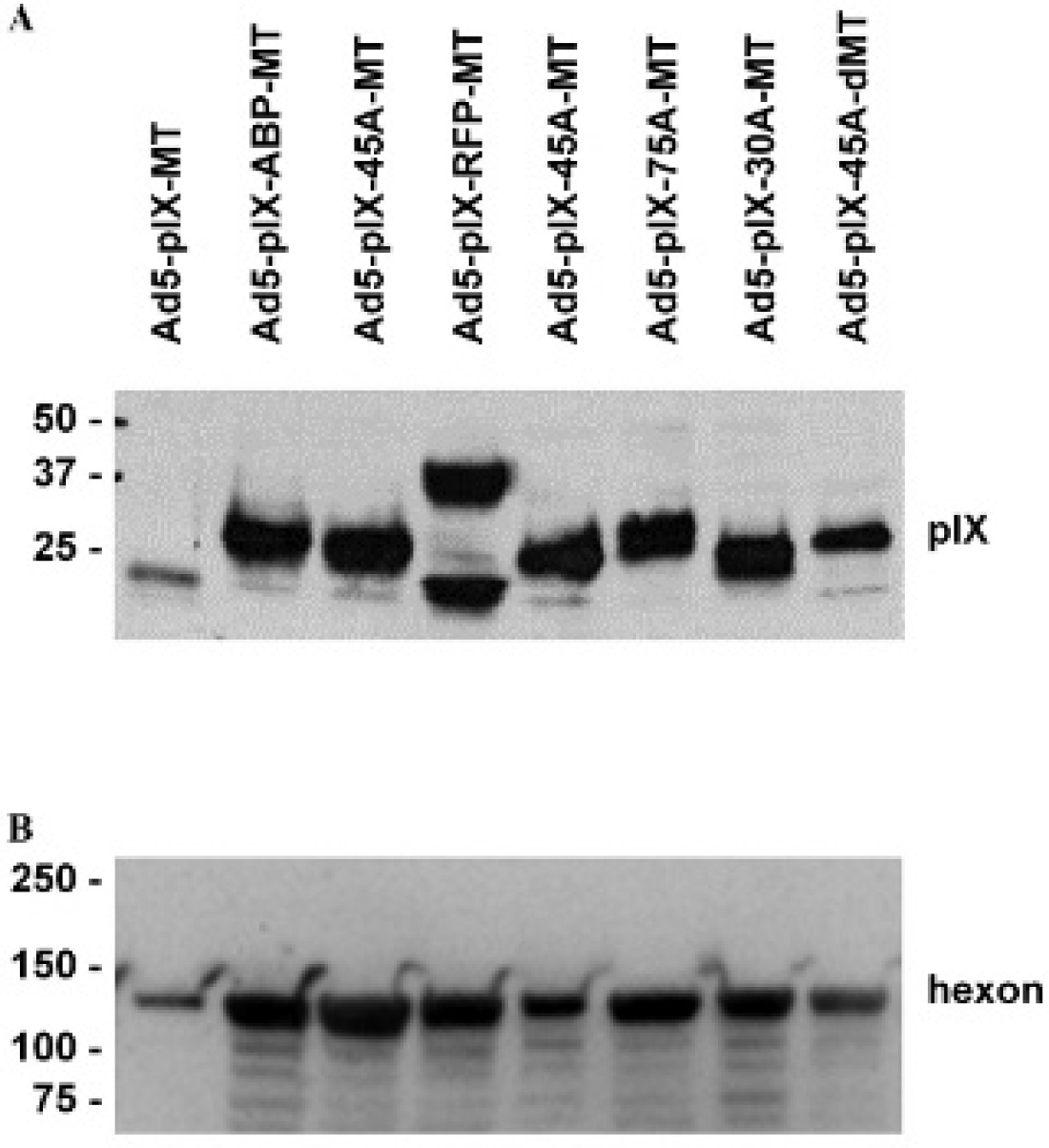
Determination of pIX-MT incorporation. Aliquots of the purified viruses were separated by SDS-PAGE, electroblotted onto nitrocellulose, and probed with (A) an anti-pIX antibody or (B) an antihexon antibody, followed by treatment with corresponding antispecies IgG HRP conjugates. Shown are representative blots after visualization by autoradiography.
Analysis of Viral DNA Replication
To investigate the replication efficacy of the spacer-modified pIX-MT fusion constructs, we compared the Ad progeny production in HEK293 cells after initial infection (Figure 3). As a control, virus production was compared to a wild-type Ad. In this experiment, 10 ifu/cell of the different Ads were used to infect the human embryonic kidney epithelial (HEK293) cell line. Aliquots of medium were collected on 1, 2, 3, and 4 days postinfection, and total DNA was extracted from the medium and analyzed for Ad5 viral E4 DNA copy number by quantitative PCR. Medium from uninfected cells was also obtained at each time point to serve as a baseline for viral replication. From days 1 through 3 postinfection, the E4 copy number for each of the Ad constructs increased logarithmically. Afterwards, from day 3 to day 4 postinfection, the E4 copy number for each of the Ad constructs reached a plateau. Taken together, these data indicate that the Ad constructs with modified pIX genes show similar replication patterns in HEK293 cells, and this replication is comparable to an Ad construct with a wildtype pIX gene.
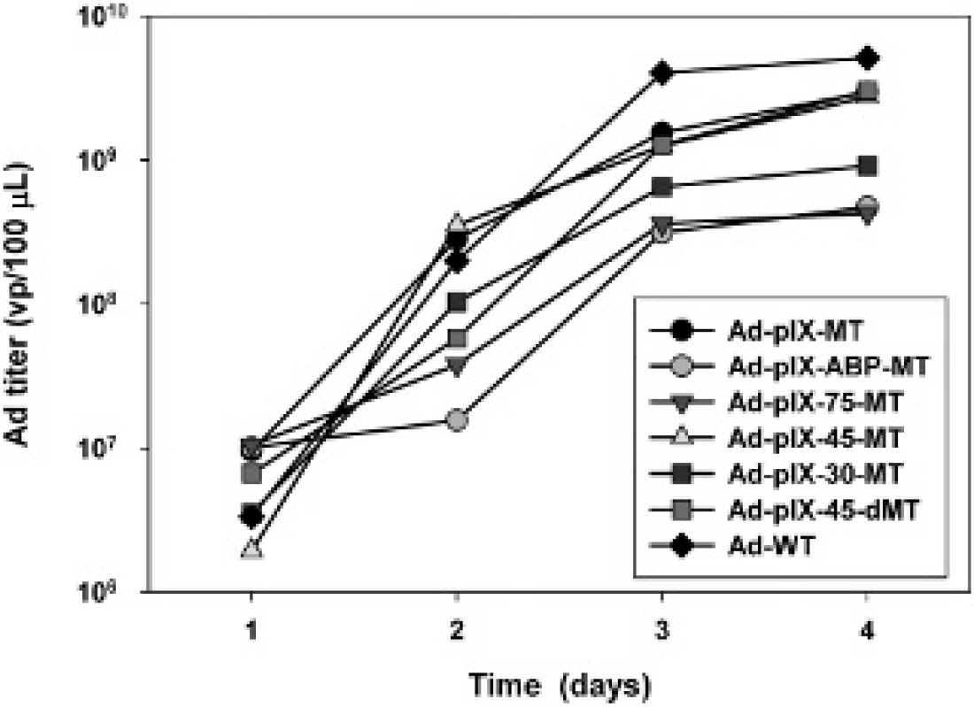
Replication of pIX-MT variants in HEK-293 cells. Viral copy number was used as an indicator of viral replication in HEK293 cells. Growth medium was collected at 1, 2, 3, and 4 days after infection with Ad constructs at an MOI of 10 ifu/cell. Each point represents the mean ± SE of three replicate samples.
Analysis of Viral Infectivity
To assay the infection efficacy of the spacer-modified pIX- MT fusion constructs, the A549 cell line was used, which is highly permissive to Ad infection due to expression of the Ad receptor CAR but is nonpermissive for replication of E1A-deleted Ads. In this experiment, 2 × 10⁵ cells were plated into 24-well tissue culture dishes and infected for 2 hours with increasing MOI of the spacer-modified pIX- MT fusion constructs from 0.1 to 100 ifu/cell. At 48 hours after infection, the percentage of infected cells was determined by quantifying GFP expression using flow cytometry analysis. All the Ad vectors had an identical backbone, the difference being the incorporation of these alternative spacer modifications in the pIX-MT. The results shown in Figure 4 indicate that the pIX-MT spacer modifications did not affect the efficiency in infecting A549 cells in vitro.

Infectivity of pIX-MT variants in A549 cells. Flow cytometric analysis of Ad-pIX-MT variants expressing green fluorescent protein marker. An analysis was performed for expression of GFP at 48 hours after infection with increasing doses from 0.1 ifu/cell to 1,000 ifu/cell. Values represent the mean ± SEM of three replicate measurements.
Radiolabeling of Ad Vectors with 99mTc
We initially compared the radiolabeling efficiency of 99mTc binding in vitro using the spacer-modified pIX-MT fusion constructs. In this experiment, 99mTc-pertechnetate was reduced in the presence of SnCl2 and weakly chelated to glucoheptonate for 15 minutes at room temperature. Afterwards, the 99mTc-glucoheptonate was added directly to the Ad samples for transchelation reactions. Incubation with each Ad sample proceeded up to 120 minutes at 37°C. Purification of 99mTc-radiolabeled Ads from 99mTc-gluco-heptonate was performed by size-exclusion chromatography using 10 mL Sephacryl S-100 columns. Collection of 0.35 mL fractions proceeded until activity peaks corresponding to Ad virions were obtained in the void volume. The fractions with the highest radioactivity between 6 and 9 also corresponded to the highest Ad particle number and infectious activity (data not shown). As shown in Figure 5, the peaks for each of the Ads containing spacer-modified pIX-MT fusion constructs showed similar radioactivity bound compared to an Ad construct lacking any spacer sequence. As a control, we also examined radiolabeling of a wild-type Ad construct containing an unmodified pIX; this virus showed that little 99mTc radioactivity was incorporated.
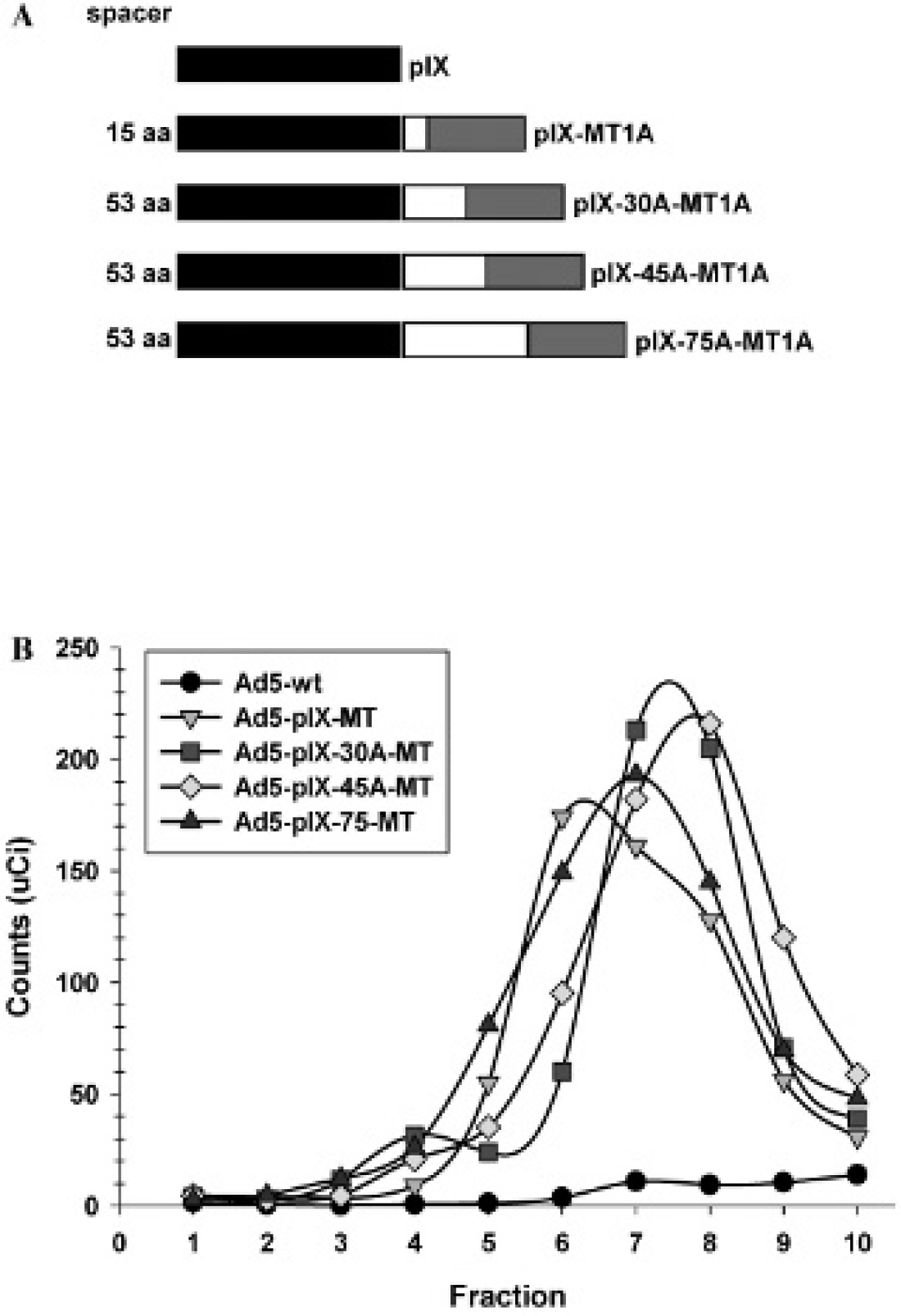
Radiolabeling of pIX-MT spacer variants with 99mTc. A, Schematic representation of the pIX-MT variants analyzed. B, After transchelation of the Ad variants containing pIX-MT fusions or an Ad5 vector containing a wild-type pIX with 99mTc glucoheptonate, the radiolabeled virions were purified from unincorporated 99mTc by gel filtration using Sephacryl S-100 columns. Each point represents the specific activity of fractions collected in microcuries.
These results demonstrate that spacer length has little or no significant effect on 99mTc binding in vitro to the pIX-MT fusion on the capsid of Ad virions.
Distinct isoforms of human MT have been identified arising from four genes that are expressed broadly in most tissues or display tissue-specific expression patterns.35,36 The relative affinity and stability constants of metal ions differ for recombinant MT isotypes, 37 as does their stability in various in vitro production systems. 38 Therefore, we sought to compare the 99mTc-binding capacity of pIX-MT fusion constructs containing MT1A, MT3, or an MT1A- MT1B dimer. As shown in Figure 6, the peaks for an Ad construct containing a pIX-MT3 fusion showed similar radioactivity bound compared to an Ad construct containing a pIX-MT1A fusion. Interestingly, we also compared radiolabeling of an Ad construct containing a pIX-MT1A- MT1A MT dimer fusion; this virus showed roughly twice the 99mTc radioactivity incorporated compared to an Ad construct containing a pIX-MT1A fusion. Quantification of specific activity showed incorporation of approximately 0.14 mBq/vp (data not shown). Based on these results, we continued with more extensive characterization of the pIX- MT dimer fusion.

Radiolabeling of pIX-MT isotype variants with 99mTc. A, Schematic representation of the pIX-MT variants analyzed. B, After transchelation of the Ad variants containing pIX-MT fusions with 99mTc glucoheptonate, the virions were purified from unincorporated 99mTc by gel filtration using Sephacryl S-100 columns. Each point represents the specific activity of fractions collected in microcuries.
Metal Competition Assay
To determine the specificity of 99mTc binding for the pIX- MT dimer fusion, we performed a metal competition assay using increasing concentrations of CdCl2, CoCl2, CuCl2, or ZnCl2. As shown in Figure 7, the bound radioactivity was competed with increasing concentrations of metal, with half-maximal concentrations required for displacement of 99mTc. Interestingly, these results showed similar ability of Co2+, Cu2+, Cd2+, and Zn2+ to displace 99mTc. Likewise, relatively high metal concentrations were required for displacement at room temperature, although this requirement was reduced when the radiolabeled virus was coincubated at 37°C. This result contrasts to the in vitro binding affinities to MT determined to be Cu2+ > Cd2+ > Zn2+ > Co2+, 39 possibly as a structural consequence of MT dimerization.
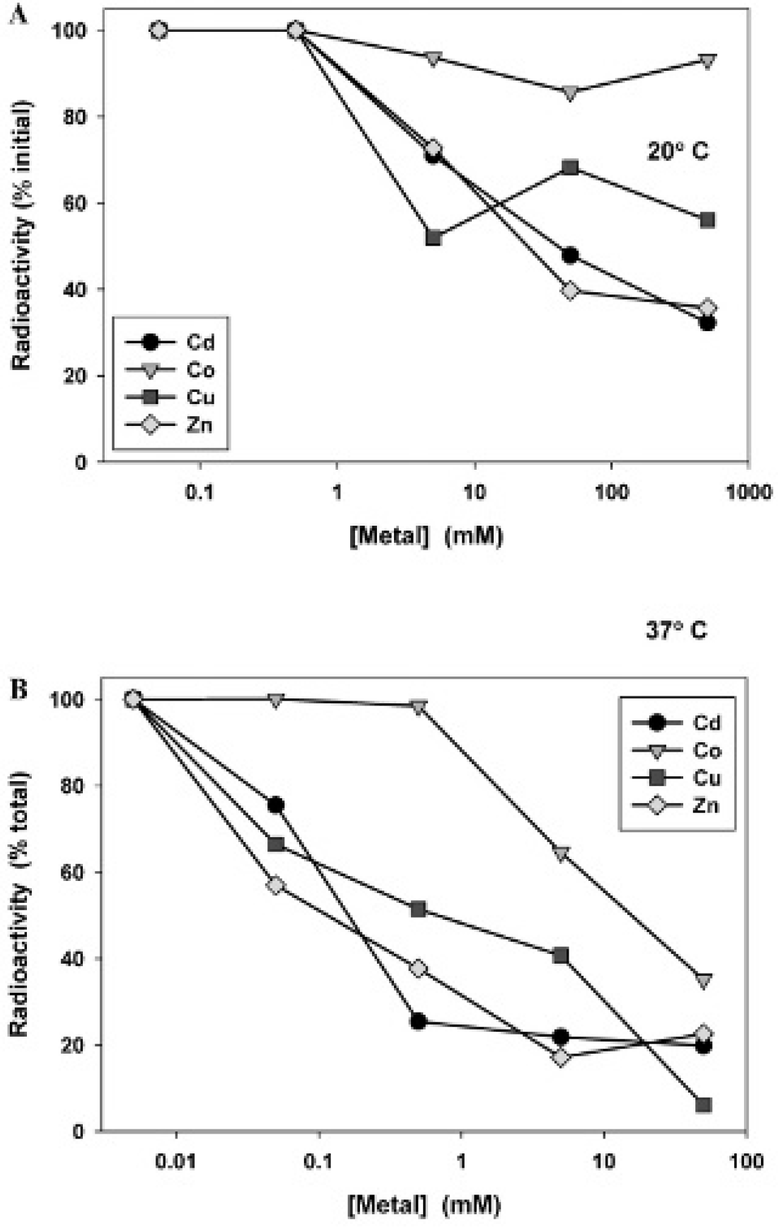
Metal competition assay of a 99mTc-radiolabeled Ad. Aliquots of 99mTc-radiolabeled Ad-pIX-MT dimer were incubated for 15 minutes at (A) 20°C or (B) 37°C with the indicated concentrations of CdCl2, CoCl2, CuCl2, or ZnCl2. Virus-bound 99mTc was determined counting radioactivity after purification through a spin column. Each point represents the percentage of radioactivity of a no-metal control.
Analysis of Ad Biodistribution In Vivo
After radiolabeling the Ad containing a pIX-MT dimer fusion, we determined whether SPECT imaging could be used to monitor Ad biodistribution and uptake in vivo. In the experiment shown in Figure 8 and Figure 9, a group of four control female C57BL6 mice were injected intravenously with 99mTc-radiolabeled Ad. A second group of mice was pretreated with the drug warfarin (an inhibitor of vitamin K–dependent coagulation factor production). This treatment should decrease coagulation factor-mediated liver transduction. 40 A third group of mice was preimmunized with wild-type Ad, a treatment that should enhance FcR-dependent uptake and phagocytosis by resident Kupffer cell populations in the liver and macrophages in the spleen.41,42 A fourth group of mice received both warfarin pretreatment and Ad preimmunization. One hour after 99mTc-radiolabeled Ad administration, the mice were imaged. As shown in Figure 8, clear localization of activity to the liver and bladder was seen, with activity also detected in the spleen, intestine, and kidneys.
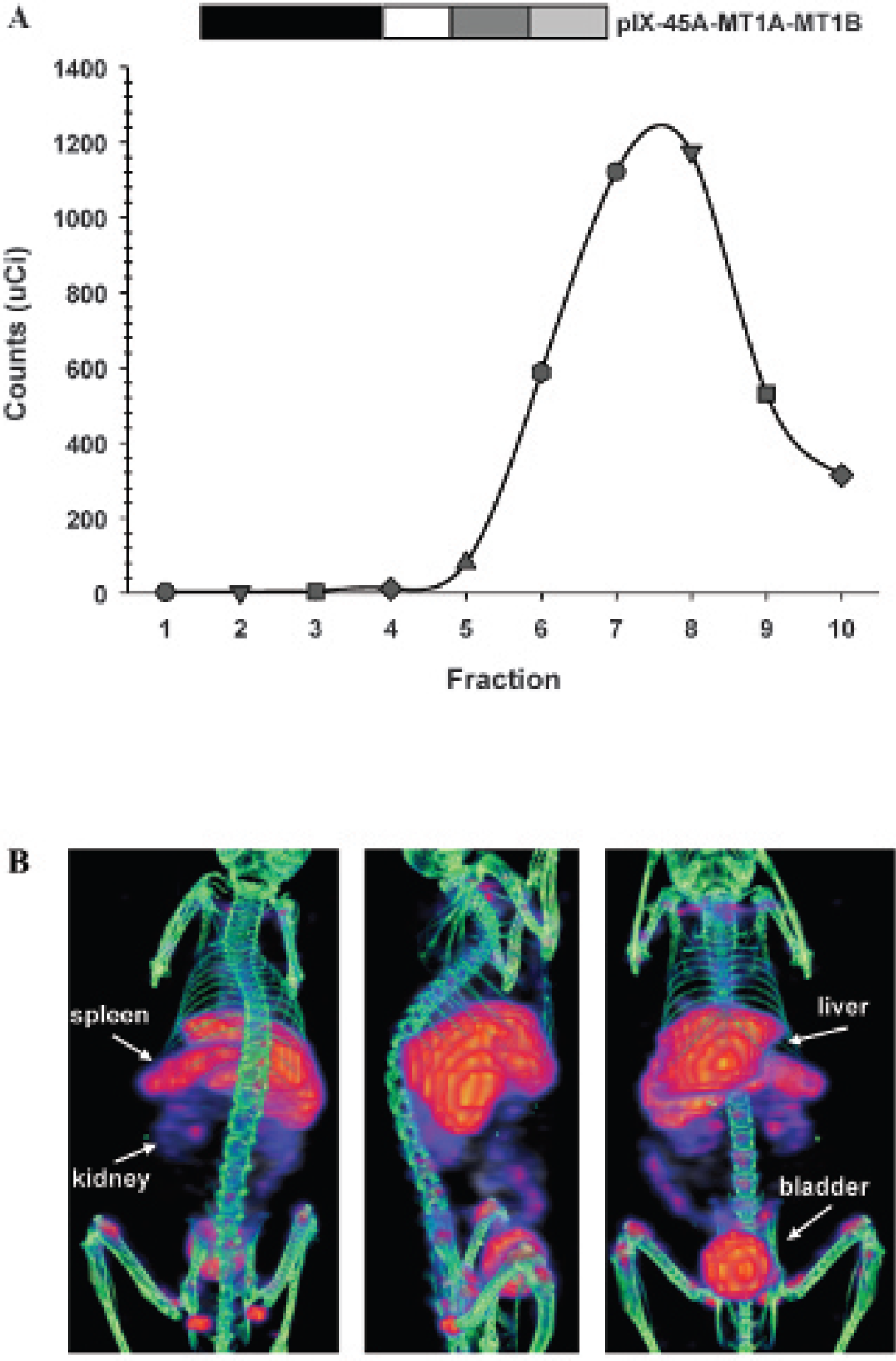
Coronal and sagittal views of fused pinhole SPECT and micro-CT images of a mouse injected with a 99mTc-radiolabeled Ad. A, Radiolabeling of the Ad-pIX-MT dimer by transchelation with 99mTc; shown is a purification from unincorporated 99mTc by gel filtration using Sephacryl S-100 columns. Each point represents the specific activity of fractions collected in microcuries. B, A representative C57BL6 mouse was injected intravenously with approximately 1 mCi of 99mTc-radiolabeled Ad-pIX-MT dimer in 0.2 mL PBS and scanned at 60 minutes after the injection. Three-dimensional rendering of fused CT and SPECT images of bony structures in white and radiolabeled adenovirus in red-yellow scale showing coronal (front) and sagittal (side) views.
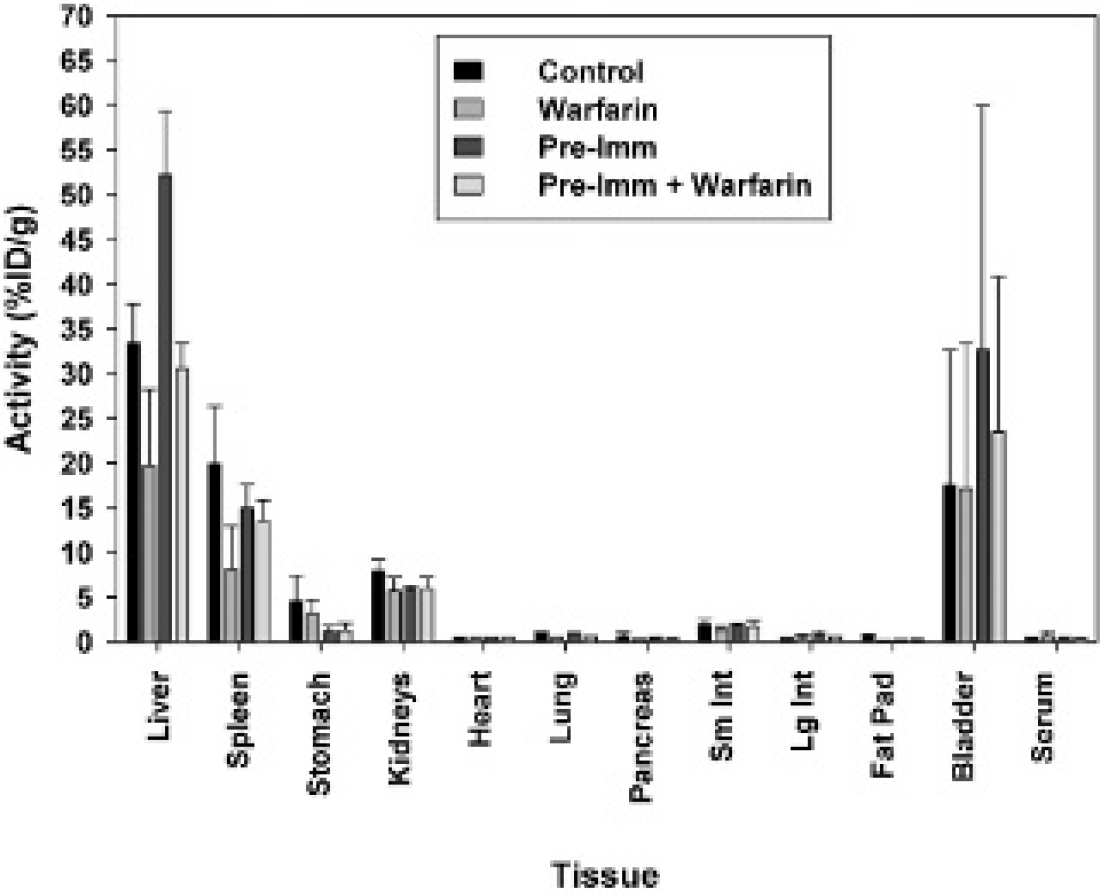
In vivo biodistribution of a 99mTc-radiolabeled Ad. Shown are the values obtained for the %ID/g of tissues from a group of four untreated female C57BL6 mice injected intravenously with approximately 1 mCi of 99mTc-radiolabeled Ad-pIX-MT dimer. These values were compared to a group of mice pretreated with warfarin, a group of mice preimmunized with wild-type Ad, or a group of mice that received warfarin pretreatment and wild-type Ad preimmunization. Each bar represents the mean ± SEM of tissues from four mice.
Immediately following the SPECT/CT imaging, the animals were euthanized and selected organs were dissected for further analyses. Radioactivity of the tissue samples was measured using a well counter and expressed as %ID/g tissue weight. The in vivo biodistribution results of the dissection analysis are summarized in Figure 9, with mean and standard error values provided in Table 1. Similar to the results observed in the SPECT images (see Figure 8), most of the radioactivity was found in all treatment groups in the liver, spleen, kidneys, and bladder. However, significant differences between the treatment groups were observed, primarily in the liver. Liver uptake of 99mTc in control (untreated) mice was significantly reduced by warfarin treatment from 33.4 %ID/g to 19.7 %ID/g (p < .05). Likewise, preimmunization with wild-type Ad resulted in a significant increase in liver uptake of 99mTc to 52.2 %ID/g compared to control (untreated) mice (p < .001). Finally, liver uptake of 99mTc in preimmunized mice was significantly reduced by warfarin treatment from 52.2 %ID/g to 30.5 %ID/g (p < .001).
Biodistribution of 99mTc-Radiolabeled Adenovirus Based on Dissection at 1.5 Hours after Injection

Discussion
We have shown herein that our novel labeling strategy for Ads yields virions capable of incorporating functional pIX-MT fusion proteins for 99mTc radiolabeling in the context of in vivo biodistribution studies. These results confirm the utility of using pIX as a platform for presenting heterologous proteins on the surface of the Ad capsid for multiple functional effects. The adenovirus pIX protein plays multiple roles, including structural virion stability as well as gene transcriptional activation and nuclear reorganization. 43 The pIX protein resides as homotrimers within the virion capsid and interacts as a connecting network of four trimers to stabilize hexon proteins. 44 Although the N-terminal domains of three pIX proteins form a triskelion structure that cements three hexons together, the C-terminal domains form a tetramer, with three domains from one icosahedral facet associating together in a parallel structure and a fourth domain from an adjacent icosahedral facet associating in an antiparallel orientation. Despite this multifunctional role in Ad capsid structure and function, all the MT variants fused to pIX were successfully incorporated into the Ad vector capsid.
In this study, imaging analysis and radioactivity determination showed localization of radioactivity primarily in the liver. On intravenous administration, Ad5- based vectors are sequestered in the liver, where resident Kupffer cells rapidly remove most of the injected dose from circulation. 45 This process depends on multiple and synergistic interactions involving different blood components, including circulating cells, blood coagulation factors, antibodies, and complement. 34 In particular, coagulation factor X (FX) binds to the Ad5 capsid protein hexon at hypervariable regions (HVR) 5 and 7 46 and mediates the majority of the Ad5 liver tropism, primarily through heparin sulfate proteoglycan-mediated pathways.
Imaging analysis and radioactivity determination showed that pretreatment with warfarin decreased liver uptake of the 99mTc-radiolabeled Ad vector. Warfarin pretreatment can deplete circulating FX levels and decrease the liver tropism of intravenously administered Ad5. 47 In addition, we showed that preimmunization with Ad vector increased subsequent liver uptake of the 99mTc-radiolabeled Ad. Beyond the role of FX, Ad inactivation and liver clearance are also mediated through anti-Ad antibody interactions, resulting in activation of the classic complement pathway. In the presence of anti-Ad antibodies, classic pathway activation occurs through direct binding of complement protein C1q to the Ad-antibody complex. 48 This pathway uses the complement component 3 (C3) protein, which has been shown to enhance liver transduction by Ads and augment the inflammatory cytokine response. In addition, the alternative complement pathway is activated by direct binding of complement protein C3 to Ads, 49 resulting in liver transduction despite a lack of complement activity.
Based on x-ray crystallography and mutation studies of the hexon HVR 5 and 7 regions, it has been possible to identify specific amino acid residues that interact with FX and to ablate this interaction. 50 In addition, serotype substitution of hexon has been demonstrated to decrease binding to FX and decrease cell transduction via FX- mediated pathways in vitro as well as decrease liver sequestration in vivo. 51 However, HVR mutations can also increase spleen uptake, 52 which is mediated through an RGD motif on Ad5 penton base with integrins. Ablation of the penton RGD motif in the context of an FX binding-ablated Ad5 vector can decrease spleen uptake as well as innate inflammatory response. 52 In addition to ablating liver uptake, improving Ad tumor targeting is also dependent on selective target infection. Ad5 infection is mediated through the Ad fiber protein, which binds to the primary CAR. We and others have shown that fiber knob modifications are effective examples of different retargeting strategies to improve tumor cell infection for a variety of cancer types and to improve antitumor efficacy of Ad5- based vectors. 53
Improvement of Ad5-based vectors to untarget liver uptake and retarget tumor infectivity is critically dependent on knowledge of biodistribution in vivo. This study demonstrates our ability to use an Ad vector, which encompasses MT for biodistribution analysis by 99mTc-based SPECT imaging. Besides SPECT imaging using 99mTc, PET imaging using 94mTc 54 could also be applied in the context of Ads containing the pIX-MT fusion protein. Likewise, 64Cu could be used for PET imaging because Cu is bound by MT with high affinity. 39 PET imaging does have some advantages over SPECT imaging due to higher spatial resolution and sensitivity and is easier to quantify than SPECT imaging. Nonetheless, SPECT radiotracers are cheaper and more widely available, without the need for an adjacent cyclotron for production of short-lived PET radiotracers. MT has also been demonstrated to bind isotopes of rhenium 55 (ie, 188Re or 186Re), which are capable of combined SPECT imaging and radionuclide therapy.56,57
The ability to view real-time molecular events in vivo in their physiologic milieu represents a significant tool to study Ad vector biology and the consequence of different vector modifications. Ultimately, the highest yield of valuable information about Ad biodistribution will come from preclinical studies and application of modified Ad vectors in patients.
Footnotes
Acknowledgments
Financial disclosure of authors: This work was supported, in part, by National Institutes of Health grant 5R01-CA154697 and funding from the Louisiana Gene Therapy Research Consortium and the Feist-Weiller Cancer Center.
Financial disclosure of reviewers: None reported.