
Abstract
Our goal is to develop multimodality imaging agents for use in cell tracking studies by positron emission tomography (PET) and optical imaging (OI). For this purpose, bovine serum albumin (BSA) was complexed with biotin (histologic studies), 5(6)- carboxyfluorescein, succinimidyl ester (FAM SE) (OI studies), and diethylenetriamine pentaacetic acid (DTPA) for chelating gallium 68 (PET studies). For synthesis of BSA-biotin-FAM-DTPA, BSA was coupled to (+)-biotin N-hydroxysuccinimide ester (biotin-NHSI). BSA- biotin was treated with DTPA-anhydride and biotin-BSA-DTPA was reacted with FAM. The biotin-BSA-DTPA-FAM was reacted with gallium chloride 3 to 5 mCi eluted from the generator using 0.1 N HCl and was passed through basic resin (AG 11 A8) and 150 mCi (100 μL, pH 7–8) was incubated with 0.1 mg of FAM conjugate (100 μL) at room temperature for 15 minutes to give 66Ga-BSA-biotin-DTPA-FAM. A shaved C57 black mouse was injected with FAM conjugate (50 μL) at one flank and FAM-68Ga (50 μL, 30 mCi) at the other. Immediately after injection, the mouse was placed in a fluorescence imaging system (Kodak In-Vivo F, Bruker Biospin Co., Woodbridge, CT) and imaged (Λex: 465 nm, Λem: 535 nm, time: 8 seconds, Xenon Light Source, Kodak). The same mouse was then placed under an Inveon microPET scanner (Siemens Medical Solutions, Knoxville, TN) injected (intravenously) with 25 μCi of 18F and after a half-hour (to allow sufficient bone uptake) was imaged for 30 minutes. Molecular weight determined using matrix-associated laser desorption ionization (MALDI) for the BSA sample was 66,485 Da and for biotin-BSA was 67,116 Da, indicating two biotin moieties per BSA molecule; for biotin-BSA-DTPA was 81,584 Da, indicating an average of 30 DTPA moieties per BSA molecule; and for FAM conjugate was 82,383 Da, indicating an average of 1.7 fluorescent moieties per BSA molecule. Fluorescence imaging clearly showed localization of FAM conjugate and FAM-68Ga at respective flanks of the mouse, whereas only a hot spot at the expected flank (FAM-68Ga injection site) was observed in microPET imaging. Our results suggest that BSA-biotin-DTPA-FAM may function as a multiprobe for PET and fluorescence imaging. Experiments are currently in progress to demonstrate cell tracking using both optical and nuclear imaging.
MULTIMODALITY IMAGING is a relatively new concept that is revolutionizing the field of experimental imaging. It is composed of more than one instrumentation technique, such as positron emission tomography (PET), single-photon emission computed tomography (SPECT), magnetic resonance imaging (MRI), and/or optical imaging (OI). Multimodality imaging requires agents that can be useful for imaging in more than one modality. Several investigators have now embarked on the development of probes for multimodality imaging. Bovine serum albumin (BSA) has been derivatized with biotin, fluorescein, and chelators for metal ions, making it suitable for use in OI and MRI studies. 1 This agent has been used to label fibroblasts for tracking of tumor-associated stroma. 2 Multimodality imaging is suited to evaluate the distribution, localization, and kinetics of these agents. 3 OI techniques are playing a major role in the development of multimodality noninvasive imaging tools.4–7 These studies are now also being applied to small nonhuman primate studies. 8
Multimodality imaging methods are currently being used for reporter gene studies because of their lower cost and the ease of validating reporter assays in vitro for translation to in vivo studies. Since OI is limited by depth of light penetration and quantitation issues, multimodality imaging along with PET or PET/computed tomography (CT) becomes an even stronger tool for noninvasive imaging. 9 Multiple applications of this imaging technique have been used. One such example is the triple labeling of albumin with biotin, fluorescein, and gadolinium–diethylenetriamine pentaacetic acid (DTPA) (BSA-biotin-GdDTPA-FAM 1 ). A positive correlation between vascular endothelial growth factor (VEGF) and lymph node metastases had been observed. To prove this finding, BSA-biotin-GdDTPA-FAM was distributed in the body of a mouse through the lymph nodes and was imaged by MRI, and a decrease in the concentration of VEGF was seen. 2 In addition, the BSA-biotin-GdDTPA-FAM helped show the directionality of the interstitial fluid of the tumor. Other recent studies have used BSA-biotin-GdDTPA-FAM imaging to track angiogenesis in tumors. 3
Our interest in this agent is for potential application in tracking transplanted cells. Thus, we propose to incorporate the PET probe in BSA-biotin-DTPA-FAM in conjunction with the OI probe. In this study, gallium trichloride was added to prepare “BSA-biotin-DTPA-FAM-Ga” and the PET radiotracer “BSA-biotin-DTPA-FAM-68Ga,” which was used as a multiprobe for PET and fluorescence imaging. Here we report our preliminary results on the radiosynthesis of BSA-biotin-DTPA-FAM-68Ga and PET and fluorescence imaging studies in a mice model.
Methods and Materials
General Methods
All chemicals and solvents were of analytical or high-performance liquid chromatography (HPLC) grade from Aldrich Chemical Co. (St. Louis, MO) and Fisher Scientific (Hampton, NH). Absorbance was measured using a Cary 50 Bio UV-Vis spectrophotometer from Varian Associates, Inc. Analytical thin-layer chromatography (TLC) was carried out on silica-coated plates (Baker-Flex, Phillipsburg, NJ). Gallium 68 chloride was obtained from the Eckert & Ziegler Isotope Products (Berlin, Germany) Ionic Gallium Generator IGG 100. High specific activity 18F-fluoride was produced in the MC-17 cyclotron using oxygen 18- enriched water (18O to 18F using p, n reaction). Gallium 68 and fluorine 18 radioactivity was counted in a Capintec (Ramsey, NJ) CRC-15R dose calibrator, whereas low-level counting was carried out in a Capintec Caprac-R well-counter. Radioactive TLCs were obtained by scanning in a Bioscan System 200 Imaging scanner (Bioscan, Inc., Washington, DC). The Kodak In-Vivo F imaging system (Bruker Biospin Co., Woodbridge, CT) was used for fluorescence imaging. All animal studies were approved by the Institutional Animal Care and Use Committee of the University of California-Irvine.
Matrix-associated laser desorption ionization (MALDI) mass spectra were obtained on a MALDI TOF/TOF 5800 mass spectrometer (AB SCIEX, Framingham, MA). As the sodium interferes with mass spectrometry detection, the small portion of the above proteins was reconstituted in 0.1 M NH4Cl solution using centrifugal filtration. The five protein samples, BSA, BSA-biotin, BSA-biotin-DTPA, BSA-biotin-DTPA-FAM, and BSA-biotin-DTPA-FAM- Ga, had their molecular weights determined by using the MALDI method using the AB SCIEX TOF/TOF 5800 System. Initially, the saturated solution of matrix, sinapic acid (Sigma D7927, St. Louis, MO) in 50% acetonitrile:- water, was prepared. The saturated matrix solution was added with diluted protein solutions in an equal volume (0.5 μL each) in the MALDI sample plate and allowed to dry. The mass spectra were obtained for all four samples. The molecular weight of the BSA sample was found to be 66,485 Da, which corresponded to the molecular weight reported by the chemical supplier, Sigma Chemicals.
Preparation of Protein PET/Fluorescence Probe
BSA-Biotin
The preparation sequence is shown in Figure 1. BSA (molecular weight ≈ 66,000 Da; Sigma A9647), 4 g, was dissolved in 60 mL of 0.1 M NaHCO3, pH 8.5 to 8.8. This solution was cooled in an ice-water bath, and 95 mg of biotin-NHSI (Sigma H1759) was then dissolved separately using 9.6 mL of dry dimethylformamide (DMF; Sigma D 4254) and cooled in an ice-water bath. The biotin solution was gradually (while vortex/stirring) added to the cooled BSA solution. The mixture was stirred in an ice-water bath for 1 hour followed by 2 hours at room temperature. After the completion of reaction, the unbound biotin was separated by dialysis against water. Dialysis was done for 6 days, with millipore water (Millipore Corporation, Bedford, MA) being changed every 24 hours. After the dialysis was completed, the content in the dialysis tube was measured. The volume of biotin-BSA was 125 mL. A 10 mL aliquot (containing ≈ 320 mg of BSA) was used for further characterization. Molecular weight using MALDI mass spectra of BSA 1 was found to be 66,485 Da, whereas that of the BSA-biotin 2 was found to be 67,116 Da (Figure 2).
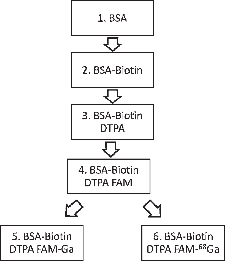
The sequence of steps in synthesizing BSA-biotin (2), BSA- biotin-DTPA (3), and BSA-biotin-DTPA-FAM (4) and, lastly, chelating with gallium chloride BSA-biotin-DTPA-FAM-Ga (5) and radiolabeled BSA-biotin-DTPA-FAM-68Ga (6) for multimodal imaging.

MALDI mass spectra peaks of BSA derivatized in daltons.
BSA-Biotin-DTPA
The BSA-biotin solution was made up to 0.1 M Hepes buffer (Sigma H3375) by adding 1:10 1.0 M Hepes buffer, pH 8.8. Subsequently, 3.6 g of DTPA-anhydride (Sigma D6148) was dissolved in 18 mL of dry dimethylsulfoxide. This DTPA-anhydride solution was gradually added (8-10 portions) to the biotin-BSA solution. The pH was checked after each addition and adjusted to 8.5 with 5 N NaOH. The content was gently stirred for 2 hours at 40°C. After completion of the reaction, the content was dialyzed (five times) against cold sodium citrate buffer 0.1 M, pH 6.5. This was performed in the cold room. The dialysis was continued using deionized water five more times. During the dialysis process, the volume of the content was decreased to ≈ 100 mL. A small dried protein sample was noticed at the end of the dialysis procedure. A small aliquot was used for further characterization. The molecular weight using MALDI mass spectra of BSA- biotin-DTPA 3 was found to be 81,584 Da (see Figure 2).
BSA-Biotin-DTPA-FAM
Buffer conversion for 2.6 mL of biotin-BSA-DTPA (100 mg) from the above dialyzed product was carried out as follows: 7.4 mL of NaHCO3, pH 8.8, was added to the protein solution and concentrated to 1.2 mL using Amicon Centriprep YM30 (Millipore Corporation, Bedford, MA) and ultracentrifuged at 25°C. This procedure was done one more time to increase the concentration of NaHCO3 solution content. The final BSA-biotin-DTPA was adjusted to 2.5 mL using NaHCO3 solution. Fluorescein (5(6)- carboxyfluorescein, succinimidyl ester; (FAM SE; Molecular Probes C1311, Grand Island, NY) was dissolved with 200 μL of DMF and added in portions (five) to the protein solution at room temperature. The content was stirred for 90 minutes. The BSA-biotin-DTPA-FAM was further purified by three times centrifugal filtration using Amicon Centriprep YM30 and NaHCO3 buffer. A small aliquot was used for further characterization. The molecular weight using MALDI mass spectra of BSA-biotin-DTPA-FAM 4 was found to be 82,383 Da (see Figure 2).
BSA-Biotin-DTPA-FAM-Ga
A solution of gallium chloride was added to 10 mg of biotin-BSA-DTPA-FAM in 1 mL of 3 M ammonium acetate at a pH of 6.5. The content was stirred at room temperature for 90 minutes. The BSA-biotin-DTPA-FAM was further purified by three times centrifugal filtration using Amicon Centriprep YM30 and buffer. A small aliquot was used for further characterization. The molecular weight using MALDI mass spectra of BSA- biotin-DTPA-FAM-Ga 5 was found to be 83,859 Da (see Figure 2).
Radiolabeling with 68Ga
The Eckert & Ziegler Isotope Products Ionic Gallium Generator IGG 100 is a closed system consisting of a borosilicate glass column containing a titanium dioxide bed on which germanium 68 is absorbed. Gallium 68 was continuously produced by decay of its radioactive parent and was eluted with 0.1 M HCI. This generator was designed to minimize both 68Ge content and metal impurities in the eluate. It therefore elutes high specific activity 68Ga as a hydrochloride without leaching out 68Ge contaminant.
Acidic 68Ga chloride 3 to 5 mCi eluted from the generator using 0.1 N HCl and 150 μCi (100 μL, pH 7–8) was incubated with 0.1 mg of FAM conjugate, BSA-biotin-DTPA-FAM (100 μL) at room temperature for 15 minutes, to give 68Ga-BSA-biotin-DTPA-FAM and then purified on a 10DG prepacked gravity flow column (Bio-Rad, Hercules, CA). Characterization and purity of BSA-biotin-DTPA-FAM-68Ga were ascertained on radio-TLC (mobile phase 0.1% citric acid). Radiolabeled biotin-BSA-DTPA-FAM-68Ga was taken up in sterile saline just prior to use.
Fluorescence Imaging
Mice (C57 black, fasted 24 hours prior to scanning) were anesthetized using 2 to 4% isoflurane. Under anesthesia, a partially shaved C57 black mouse was injected (subcutaneously) with FAM-Ga conjugate 5 (50 μL) and FAM-68Ga conjugate 6 (50 μL; 0.03 to 0.05 mCi) on opposite sides in each thigh. Immediately after the mouse was injected, it was placed in the fluorescence imaging system, Kodak In-Vivo F, and imaged. FAM was visualized at an excitation wavelength of 465 nm and an emission wavelength of 535 nm using a Xenon Light Source under the settings of luminescence. Analysis was done using the Carestream software (Bruker Biospin Co., Woodbridge, CT) in photons/second. Next, the same mouse was placed under the Inveon microPET scanner (Siemens Medical Solutions, Knoxville, TN) and was imaged for 30 minutes.
MicroPET Imaging
A preclinical Inveon dedicated microPET scanner (Siemens Medical Solutions) with a transaxial full width at half maximum (FWHM) of 1.46 mm and an axial FWHM of 1.15 mm10 was used for the PET studies. After fluorescence imaging, the mouse was positioned on the scanner bed using a mouse holder under 4% isoflurane. A transmission scan was subsequently acquired. To visualize skeletal uptake, sodium 18F-fluoride (0.025 mCi) was injected intravenously into the tail vein of the mouse. Isoflurane was reduced and maintained at 2.5% following injection. Scans were carried out for 30 minutes and acquired by the Inveon microPET scanner. The images were reconstructed using two-dimensional filter back-projection using a Hanning filter with a Nyquist cutoff at 0.5 and corrected for attenuation using the 57Co attenuation scan data. Calibration was conducted to Bq/cc units using a 68Ge phantom, which was scanned in the Inveon microPET scanner and reconstructed under the same parameters as the subjects. Analyses of all data were carried out using Acquisition Sinogram Image Processing IDL's virtual machine (ASIPro VM, Siemens Medical Solutions).
Results and Discussion
The molecular weights of all compounds were confirmed by MALDI. A commercial sample of BSA 1 showed a mass of 66,485 Da, consistent with the reported molecular weight from Sigma. BSA-biotin 2 displayed a mass of 67,116 Da, suggesting that 2.6 moieties of biotin were added per BSA molecule. BSA-biotin-DTPA 3 had 36.8 moieties of DTPA to make it to a mass of 81,584 Da, as shown in Table 1. The compound BSA-biotin-DTPA-FAM 4 added 1.7 more moieties of FAM and showed a mass of 82,383 Da. This confirmed that there was an average of 1.7 fluorescent moieties per BSA molecule. The final compound of BSA-biotin-DTPA-FAM-Ga 5 had a mass of 83,589 Da with 21 moieties of gallium.
Mass Spectra of BSA Derivatives

BSA = bovine serum albumin; DTPA = diethylenetriamine pentaacetic acid.
For further characterization studies, ultraviolet-visible absorption spectra of BSA and BSA conjugates were taken (Figure 3). At a wavelength of 280 nm (absorbance from the aromatic rings of amino acids), an absorbance of approximately 0.4 AU was seen in BSA, BSA-biotin, BSA- biotin-DTPA, and BSA-biotin-DTPA-FAM and 0.18 AU was seen in BSA-biotin-DTPA-FAM. At a wavelength of 490 nm (absorbance by FAM), an absorbance of 0.43 AU was seen in BSA-biotin-DTPA-FAM and of 0.12 AU in BSA-biotin-DTPA-FAM-Ga, thus confirming the presence of the FAM moiety. This absorbance was absent in BSA, BSA-biotin, and BSA-biotin-DTPA, thus confirming the presence of FAM in the assigned complexes.
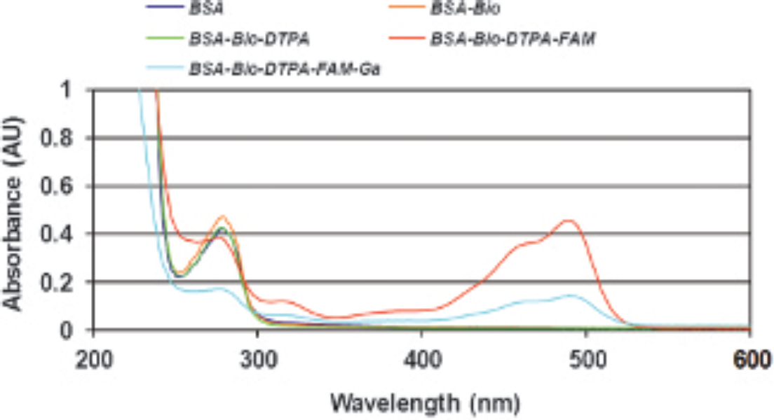
The ultraviolet-visible absorption spectra of BSA and BSA conjugates.
Radiolabeling Results
Radiolabeling of BSA-biotin-DTPA-FAM with 68Ga was verified with radio-TLC as shown in Figure 4. The BSA- biotin-DTPA-FAM-68Ga (Figure 5) was obtained in approximately 20% radiochemical yield starting with 68GaCl3. The radiolabeled product was found to be stable. The BSA-biotin-DTPA-FAM-68Ga was used in sterile saline solution for in vivo experiments.

Radio-TLC of (A) 68GaCl3 eluted from the generator and (B) BSA- biotin-DTPA-FAM-68Ga.

The structure of BSA-biotin-DTPA-FAM during the process of radiochelation with 68GaCl3 to form BSA-biotin-DTPA-FAM-68Ga.
Fluorescence Results
In the fluorescence image, bright spots at both thigh flanks of the mouse were seen (Figure 6A); one was for the BSA- biotin-DTPA-FAM conjugate 5, and the other was for the BSA-biotin-DTPA-FAM-68Ga conjugate 6. The mean value intensities were analyzed for both regions of interest (ROI) using the Carestream software and resulted in a mean intensity value of 4,730 photons/s for the BSA- biotin-DTPA-FAM conjugate 5 and 5,150 photons/s for the BSA-biotin-DTPA-FAM-68Ga conjugate with a 10% error, thus achieving similar intensities with the same quantity of each conjugate injected.
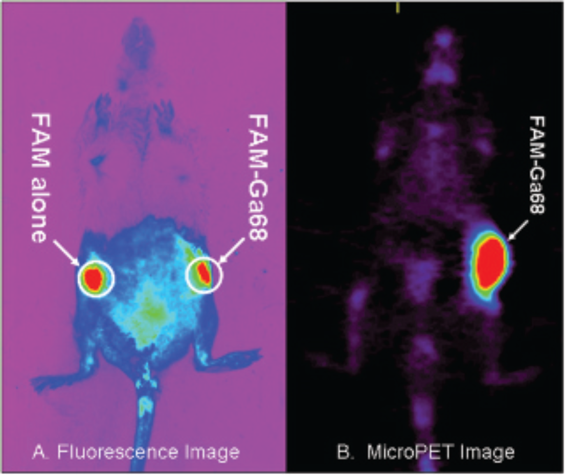
A, Fluorescence image: bright spots at both flanks: one for FAM conjugate (50 μL) and the other for FAM-68Ga conjugate (50 μL). B, MicroPET image: only one hot spot at the expected flank (FAM-68Ga conjugate 30 mCi; 25 mCi of 18F was also injected intravenously for skeletal uptake).
It must be noted that the emission of FAM at 535 nm is likely to limit its in vivo use for deeper tissues/organs due to the greater absorption and scattering of light by living tissue. Thus, incorporation of a fluorophore in the near-infrared region (700–900 nm) may be more optimal for in vivo imaging.11,12
MicroPET Results
In the microPET image, there was only one hot spot at the expected flank, demonstrating localization of the BSA- biotin-DTPA-FAM-68Ga (Figure 6B). This indicated that the BSA-biotin-DTPA-FAM-68Ga was visible in both the microPET and the fluorescence imaging. To visualize the remainder of the mouse body, 18F-fluoride was administered, which highlighted the skeleton of the mouse.
Our results indicate that we were able to synthesize BSA-biotin-FAM-DTPA68Ga successfully and it was found to be regionally localized in vivo. 68Ga radioactivity was predominantly present at the injection site, with little leakage into the vasculature, as evidenced in the PET image. However, more detailed blood work, including different routes of administration, will have to be done to confirm the in vivo stability of the complex. This is similar to the findings of BSA derivatized with gadolinium for use in optical imaging and MRI studies.1,2 The fluorescence of the BSA-biotin-DTPA-FAM-68Ga conjugate 6 in both OI and microPET indicates that it could be useful in developing a multimodal imaging agent.13,14 In future experiments, we hope to inject intravenously to analyze the effectiveness of BSA-biotin-DTPA-FAM-68Ga in detecting tumors using fluorescence and PET.
Summary
A new PET/fluorescence probe, BSA-biotin-DTPA-FAM-68Ga, has been successfully synthesized, and preliminary in vivo studies indicate its stability for carrying out fluorescence and PET imaging studies. Experiments are currently under way to demonstrate cell tracking using both optical and nuclear imaging. This “BSA-biotin-DTPA-FAM” may function as a multiprobe when appropriately chelated with metal ions for nuclear imaging and MRI.
Footnotes
Acknowledgments
We would like to thank Sneha Panchal for technical assistance. Financial disclosure of authors: This research was funded by the National Institute of Diabetes and Digestive and Kidney Diseases (NIDDK), with award numbers RC1DK087352 and R21DK092917.
Financial disclosure of reviewers: None reported.