
Abstract
Esophageal tumors provide unique challenges and opportunities for developing and testing surveillance imaging technology for different tumor microenvironment components, including assessment of immune cell modulation, with the ultimate goal of promoting early detection and response evaluation. In this context, accessibility through the lumen using a minimally invasive approach provides a means for repetitive evaluation longitudinally by combining fluorescent endoscopic imaging technology with novel fluorescent nanoparticles that are phagocytized by immune cells in the microenvironment. The agent we developed for imaging is synthesized from Feraheme (ferumoxytol), a Food and Drug Administration-approved monocrystaline dextran-coated iron oxide nanoparticle, which we conjugated to a near-infrared fluorochrome, CyAL5.5. We demonstrate a high level of uptake of the fluorescent nanoparticles by myeloid-derived suppressor cells (MDSCs) in the esophagus and spleen of L2Cre;p120ctnflox/flox mice. These mice develop esophageal dysplasia leading to squamous cell carcinoma; we have previously demonstrated that dysplastic and neoplastic esophageal lesions in these mice have an immune cell infiltration that is dominated by MDSCs. In the L2Cre;p120ctnflox/flox mice, evaluation of the spleen reveals that nearly 80% of CD45+ leukocytes that phagocytized the nanoparticle were CD11b+Gr1+ MDSCs. After dexamethasone treatment, we observed concordant decreased fluorescent signal from esophageal lesions during fluorescent endoscopy and decreased CyAL5.5-fluorescent-positive immune cell infiltration in esophageal dysplastic lesions by fluorescence-activated cell sorting analysis. Our observations suggest that this translatable technology may be used for the early detection of dysplastic changes and the serial assessment of immunomodulatory therapy and to visualize changes in MDSCs in the esophageal tumor microenvironment.
THE ROLE OF THE TUMOR microenvironment during the initiation and progression of carcinogenesis is increasingly recognized to be of critical importance, for both enhanced understanding of fundamental cancer biology and implementation in molecular cancer diagnostics and treatment strategies. 1 The tumor microenvironment is composed of an array of diverse cell types that cooperate to promote tumor cell survival, migration, and invasion. 2 The extracellular matrix is altered in cancer and infiltrated by numerous invading tumor cells and stromal cell types, including fibroblasts, endothelial cells, pericytes, immune cells, neurons, adipocytes, and likely other cell types.3–5 In this context, Gr-1+CD11b+ immature myeloid cells, also known as myeloid-derived suppressor cells (MDSCs), have been shown to have a prominent role in tumor progression by suppressing the antigen-specific T-cell responses and inhibiting T-cell activation.2,6,7 MDSCs have also been shown to enhance inflammation-associated carcinogenesis. 8 MDSCs have other nonimmune functions, such as promoting angiogenesis and perhaps the activation of fibroblasts.
Our group has developed the technology of mouse endoscopy, which has facilitated the visualization of esophageal inflammation and neoplasia in transgenic mouse models using minimally invasive white light imaging. 9 We have also generated a mouse model for esophageal dysplasia that ultimately leads to squamous cell carcinoma based on conditional p120 catenin (p120ctn) knockout (L2Cre;p120ctnflox/flox), which phenocopies human disease. 2 The complex between E-cadherin and p120-ctn is critical for the formation and maintenance of the adherens junctions. Disruption of this complex is a hallmark feature of many epithelially derived cancers.2,10,11 The esophageal lesions (dysplasia) in this model have been shown to have a significant inflammatory component. There is a marked increase in splenic, circulating, and esophageal MDSCs in the L2Cre;p120ctnflox/flox mouse, making it well suited for the study of imaging this component of the immune infiltrate given our previous findings that the recruitment of MDSCs occurs early and is highly predictive of later cancer development. 8 We observed previously that dexamethasone treatment in these mice results in a marked attenuation of esophageal tumor invasion that was associated with decreased Gr1+CD11b+ MDSCs in the esophagus, suggesting that inflammatory cells, including the MDSC population, foster tumorigenicity. Imaging of changes in subpopulations of leukocytes, in particular the MDSC population, in the murine esophagus may provide a method for early detection of dysplastic changes and assessing how such interventions affect carcinogenesis. Although the obtained images are much lower resolution than the cellular scale, the image signal intensity modulation reflects changes in cell population concentration within the esophageal lesions.
Clinically, nanoparticles with a superparamagnetic iron oxide core and overall diameters of 20 to 40 nm (overall diameter = core + polymeric coating) can be used to image phagocytic leukocytes, including cells at sites such as normal lymph nodes and inflammatory atherosclerotic plaques,12,13 based on the magnetic resonance (MR)- detectable nanoparticle properties. Currently, only one superparamagnetic nanoparticle, Feraheme (FH; ferumoxytol), is approved for parenteral use; FH is approved for the treatment of iron anemia in the United States and European countries. We therefore conjugated the fluorochrome CyAL5.5 to Feraheme; the near-infrared (NIR) fluorescence allows simultaneous full-color spectrum white light imaging and correlative fluorescence imaging (given the nonoverlapping wavelengths of the two), low background signal, and decreased tissue autofluorescence compared to fluorescence imaging at shorter wavelengths. The CyAL5.5-FH is a superparamagnetic, fluorescent nanoparticle that can be used to understand immune cell function/tumor microenvironment by fluorescence imaging in research settings while offering the potential for clinical translation by imaging immune cell populations through the use of FH as an MR contrast agent. A second option would be to employ FH as a safe, metabolized nanoparticle for CyAL5.5 attachment and employ CyAL5.5-FH as a new drug. With either approach, FH could serve as the basis for understanding the tumor microenvironment in clinical settings. For the current study and imaging precancerous states such as dysplasia, fluorescent properties of these particles can be used, as imaging very thin layers of dysplastic cells in vivo using magnetic resonance imaging (MRI) is not feasible. However, in later stages, such as with metastases to lymph nodes, the superparamagnetic properties of these particles may be employed for MRI.
Although populations such as macrophages have been known to phagocytize such particles, we hypothesized that MDSC populations abundant in some tumor microenvironments would likewise demonstrate increased nanoparticle uptake in this setting. Furthermore, modification of the nanoparticles could allow multimodal detection. In particular, for local assessment of nanoparticle phagocytosis and accumulation in microenvironments near a luminal surface, fluorescent imaging is ideal given the high sensitivity, quantitative imaging capabilities, direct anatomic correlation, and ability to serially follow interventions. We constructed an endoscopic imaging system to interrogate the fluorescent signal from the esophagus of mice. In the current study design, single time point imaging (without repeat interrogation) was employed to allow for acquisition of correlative immunohistochemistry and flow cytometry data. We evaluated the combination of optical imaging of nanoparticles with the fluorescent imaging system in the L2Cre;p120ctnflox/flox mice and correlated the imaging assessment with histologic analysis and fluorescence-activated cell sorting (FACS) analysis with dexamethasone treatment to follow changes in leukocyte infiltration in the tumor microenvironment.
Materials and Methods
CyAL5.5-FH Synthesis and Purification
FH (AMAG Pharmaceuticals, Lexington, MA) was aminated through reacting with ethylenediamine. Sixty milligrams of FH (30 mg Fe/mL) was transferred to 0.1 M MES (2-(N-morpholino) ethanesulfonic acid buffer, pH 5.5) buffer using PD-10 columns (GE Healthcare, Little Chalfont, Buckinghamshire, UK). Ethylenediamine (Thermo Scientific, Rockford, IL) was added to the buffer-exchanged FH to bring its final concentration to 1 M. EDC (1-ethyl-3-(3-dimethyl-aminopropyl) carbodiimide was also added to the FH buffer at a final concentration of 2 mg/mL. The mixture was maintained for 1 hour at room temperature for the completion of reaction. The amine-modified FH was purified by gel filtration using a PD-10 column, followed by dialysis against phosphate-buffered solution (PBS).
To prepare CyAL5.5-FH, CyAl5.5, a fluorochrome with optical properties similar to those of Cy5.5, was synthesized as described before 14 and converted to an NHS (N-hydroxysuccinimide) ester. Then 1.2 mg of NHS-CyAl5.5 was added to amine-FH (500 μg Fe in 10 mL PBS), and the mixture was gently mixed on a shaker for 1 hour at room temperature. Nanoparticles were concentrated (Amicon Ultra 50K, Millipore, Billerica, MA), and excess CyAL5.5 was removed using a PD-10 column.
Characterization of CyAL5.5-FH
CyAL5.5 concentrations were determined using an extinction coefficient of 130,000 mM−1cm−1 at 674 nm, 14 whereas iron concentrations were determined spectrophotometrically (300 nm) using an FH standard. The number of CyAL5.5s per nanoparticle was determined from CyAL5.5 and iron concentrations, with the value of 5,874 Fe atoms per nanoparticle supplied by the manufacturer. The number of CyAl5.5s per FH particle used in this study is 9.94. Zeta potentials were determined by Nano-ZS Zetasizer (Malvern, Marlboro, MA) after exchange into 40 mM Tris-HCl buffer at a pH of 8. All other measurements were performed in PBS. For blood half-life measurements, anesthetized mice were injected with 10 mg Fe/kg by tail vein, and 50 μL blood samples were collected via the tail vein. The blood samples were obtained at 5, 30, 60, 120, and 240 minutes following injection. Blood half-life was determined from the T2 measurements by 20 MHz MiniSpec (Bruker Systems, Billerica, MA) as increased concentration of iron in tissues is associated with shorter T2 relaxation times and there is a linear correlation between iron concentration and 1/T2 (Figure 1B). 15 Data were analyzed by GraphPad Prism software (GraphPad Software, San Diego, CA).

A and C, Synthesis and structure of CyAL5.5-FH. Feraheme (FH) is aminated and reacted with an N-hydroxysuccinimide ester of the CyAL5.5 fluorochrome (NHS-CyAL5.5). B, Determination of blood half-lives from 1/T2 magnetic resonance relaxation values. Black lines are fits using the clearance equation. D, Fluorescence excitation and emission spectra showing the optical properties of the CyAl5.5 before and after conjugation. CDI = carbodiimide; EDA = ethylene diamine; HOBT = hydroxybenzotriazole.
Multichannel Endoscope System
Based on previous designs, 16 we constructed a dual-channel endoscope system suitable for murine esophageal evaluation that can simultaneously acquire full-spectrum white light and NIR images optimized for the conjugated nanoparticle CyAL5.5-FH. The optical acquisition module was coupled to a 0.8 mm diameter flexible fiberoptic bundle with extrinsic excitation fibers (Zibra Corporation, Westport, MA). A schematic image of the light source and acquisition module is shown in Figure 2, A and B. As shown in the figure, we modified the filters and excitation laser compared to the original design to optimize the imaging system for the nanoparticle excitation and emission wavelength used in the current study. Concentration dependence phantom studies were conducted on CyAL5.5-FH to assess imaging signal correlation. Dilutions of 1 to 20 μM CyAL5.5, representing a realistic tissue concentration range, were used. The catheter tip was placed 1 mm from the surface of the solution. For each experiment, five repeated measurements were used to generate each data point.

A and B, Schematic design of the multichannel endoscope system. Excitation light source for the system (A). White light (WL) is combined with laser illumination for fluorochrome excitation. Longer wavelengths are removed before transmission through the fiberoptic bundle to prevent crosstalk with excitation and emission channels. The recording system (B) is composed of near-infrared (NIR) and WL cameras attached to a wavelength-specific dichroic mirror, which transmits NIR or visible light to the respective cameras from the signal transmitted from the fiberoptic catheter. C, Concentration dependence phantom studies for CyAL5.5 demonstrate that imaged signal intensity monotonically increases with increasing fluorochrome concentration.
Detection and Immunophenotyping of Nanoparticle-Positive Immune Cells in Mice
For these experiments, nanoparticles (CyAL5.5-FH, 10 mg/kg) were injected into the tail vein and mice were sacrificed after a 3-hour incubation. Single-cell suspensions were generated from harvested esophagi following dissociation via rat tail collagenase and trypsin/ethylenediaminetetraacetic acid (EDTA) digestion. Similarly, spleens were mechanically dissociated and red blood cells were lysed to generate single-cell suspensions of leukocyte populations. 2 Staining of cells was performed in PBS + 2% fetal bovine serum. The following antibodies were used for immunophenotyping: CD45 (clone 30-F11), CD19 (clone 6D5), CD3E (clone 145-2C11), CD49b (clone DX5), CD11c (clone N418), F4/80 (clone CI:A3-1), Gr-1 (clone RB6-8C5), and CD11b (clone M1/70), all from BioLegend (San Diego, CA). Nanoparticles were detected via the conjugated CyAL5.5 fluorochrome. Flow cytometry was performed on a BD LSR II instrument (BD Biosciences, San Jose, CA). Data were analyzed using FlowJo version 9.4 software (Tree Star, Ashland, OR). For simulating the modulation of immune cells in response to treatment, 9-month-old L2-Cre;p120flox/flox mice were treated with dexamethasone (10 mg/kg/day) or PBS (control) once a day for 7 days. Twenty-four hours following treatment, all mice received nanoparticle injection followed by immunophenotyping of CyAL5.5-positive cells as described above.
Animals
Nine-month-old L2Cre;p120ctnflox/flox mice in a C57BL/6 genetic background were used for the in vivo imaging experiments. L2Cre;p120ctnflox/flox mice with or without dexamethasone (10 mg/kg/day, once a day for 7 days) treatment and a group of control L2-cre;p120ctnflox/+mice were used for the experiments (three groups, n = 3 each group). Control L2Cre;p120ctnflox/flox mice not receiving dexamethasone were treated with PBS instead. All animals were fed a nonfluorescent diet for 1 week before imaging (D10001, Research Diets Inc, New Brunswick, NJ). The mice were housed in a specific pathogen-free facility in microisolator, solid-bottomed polycarbonate cages, fed a commercially prepared pelleted diet, and given water ad libitum. The mice were all maintained in a facility under barrier conditions as virus antibody-free mice for the duration of the experiment (approved by the Association for Assessment and Accredition of Laboratory Animal Care). The protocol was approved by the Animal Care Committee of the Massachusetts General Hospital.
Multichannel Upper Gastrointestinal Endoscopy
Each mouse was injected with 10 mg/kg of the imaging probe (CyAL5.5-FH), and endoscopy was performed at approximately 180 minutes, after blood-pool clearance of the probe (see Figure 1B). Anesthesia was induced with isofluorane, and the animals were maintained at 37°C. The animals were then intubated using a 22-gauge plastic catheter that was connected to a small-animal respirator (Harvard Apparatus, Holliston, MA), and the anesthesia was maintained by 1.5% isoflurane.
The upper endoscopy was performed using the custom multispectral endoscopy system described above. The imaging catheter was introduced through the oropharynx. Simultaneous white light and NIR images from the esophagus were acquired with slow advancement of the fiberscope and recorded for analysis. After endoscopy, the animals were immediately sacrificed using a CO2 chamber, the thorax and abdomen were opened, and esophagi were dissected and preserved for further processing.
Quantitative measurements were performed on the acquired raw NIR images with full 12-bit dynamic range. The mean signal intensity and standard error of the mean (SEM) of esophageal tumor and an adjacent esophageal normal mucosa, as well as a region outside the specimen (noise), were measured on 12-bit grayscale images using previously developed software. 17 The target (esophageal tumor [ET]) to background (normal esophagus [NE]) ratio (TBR) was calculated as follows: TBR = (ET — Noise)/(NE — Noise). The mean signal intensity in regions of interest (ROI) of constant size comprising 10 × 10 units of width and height was measured. The tumor-bearing ROI were placed with the center over the respective brightest fluorescence in the lesions; the normal esophagus ROI (background) were placed over normal esophagus tissue at a distance of three ROI diameters from the tumor center in adjacent normal mucosa. To minimize the distance effect on fluorescent signal measurements, luminal diameter was used for distance estimation, and signal intensity measurements were performed at the same distance. Moreover, normal esophagus signal in the TBR formula was measured in the normal esophagus mucosa adjacent to the lesion (at a distance of three ROIs) in the same circumferential plane, further ensuring the same distance effect on tumor and background. For consistency, the same investigator placed all ROI. The data are shown as mean ± SEM. A two-tailed Student t-test for independent samples was used for comparison, and p values less than .05 were considered significant.
Histologic Confirmation and Confocal Microscopy
The extracted esophageal samples were immediately transferred into optimal cutting temperature compound (Fisher Scientific, Allentown, PA) and liquid nitrogen for fixation and further processing. The frozen blocks were kept at −80°C, away from light for preserving the CyAL5.5 fluorochrome. Hematoxylin-eosin (H&E) staining was used for the confirmation of tumor presence.
For fluorescent staining, frozen slides were fixed by incubating in ice-cold acetone for 5 minutes, and tissue sections were incubated with 5% normal rabbit serum (Vector Laboratories, Burlingame, CA) in PBS (30 minutes). Primary antibody against Gr-1 (Abcam, Cambridge, MA) (1:500 dilution) was applied, and the slides were maintained at 4°C overnight, followed by washing in phosphate buffer saline-Tween (PBST) (three times, each for 5 minutes). Propidium iodide (PI) counterstaining was performed by incubating the samples for 5 minutes in 500 nM PI solution and washing (PBST, three times, each for 5 minutes). Gold antifade reagent (Invitrogen, Carlsbad, CA) was used as mounting medium, and the slides were studied immediately using a Zeiss LSM 5 Pascal laser confocal microscope with a Zeiss RGB vario laser module (458/488/514/543/633 nm) (Zeiss, Germany).
Immunohistochemistry was used for determination of the presence of CD11b-positive cells before and after dexamethasone treatment. Briefly, after antigen retrieval (boiling in 10 mM citric acid buffer [pH 6.0] for 15 minutes in a microwave oven) and endogenous peroxidase blockage (15-minute incubation in 3% H2O2), sections were rinsed in deionized H2O (diH2O), blocked (Avidin D, biotin, Sigma-Aldrich, St. Louis, MO; protein blocking agent, Thermo Fisher Scientific, Waltham, MA), and washed in PBS. After blocking, slides were incubated overnight in anti-CD11b primary antibody (rabbit polyclonal, Abcam ab75476, 1.1 μg/mL), followed by PBS wash (2 × 5 minutes). Incubation in biotinylated secondary antibody (goat antirabbit, 1:200, Vector Laboratories) was performed for 30 minutes at 37°C, followed by PBS rinse and incubation in horseradish peroxidase (HRP)- conjugated ABC reagent (Vector Elite, Vector Laboratories). Slides were washed in diH2O, counterstained in hematoxylin (3 seconds), rerinsed, and incubated in ethanol (70%, 95%, 100%) and xylene before coverslips were added. Images were taken using Nikon Eclipse E600 and IPLab software (Scanalytics, Milwaukee, WI).
Results
CyAL5.5-FH Synthesis, Purification, and Characterization
To generate a fluorescent nanoparticle that would be phagocytized by MDSCs, we conjugated FH to the fluorochrome CyAL5.5. Figure 1, A and C, shows a schematic view of CyAL5.5-FH structure and synthesis, which was achieved by FH amination followed by reaction with an N-hydroxysuccinimide ester of the CyAL5.5 fluorochrome (NHS-CyAL5.5). As can be seen in Figure 1B, in 3 hours following probe injection, almost all particles are cleared from the blood pool, and the addition of the fluorochrome to FH does not result in a significant change in blood half-life (33.1 minutes for CyAL5.5-FH vs 39.2 minutes for FH). Properties of CyAL5.5-FH compared to free FH are summarized in Table 1. Fluorescence excitation and emission spectra for CyAL5.5 before and after conjugation to FH are shown in Figure 1D. CyAl5.5 excitation and emission spectra have a very comparable pattern before and after conjugation to FH despite decreased absorbance after conjugation.
Properties of CyAL5.5-FH and Feraheme
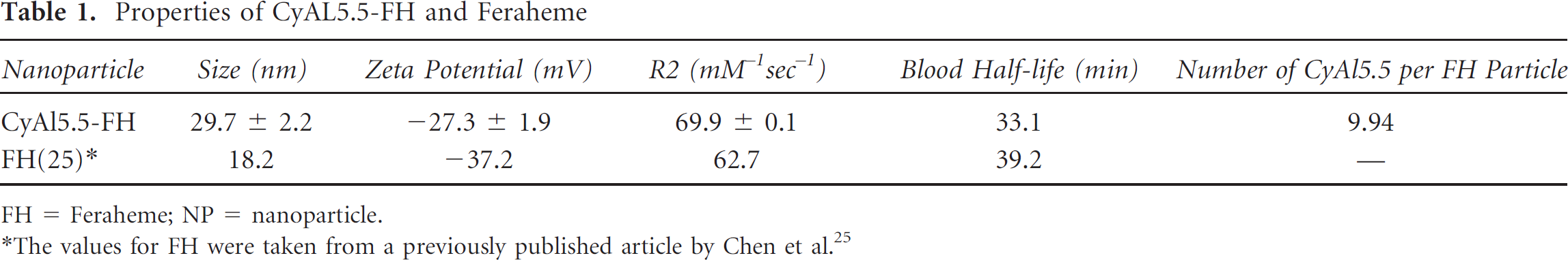
FH = Feraheme; NP = nanoparticle.
The values for FH were taken from a previously published article by Chen et al. 25
Multichannel Endoscope System
To image the CyAL5.5-FH probe in the mouse esophagus, we modified our previously developed endoscope and imaging system (see Figure 2, A and B). The system was able to separate the CyAL5.5 emission wavelength from the filtered white light spectrum. Phantom studies demonstrate a monotonic increase in NIR pixel counts per second as the concentration of the fluorochrome increases. The imaging approach is able to quantitatively assess the fluorochrome intensity in the field of view. Figure 2C shows the results of the phantom studies.
Modulation and Immunophenotyping of Nanoparticle-Positive Immune Cells in Mice
To determine the utility of CyAL5.5-conjugated nanoparticles for imaging Gr-1+ CD11b+ MDSCs, we performed flow cytometric immunophenotyping of splenocytes from 9-month-old L2-Cre;p12Cflox/flox mice following nanoparticle injection. Spleens were isolated at 180 minutes following nanoparticle injection, and leukocytes were sorted for fluorochrome (CyAL5.5) expression and stained for CD11b+ and Gr-1+. Nearly 80% of CyAL5.5+ leukocytes were Gr-1+CD11b+ MDSCs (Figure 3A). Thus, the fluorescent signal from leukocytes is predominantly derived from MDSCs. Note that a similar percentage of CyAL5.5+ leukocytes also expressed F4/80, whereas a smaller fraction expressed CD11c, a dendritic cell marker.

Dexamethasone treatment reduces nanoparticle-positive leukocyte population in the esophagus. A, Immunophenotyping of CD45+CyAL5.5+ splenocytes from a L2-Cre;p120flox/flox mouse by flow cytometry. Note that approximately 80% of the Gr-1+CD11b+ myeloid-derived suppressor cells (MDSCs) have accumulated the nanoparticles. Analysis and quantification were performed using FlowJo software. CD45+CyAL5.5+ cells were gated and positivity for additional markers was determined by comparison with isotype controls. B, Splenic MDSC totals following dexamethasone (DEX; n = 2) or phosphate-buffered saline (PBS) (control; n = 1) treatment. Dexamethasone treatment reduced Gr-1+CD11b+ MDSC splenocytes by eighfold. The mean MDSC total for dexamethasone-treated mice is shown (total = percentage × total splenic cell count). C, Flow cytometric analysis of an L2-Cre;p120fox/fox esophagus after dexamethasone or PBS control treatment (n = 1 for each). The percentage of CD45 +CyAL5.5+ (nanoparticle-positive leukocytes) is reduced fivefold after dexamethasone treatment. Flow cytometric analysis was used to determine the percentage of splenic Gr-1+CD11b+ cells.
To determine whether CyAL5.5 imaging could be predictive of a therapeutic response in this animal model, we treated L2-Cre;p120flox/flox mice with dexamethasone, which has previously been shown to reduce MDSCs. 2 Thus, we employed dexamethasone treatment to determine whether the CyAL5.5 signal would diminish as MDSC numbers declined. As expected, dexamethasone treatment of 9-month-old L2-Cre;p120flox/flox mice resulted in an eightfold reduction in MDSCs in the spleen as measured by flow cytometry (Figure 3B). Similarly, dexamethasone treatment resulted in a fivefold reduction in the CyAL5.5+ leukocytes within the esophagus by flow cytometry (Figure 3C). The parallel between loss of CyAL5.5+ leukocytes within the esophagus and MDSC reduction following dexamethasone treatment, coupled with the immunophenotype data, suggests that the CyAL5.5-conjugated nanoparticles are preferentially scavenged by MDSCs in the L2-Cre;p120flox/flox mouse.
Multichannel Upper Gastrointestinal Endoscopy
In line with the flow cytometry findings, multichannel upper gastrointestinal endoscopy showed significantly higher NIR signal in the esophagus of 9-month-old L2-Cre;p12Cflox/flox mice treated with PBS (Figure 4, A-C, PBS group) compared to dexamethasone-treated animals (Figure 4, E-G, dexamethasone group). Additionally, almost no NIR signal was detected in the control animals with heterozygous L2-Cre;p120flox/+ genotype (Figure 4, I-K, control group). Representative endoscopic images are shown in Figure 4, and the results for the image analysis are quantitated in Figure 5. TBRs are calculated for both white light and NIR images to compare the relative signal intensity contrast between the esophageal lesion and background and to highlight the additional benefit of fluorescent imaging. The mean TBR is significantly higher in PBS-treated L2-Cre;p120flox/flox mice compared to dexamethasone-treated animals, whereas the TBR in the control animals is approximately 1, which reflects a background-level signal.

White light (WL), near-infrared (NIR), and overlay images from upper gastrointestinal endoscopies performed on three cohorts of mice (all approximately 9 months old) (top row, untreated L2-Cre;p120flox/flox; middle row, dexamethasone-treated L2-Cre;p120flox/flox; and bottom row, untreated control L2-Cre;p12flox/+) with corresponding hematoxylin-eosin (H&E) images. The fluorescent signal intensity decreases following dexamethasone treatment in L2-Cre; p120flox/flox mice. H&E staining (× 200 original magnification) showed significant dysplasia in L2-Cre; p120flox/flox mice (phosphate-buffered saline [PBS] and dexamethasone treated) but not in the control group. Arrows indicate the target lesion on endoscopy images.

Quantification of fluorescent signal from the CyAL5.5-FH nanoparticle imaged during upper gastrointestinal endoscopies, performed on mice from the three cohorts in Figure 4 (n = 3 from each cohort). A target to background ratio of 1 reflects a background level of fluorescent signal, comparable to normal esophagus. The bar represents target to background ratios ± SEM.
To confirm the presence of esophageal lesions, histologic studies were performed on the samples obtained after upper gastrointestinal endoscopies. Severe dysplasia was seen in all samples obtained from 9-month-old L2-cre;p120ctnLoxP/LoxP mice, whereas the samples from heterozygous L2-cre;p120ctnLoxP/+ control mice did not show dysplastic changes. Sample H&E images correlating with the endoscopies are also shown in Figure 4. For histologic localization of the nanoparticles, staining with fluorescein isothiocyanate (FITC)-Gr-1 was performed on slides from mice treated with PBS that received CyAL5.5- FH injections followed by confocal microscopy. These studies (Figure 6A) confirmed the colocalization of CyAL5.5 and FITC within these samples. Further studies with Gr-1 and CD11b antibodies (Figure 6, B and C) on samples obtained from dexamethasone- and PBS-treated L2-Cre;p120flox/flox animals confirmed the decreased number of Gr-1- and CD11b-positive cells in the stroma of esophagi of the dexamethasone-treated animals, all in agreement with the flow cytometry and endoscopy results.

Ex vivo confirmation of nanoparticle uptake in specific leukocyte populations in the esophagus. A, Confocal microscopy confirmed colocalization of CyAL5.5-FH and FITC-Gr-1 antibodies in L2Cre;p120flox/flox mice. Propidium iodide (PI) was used for nuclear stain and is shown in red. Gr-1 and CyAL5.5 are shown in blue and green, respectively. B, A decrease in Gr-1 signal following dexamethasone treatment is noted. Note that residual green fluorescence in the dexamethasone-treated mouse corresponds to background fluorescent dye precipitation. White arrows point to a sample of Gr-1-positive cells in the image. C, Immunohistochemistry (X200 original magnification) results for CD11b confirmed a decreased number of CD11b cells in the stroma of dexamethasone-treated animals compared to the phosphate-buffered saline (PBS)- (control) treated mice. The dark arrow points toward the CD11b-positive inflammatory cells in the stroma.
Discussion
MDSCs have a pivotal role in tumorigenesis and progression2,8,18,19 and can be functionally defined by their capability to suppress T cell-dependent responses. 20 In addition, MDSCs are sometimes induced by chemotherapy and can predict resistance to or failure of the chemotherapeutic agent. 21 The cells are found in marrow, spleen, circulation, and tumor sites. Tracking the temporal course in the tumor microenvironment allows evaluation of the natural time course of MDSC infiltration in tumors and assessment of therapeutic intervention. Imaging techniques potentially targeting MDSCs would be able to detect the early stages of the malignancy and to predict the tumor response to therapy. We reported previously the first genetically engineered mouse model for esophageal squamous cell cancer. 2 Conditional deletion of the p120 gene in the L2Cre;p120flox/flox model results in severe dysplasia and cancer in the mouse distal esophagus within 7 to 9 months of age. These lesions are associated with a large inflammatory component and a splenomegaly. Flow cytometry analysis of the CD45+ population (leukocytes) extracted from the tissues of these mice revealed that MDSCs (Gr-1+CD11b+ population) are excessively present within both the esophageal lesions and spleens. Thus, these mice are ideally suited to evaluate imaging assessment of MDSCs in the tumor microenvironment.
We developed a multimodal imaging nanoparticle that is detectable by both optical fluorescent imaging and MRI. Nearly 80% of the leukocytes that accumulated the nanoparticle in the L2Cre;p120flox/flox model were Gr- 1+CD11b+ MDSCs. Thus, endoscopically measured fluorescent imaging signal intensity is dominated by the MDSC contribution. Additional insight regarding MDSC behavior should be gained in the future by studying the nanoparticle phagocytic behavior of different subpopulations of MDSC cells. We demonstrated that modulation with a 1-week treatment with dexamethasone results in a significant decrease in nanoparticle-based fluorescence in esophageal lesions, which corresponds to decreased MDSCs globally (measured with splenocytes) and decreased leukocytes in the esophagus. Moreover, confocal imaging of excised esophageal lesions confirmed that the nanoparticle fluorescence colocalized with Gr-1+ cells and that the number of Gr-1+ cells decreased in the esophageal dysplastic lesions with dexamethasone therapy. Thus, the developed nanoparticle and endoscopy system allows minimally invasive assessment of leukocyte changes in the esophageal tumor microenvironment that is dominated by MDSC modulation. Furthermore, the system allows visual reporting of the tissue, allowing changes in tumor volume to be assessed over time along with the cell-reporting NIR fluorescence. Additionally, the system can be further combined with other fluorescent reporters at distinct NIR wavelengths to simultaneously reveal separate 22 microenvironment parameters. 23
Finally, the developed paradigm for assessment of MDSCs in the esophageal tumor microenvironment is translatable. The base nanoparticle, FH, is approved by the Food and Drug Administration for patients with iron deficiency anemia. Fluorochromes similar to that used in this study for conjugation to the nanoparticle have been brought into human testing. 23 We have developed and tested versions of the imaging system for fluorescent human colonoscopy using nonspecific fluorescent blood-pool agents. 24 Thus, this technique can be used for the detection of early stages of esophageal carcinogenesis highlighted by the aggregation of MDSCs. Therapeutic approaches developed preclinically for modulation of immunoinfiltration, in particular with MDSC targeted therapy, can potentially be further assessed in appropriate patient populations to optimize immunomodulatory treatment.
Footnotes
Financial disclosure of authors: This research was supported in part by National Institutes of Health (NIH) grants U01CA143056 (to U. Mahmood, T.C. Wang, A.K. Rustgi), U01CA084301 (to U. Mahmood), and P50CA127003 (to U. Mahmood). This work was also supported by NIH/National Institute of Diabetes and Digestive and Kidney Diseases (NIDDK) T32-DK007066 (to T.C. Wang), NIH F32-CA162719 (to T.C. Wang), NIH/National Cancer Institute P01-CA098101 (to T.C. Wang, A.K. Rustgi), and the NIH/NIDDK P30DK050306 Center for Molecular Studies in Digestive and Liver Diseases (and its Molecular Pathology and Imaging, Molecular Biology, Cell Culture, and Mouse Core facilities).
Financial disclosure of reviewers: None reported.