
Abstract
The role of apoptosis imaging for monitoring treatment response in ischemic limbs has not been properly explored. In this study, we investigated the ability of annexin V (AnxV) imaging to assess the efficacy of antiapoptotic treatment in ischemic limbs of diabetic mice. Normal C57BL/6 mice and streptozotocin-induced diabetic mice were subject to hindlimb ischemia. AnxV-conjugated fluorescent streptavidin probes were intravenously injected, and optical imaging was performed. Tissue apoptosis was quantified by histochemistry and Western blotting. The AnxV probes showed specific targeting to apoptotic cells on confocal microscopy and flow cytometry. Intravenous AnxV probes displayed substantially greater accumulation in ischemic limbs of diabetic mice. Benfotiamine (BFT) treatment of diabetic mice led to better perfusion recovery on laser Doppler imaging and reduced AnxV binding on optical imaging. TUNEL staining and cleaved caspase-3 Western blots confirmed accelerated apoptosis by diabetes and its suppression by BFT treatment. Furthermore, AnxV-SAv-PEcy5.5 uptake in the ischemic limbs closely correlated to cleaved caspase-3 expression. Thus, AnxV imaging may be useful for monitoring the efficacy of therapeutic agents designed to suppress ischemia-induced apoptosis.
PERIPHERAL ARTERY DISEASE is the most common diabetic vasculopathy and is associated with increased morbidity and the risk of limb amputation in diabetic patients. 1 Programmed cell death plays a key role in the pathogenesis of various cardiovascular diseases,2–4 and several lines of evidence indicate that it may also be a major contributor to poor recovery of diabetic limb ischemia.5–7 Monitoring apoptosis in ischemic limbs of diabetic subjects could therefore be useful to assess the efficacy of newer treatments designed to accelerate recovery.
Apoptotic cells are identified by phosphatidylserine externalized to the outer plasma membrane. Annexin V (AnxV), a 35 kDa protein with strong affinity for phosphatidylserine, can be coupled to contrast agents and used to image apoptosis in vivo. AnxV-based apoptosis imaging has been extensively studied in myocardial disease.3,4 However, it has not been properly explored in limb ischemia, which can also increase apoptosis,5,6 and it is not known whether AnxV imaging can monitor treatment efficacy in this disease setting.
In this study, we investigated the ability of AnxV imaging to detect accelerated apoptosis in ischemic limbs of diabetic mice and further assessed its ability to monitor the beneficial effect of treatment with the synthetic thiamine derivative benfotiamine (BFT).7,8
Methods
Preparation of Imaging Probe
AnxV-biotin (Biovision, Milpitas, CA) was attached to SAv-fluorescein isothiocyanate (FITC) or SAv-PEcy5.5 (Invitrogen, Carlsbad, CA) by incubation at a 4:1 molar ratio in phosphate-buffered saline at room temperature (RT) for 30 minutes (Figure 1A). Unreacted reagents were eliminated by a 100 K Amicon filter.

Apoptotic cell targeting of annexin-V (AnxV) probe. A, Scheme for AnxV attached to fluorophoreconjugated streptavidin (AnxV-SAv-FITC or AnxV-SAv-PECy5.5). B, Confocal microscopy showing AnxV-SAv-FITC (green) bound to the surface of CT-26 cells treated with 1 μM staurosporine for 24 hours. C, Fluorescence-activated cell sorting analysis showing targeting of staurosporine-treated (1 μM for 24 hours) CT-26 cells by AnxV-SAv-FITC and commercial AnxV-FITC probes.
Confocal Microscopy and FACS Analysis
CT-26 mouse colon cancer cells were maintained in RPMI 1640 medium with 10% fetal bovine serum and antibiotics.
To visualize binding to apoptotic cells, cells grown on an eight-well chamber slide were treated with 1 μM staurosporine for 24 hours, incubated with AnxV-SAv-FITC at 4°C for 30 minutes, and inspected under a confocal microscope. For fluorescence-activated cell sorting (FACS) analysis, CT-26 cells treated with 0.1 μM staurosporine for 24 hours were washed, incubated with AnxV-SAv-FITC or commercial AnxV-FITC (BD Biosciences, Franklin Lakes, NJ) for 15 minutes at RT, and analyzed on a BD Caliber flow cytometer (BD Biosciences).
Diabetic Mouse Model and BFT Treatment
All animal experiments were in accordance with the National Institutes of Health's Guide for the Care and Use of Laboratory Animals and approved by the Institutional Animal Care and Use Committee (IACUC). Twenty-seven wild-type male C57BL/6 mice (8 weeks old) were used for our experiments, and the overall study design is illustrated in Figure 2. Diabetes was induced by daily intraperitoneal injection of 40 mg/kg streptozotocin (Sigma, Milan, Italy) for 5 days. Two weeks later, mice with fasting blood glucose > 300 mg/dL were randomly assigned as BFT- treated or untreated diabetic groups, whereas control mice were included as the nondiabetic group (n = 9 per group). Animals received 80 mg/kg per day of BFT (Sigma) or vehicle in drinking water for 14 days before ischemia induction.
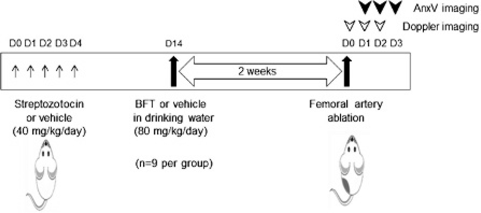
Illustration showing the overall study design. BFT = benfotiamine.
Hindlimb Ischemia
Mice were anesthetized with xylazine/ketamine, and a vertical skin incision was applied to expose the left femoral artery, which was carefully separated, ligated, and excised. Hindlimb perfusion was measured with a laser Doppler flow meter (Moor Instruments, Milwey, Axminser, Devon, UK) immediately after surgery (day 0) and on days 1 and 2. Regions of interest (ROI) were drawn along the limb outlines, and average perfusion levels were measured using Moor Instruments LDI software version 5.3.
In Vivo and Ex Vivo Optical Imaging
On day 1, 2, or 3 of limb ischemia (n = 3 per group on respective days), animals were injected in the tail vein with 2.5 μg of AnxV-SA-PEcy5.5 and imaged under isofluorane anesthesia on an IVIS Spectrum imaging system (Xenogen, Hopkinton, MA) using 500 nm excitation and 700 nm emission filters. An imaging time of 16 hours postinjection was selected based on preliminary experiments. Animals were sacrificed immediately after in vivo imaging. Bilateral limb tissues were extracted, were eliminated of overlying skin, and underwent ex vivo optical imaging to quantitate the level of probe accumulation.
TUNEL Staining for Apoptotic Cells
Limb tissue frozen at −20°C embedded in Tissue-Tek O.C.T. Compound (Sakura, Alphen aan den Rijn, the Netherlands) were sectioned at a thickness of 10 μm. Apoptotic cells were visualized by TUNEL staining using an ApopTag Fluorescein In Situ Apoptosis Detection Kit (Millipore, Billerica, MA). Slides were visualized under a fluorescent microscope, and apoptotic cells were counted using Image J version 1.34s software (National Institutes of Health, Bethesda, MD).
Western Blotting for Cleaved Caspase-3 Expression
Limb tissue snap-frozen in liquid nitrogen was homogenized by mortar grinding, and protein was extracted in tissue extraction solution supplemented with a protease inhibitor cocktail (Sigma-Aldrich, St. Louis, MO). Extracted protein (50 μg) was separated by 15% SDS-PAGE and transferred to a polyvinylidene fluoride membrane (Pall Corporation, Port Washington, NY). The membrane was incubated overnight at 4°C with an antibody against cleaved caspase-3 (1:500; Cell Signaling Technology, Beverly, MA), followed by a secondary antibody at RT for 1 hour. Immune reactive protein was detected by chemiluminescence. An antibody against β-actin (1:5,000, Cell Signaling Technology) was used for loading control. Protein band intensities were quantified using a GS-800 densitometer and Quantity One software (Bio-Rad Laboratories, Berkeley, CA), and cleaved caspase-3 to β-actin protein ratios were calculated.
Assays for NOX4 Level and Nitric Oxide Concentration
Nicotinamide adenine dinucleotide phosphate (NADPH) oxidase-4 (NOX4) was measured with a competitive enzyme-linked immunosorbent assay (ELISA) kit (BlueGene Biotech, Shanghai, China) containing a monoclonal anti-NOX4 antibody. Briefly, diluted tissue homogenates were incubated with NOX4-horseradish peroxidase conjugate in a microtiter plate for 1 hour at 37°C. After washing, substrates A and B were added, and plates were covered and incubated for 15 minutes at 37°C. After reaction termination by stop solution, enzyme activity was determined by 450 nm absorbance.
Nitric oxide (NO) concentration in tissue homogenate was measured by adding an equal volume of Griess reagent containing 1% sulfanilamide, 0.1% N-(1-naphthyl)ethylenediamine dihydrochloride, and 2.5% phosphoric acid. After incubation for 10 minutes, NO concentration was measured by absorbance at 540 nm.
Statistical Analysis
All data are presented as mean ± SD. Significance of difference between multiple groups was determined by one-way analysis of variance (ANOVA) with post hoc Bonferroni multiple comparison tests and with t-tests. Correlation was evaluated by linear regression analysis.
Results
AnxV Probe Targeting to Apoptotic Cells
Confocal microscopy confirmed specific binding of AnxV-SAv-FITC to staurosporine-treated apoptotic CT-26 cells but not to control cells (Figure 1B). Furthermore, FACS analysis displayed better targeting of apoptotic cells with AnxV-SAv-FITC (50.6% positive cells) compared to commercial AnxV-FITC (38.1%; Figure 1C).
Effect of BFT on Perfusion Recovery in Ischemic Limbs
All groups of mice showed severe reduction of hindlimb perfusion immediately after left femoral artery ablation (14.7-22.3% of contralateral side; 53.5-91.0 Flux-perfusion unit [Flux-PU]; Figure 3), and there was no significant difference between groups. On the following day, perfusion recovery was greatest for BFT-treated diabetic mice (142.2 ± 10.3 Flux-PU) and lowest for untreated diabetic mice (0.5 ± 10.6 Flux-PU; see Figure 3).

Laser Doppler imaging of mouse models of hindlimb ischemia. A, Laser Doppler images of mice in the supine position. Regions of interest are drawn on the ischemic left hindlimbs. B, Blood flow measurements of ischemic left hindlimbs immediately after and days 1 and 2 following femoral artery ablation. Data are mean ± SD of results obtained from three animals per group for each time point (n = 2 for day 2 benfotiamine [BFT]-treated group due to loss of one animal). †p < .005, compared to untreated diabetic mice. Flux-PU = Flux-perfusion unit.
AnxV Imaging Detects Accelerated Apoptosis by Diabetes and BFT Treatment Effect
In vivo optical imaging of mice intravenously injected with AnxV-SAv-PEcy5.5 displayed increased probe accumulation in the ischemic limbs for all groups (Figure 4A). Since in vivo images can be influenced by activity from surgical wounds in the overlying skin, a closer inspection of probe uptake was performed by ex vivo measurement of fluorescent signals from extracted tissue. This revealed substantially increased probe uptake in ischemic limbs of untreated diabetic mice compared to nondiabetic mice at day 3 (Figure 4B). Hence, ischemic to contralateral limb fluorescent intensity ratios were 4.53 ± 1.77 for diabetic mice and 1.27 ± 0.47 for nondiabetic mice. However, the ratio for BFT-treated diabetic mice recovered to the level of nondiabetic mice (1.16 ± 0.18; Figure 4C).
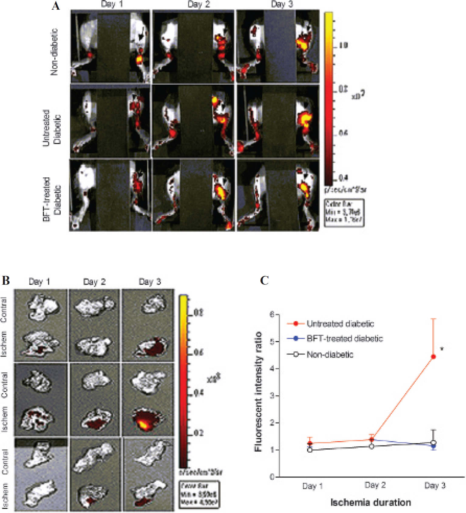
In vivo and ex vivo AnxV imaging of ischemic and contralateral hindlimbs. A, In vivo optical imaging of mice in the supine position following intravenous injection with AnxV-SAv-PEcy5.5. Arrows indicate ischemic left hindlimbs. B, Ex vivo optical imaging of extracted hindlimb tissue. C, Ex vivo measurements of ischemic to contralateral limb fluorescent intensity ratios. Data are mean ± SD of results obtained from three animals per group for each time point (n = 2 for day 2 benfotiamine [BFT]-treated group). *p < .05 by ANOVA as well as by t-tests, compared to nondiabetic and BFT- treated diabetic mice.
Correlation of AnxV Binding to Apoptotic Response in Limb Tissue
Microsections of hindlimb tissues showed that on day 3, TUNEL-positive cells in the ischemic limb were 4.6 ± 1.6- fold of the contralateral limb in nondiabetic mice. In untreated diabetic mice, this was substantially increased to 22.2 ± 5.2-fold. BFT treatment of diabetic mice reduced the ischemic to contralateral TUNEL-positive cell ratio to 1.4 ± 0.5-fold (Figure 5). Tissue levels of cleaved caspase-3 on day 3 were also significantly increased in untreated diabetic mice compared to nondiabetic and BFT-treated mice (Figure 6A). Moreover, tissue levels of AnxV-SAv-PEcy5.5 binding in ischemic limbs (expressed as I/C ratios of fluorescent intensity) showed close correlation to amounts of activated caspase-3 (Figure 6B). In addition, BFT treatment was shown to reduce NO concentration (Figure 6C) and NOX4 expression (Figure 6D) in ischemic limbs of diabetic mice, which may partly contribute to its ability to suppress apoptosis.
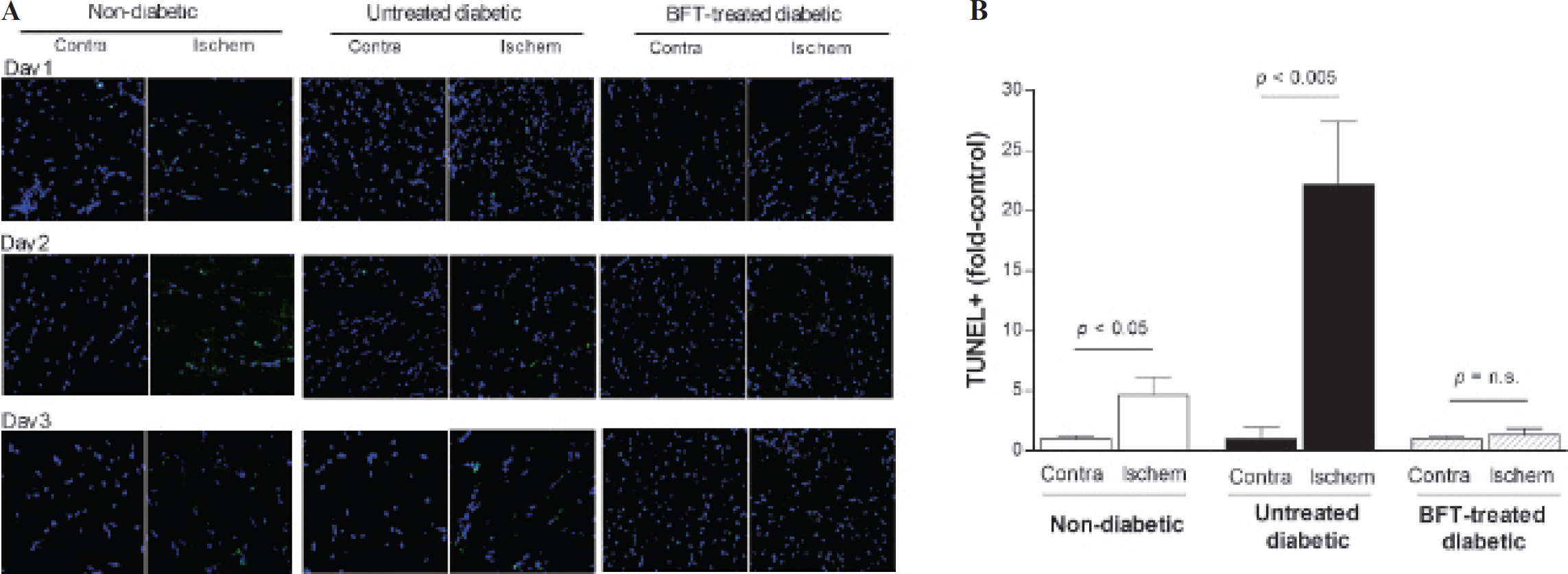
TUNEL staining of apoptotic cells in ischemic and contralateral hindlimb tissue at day 3. A, TUNEL-stained apoptotic cells (green) in microsections of ischemic and contralateral limb tissue. B, Relative number of TUNEL-positive cells in tissues between groups. Bars are mean ± SD of data from three animals per group. BFT = benfotiamine.

Tissue levels of caspase-3, nitric oxide (NO), and NADPH oxidase-4 (NOX4). A, Western blot of cleaved caspase-3 protein in ischemic limb tissue at day 3. *p < .05, compared to nondiabetic and benfotiamine (BFT)-treated diabetic mice. B, Correlation between cleaved caspase-3 band intensity and AnxV-SAv-PEcy5.5 accumulation level for ischemic limb tissue between days 1 and 3. Data points represent data from animals pooled from all groups (n = 26). C and D, Levels of NO (C) and NOX4 (D) in ischemic limb tissue at day 3. All bars are mean ± SD of data from three animals per group.
Discussion
This study demonstrates the ability of AnxV imaging to detect diabetes-induced acceleration of apoptosis in ischemic limbs and to monitor the efficacy of antiapoptotic treatment with BFT.
The AnxV probe used in this study was prepared by linking AnxV-biotin to fluorescent SAv as a means to improve in vivo pharmacokinetics and target tissue delivery of the peptide without affecting its binding affinity. 9 In vitro experiments confirmed specific and high-affinity targeting of the AnxV probe to apoptotic cells.
In our study, BFT treatment of diabetic mice led to earlier improvement in ischemic limb perfusion compared to untreated mice. This is consistent with previous observations that BFT improves vascularization of ischemic myocardia 7 and hindlimbs 8 in diabetic mice. Therefore, our finding is likely contributed to by induction of collateral circulation by BFT.
Ischemic insult from peripheral artery disease induces programmed cell death of vascular cells and skeletal muscles in the involved limb. The presence of diabetes is believed to impair adequate tissue response to ischemia and further promote apoptotic cell death.10,11 Our results showed that intravenously administered AnxV-SAv-PEcy5.5 accumulated in greater amounts in ischemic limbs of diabetic compared to nondiabetic mice. This was associated with an increased promotion of apoptosis in ischemic limbs by diabetes, as shown by greater TUNEL staining and caspase-3 activation. Thus, in untreated mice, although limb perfusion later recovered to levels of BFT-treated mice, ischemic injury to the tissue appears to have already ensued, resulting in greater apoptotic change at day 3.
BFT is a synthetic derivative of thiamine that alleviates multiple diabetic complications by inhibiting formation of advanced glycation end products. 7 , 8 The agent may also reduce oxidative stress and prevent vascular endothelial dysfunction. A previous study showed that BFT treatment accelerated healing of ischemic limbs of diabetic mice, likely through inhibition of apoptosis. 8 Our results showed that BFT treatment restored apoptosis in ischemic limbs to a nondiabetic level and that this was accompanied by reduced accumulation of AnxV-SAv-PEcy5.5 probes. Furthermore, the magnitude of probe uptake in ischemic limbs closely correlated with the level of caspase-3 activation. These results indicate that AnxV imaging might be a reliable marker of the apoptotic response in ischemic limbs.
Although the beneficial effect of BFT was also detected by Doppler imaging in our study, it should be stressed that although perfusion imaging may offer important hemodynamic information, it does not allow assessment of programmed cell death. In this respect, AnxV imaging may potentially have a unique role for direct visualization and quantification of apoptosis and for monitoring therapeutic response in subjects with limb ischemia.
A major limitation of our study is that the number of animals was small. Although the large effect size allowed the results to reach statistical significance despite the small number of animals per group, further studies with larger sample size will be required to more clearly elucidate the role of AnxV imaging for monitoring apoptotic response in ischemic limbs.
Conclusions
AnxV imaging can detect diabetes-induced acceleration of apoptosis in ischemic limbs and may be useful for noninvasive monitoring of the efficacy of antiapoptotic treatments.
Footnotes
Financial disclosure of authors: This study was supported by a grant from the National R&D Program for Cancer Control, Ministry for Health and Welfare, Republic of Korea (# 1120380), and Samsung Biomedical Research Institute grant # GL1-B2-191-1.
Financial disclosure of reviewers: None reported.