
Abstract
Poly(HPMA)-c(RGDyK)-DOTA-64Cu copolymers were synthesized and characterized for tumor localization in vivo as a theranostic scaffold for cancer imaging and anticancer drug delivery targeting tumor angiogenesis. Tumor localization of the poly(HPMA)-c(RGDyK)-DOTA-64Cu copolymers was visualized in mice bearing human prostate cancer xenografts by positron emission tomography (PET) using a microPET scanner. PET quantitative analysis demonstrated that tumor 64Cu radioactivity (2.75 ± 0.34 %ID/g) in tumor-bearing mice 3 hours following intravenous injection of the poly(HPMA)-c(RGDyK)-DOTA-64Cu copolymers was significantly higher than the tumor 64Cu radioactivity (1.29 ± 0.26 %ID/g) in tumor-bearing mice injected with the nontargeted poly(HPMA)-DOTA-64Cu copolymers (p = .004). The poly(HPMA)-c(RGDyK)-DOTA-64Cu copolymers hold potential as a theranostic scaffold for cancer imaging and radiochemotherapy of prostate cancer targeting tumor angiogenesis by noninvasive tracking with PET.
TUMOR ANGIOGENESIS is extensively explored as a target for cancer imaging and therapy. The cell adhesion molecule integrin αvβ3 is highly expressed in tumors and has been found to serve as a receptor for a variety of proteins and small peptides with the exposed arginine-glycine-aspartic acid (RGD) sequence.1–3 The integrin αvβ3 plays an important role in prostate cancer progression. 4 RGD peptide binds specifically and with high affinity to integrin αvβ3 receptors that are known to be overexpressed in tumor vessels.5,6 RGD peptide was tested for anticancer drug delivery targeting tumor neovasculature.7–9 The radiolabeled cyclic RGD peptide was tested as a probe for imaging tumor angiogenesis with a positron emission tomography (PET) or single-photon emission computed tomography (SPECT) scanner.10–12
Polymers have been extensively investigated as carriers for cancer drug delivery based on the enhanced permeability and retention (EPR) effect in tumor tissues. N-(2-Hydroxypropyl) methacrylamide (HPMA) copolymers are hydrophilic, biocompatible, and nonimmunogenic.13,14 HPMA copolymers have been successfully applied in the delivery of many different drugs, such as doxorubicin, 15 paclitaxel, 16 camptothecin, 17 and TNP-470. 18 In the context of targeting tumor angiogenesis, HPMA copolymers conjugated with cyclic RGD were used as a vehicle for targeted delivery of anticancer drugs19–21 and as carriers of radionuclides for cancer imaging and radiotherapy.22–24
Copper-64 radionuclide emits both β+ and β− particles and is potentially useful for both cancer imaging and radionuclide cancer therapy based on its desirable, relatively long half-life of 12.7 hours.25,26 Prolonged tumor retention of N-(3-aminopropyl)methacrylamide-1,4,7,10-tetraazacyclododecane-1,4,7,10-tetraacetic acid (APMA-DOTA)-64Cu copolymers was visualized by microPET after intratumoral injection for interventional radionuclide therapy of human prostate cancer xenografts in mice. 27 Additionally, HPMA-DOTA copolymer was tested as a carrier for systemic delivery of copper-64 radionuclide. 28 More recently, multivalent, multifunctional polymeric nanoparticles containing functionalized Comb copolymers with copper-64 radionuclide were successfully synthesized for imaging applications. 29 In the present study, we aimed to synthesize new theranostic poly(HPMA)-c(RGDyK)-DOTA-64Cu copolymers for prostate cancer PET imaging and imaging-guided radiochemotherapy. Using DOTA as a chelator for 64Cu, HPMA copolymer was used as an anticancer drug carrier, whereas c(RGDyK) peptide ligand was used as a tumor-targeting ligand for targeting αvβ3 integrin in tumor neovasculature. Poly(HPMA)-c(RGDyK)-DOTA-64Cu copolymers were synthesized and tumor localization of this new copolymer was assessed in vivo by PET. The findings from this preclinical study will provide useful information to determine the feasibility and utility of poly(HPMA)-c(RGDyK)-DOTA-64Cu copolymers as a theranostic scaffold for PET imaging and imaging-guided radiochemotherapy of prostate cancer.
Materials and Methods
Reagents and Analytical Equipments
HPMA and APMA were purchased from Polyscience Inc (Warrington, PA). Methacryloyl chloride (MA), N,N′-dicyclohexylcarbodiimide (DCC), 4-nitrophenol (HONp), 2,2′-azobisisobutyronitrile (AIBN), thionyl chloride, and DOTA were purchased from Fluka (Sigma-Aldrich, St. Louis, MO). The peptide c(RGDyK) was purchased from Peptides International, Inc. (Louisville, KY). Dimethyl sulfoxide (DMSO) and acetone were predried with 4 A molecular sieves and distilled over CaH2 under dry nitrogen. Radioactive 64CuCl2 was obtained from the Mallinckrodt Institute of Radiology, School of Medicine, Washington University (St. Louis, MO). Monomers and poly(HPMA)-Cu-64 and poly(HPMA)-c(RGDyK)-Cu-64 copolymers were synthesized according to methods reported from the literature. 28 The reaction temperatures were controlled by oil bath (above room temperature), ice bath (0°C), ice-NaCl (–10°C) bath, and dry ice/isopropyl alcohol bath (– 78°C).
The weight- and number-average molecular weights (Mw, Mn) and molecular weight polydispersity index (PDI = Mw/Mn) were determined by size exclusion chromatography (SEC) using a Polymer Labs PL gel 5 μm mixed C column run in dimethylformamide (DMF). The SEC system was equipped with a seven-angle BIMwA multiangle static light scattering detector and BIDNDC differential refractometer (Brookhaven Instruments, Holtsville, NY). DMF was used as the eluent, at a flow rate of 1.0 mL/min at 30°C. The BIMwA detector was equipped with a 30 mW vertically polarized solid-state laser (660 nm) as a light source. SEC data were analyzed using PSS WinGPC Unity software (Polymer Standards Services, Mainz, Germany). 1H-Nuclear magnetic resonance (NMR) spectra were recorded using a Varian AC400 NMR spectrometer (Varian NMR Systems, Palo Alto, CA).
Synthesis and Characterization of Monomers and Poly(HPMA) -DOTA and Poly(HPMA)-c(RGDyK)-DOTA Copolymers
Synthesis of APMA-DOTA
The procedures were carried out under N2 atmosphere using standard Schlenk techniques. Thionyl chloride (0.12 g, 1.02 mmol) was slowly added to a stirred solution of DOTA (0.45 g, 1.12 mmol) in DMSO (2 mL) at room temperature. The mixture was stirred for 5 hours at 60°C, cooled, and precipitated with CH2Cl2 (20 mL). The precipitate was separated by filtration and dried under vacuum at room temperature to give the activated DOTA. The activated DOTA was dissolved in DMSO (1 mL), and APMA (0.14 g, 1.0 mmol) was added. The mixture was stirred for 48 hours at room temperature. The solution was added slowly to CH2Cl2 (20 mL) cooled to − 10°C, using an ice-NaCl bath with stirring. The precipitate was filtered off, washed thoroughly with CH2Cl2, and dried at room temperature under vacuum. The precipitate was recrystallized from methanol, washed with cold methanol, and dried under vacuum to give monomer 1. Yield: 0.33 g, 64%. 1H NMR (400 MHz, D2O): δ 5.85 (s, 1H, =CH2), 5.61 (s, 1H, =CH2), 3.48 (s, 8H, -CH2COOH of DOTA), 3.25 (m, 2H, CH3-C-CO-NH-CH2CH2CH2-NH- of APMA), 3.02 (t, 16H, -N-CH2CH2-N- of DOTA), 2.84 (m, 2H, CH3-C-CO-NH-CH2CH2CH2-NH- of APMA), 1.80 to 2.00 (m, 5H, -CH3 and -NH-CH2CH2CH2-NH- of APMA).
Synthesis of MA-GG-ONp
MA (0.64 g, 6.20 mmol) was slowly added to a stirred solution of 2-(2-aminoacetamido)acetic acid (GG; N-glycylglycine 0.79 g, 6.00 mmol) in 4 N NaOH (20 mL) cooled with an ice bath. The mixture was stirred with the ice bath for 2 hours. The pH of the solution was adjusted to 2 using concentrated hydrochloric acid. The resulting precipitate was filtered off and dried at room temperature under vacuum. The precipitate was recrystallized from ethanol/water (1:1, v/v) and dried under vacuum to give 2-(2-methacrylami-doacetamido) acetic acid (MA-GG-OH). Yield: 1.06 g, 75%. 1H NMR (400 MHz, DMSO-d6): δ 5.87 (s, 1H, =CH2), 5.63 (s, 1H, =CH2), 4.14 (s, 2H, -CH2COOH), 3.85 (s, 2H, -NHCH 2 CONHCH2COOH), 1.95 (s, 3H, -CH3).
DCC (0.57 g, 2.70 mmol) and HONp (0.38 g, 2.7 mmol) were added to a stirred solution of MA-GG-OH (0.5 g, 2.5 mmol) in DMF (20 mL) at − 10°C. The mixture was stirred for 3 hours at − 10°C and for 8 hours at room temperature. The resulting precipitate was removed by filtration. The filtrate was added slowly to ethyl acetate (20 mL) with stirring. The precipitate was filtered off, washed with ethyl acetate thoroughly, and dried at room temperature under vacuum. The precipitated 4-nitrophenyl (ONp) ester 2 was recrystallized from ethanol/ether (1/1, v/v), washed with cold ethanol/ether, and dried under vacuum. Yield: 0.55 g, 68%. 1H NMR (400 MHz, DMSO- d6): 5 8.19 (d, 2H, protons of ONp near -NO2), 7.36 (d, 2H, protons of ONp near -O-), 5.86 (s, 1H, =CH2), 5.62 (s, 1H, =CH2), 4.12 (s, 2H, -CH2COONp), 3.82 (s, 2H, -NHCH2- CONHCH2COONp), 1.91 (s, 3H, -CH3).
Synthesis of Poly(HPMA)-APMA-DOTA Copolymer
Acetone (3 mL), DMSO (3 mL), HPMA (143 mg, 1.0 mmol), monomer 1 (63 mg, 0.12 mmol), and AIBN (10 mg, 0.006 mmol) were introduced into a 10 mL ampule and stirred at room temperature to give a homogeneous solution. The reaction mixture was sealed under vacuum after deoxygenation by three cycles of nitrogen/vacuum in a dry ice bath. The polymerization was carried out at 60°C for 24 hours. Then the reaction mixture was cooled to room temperature and filtered. The resulting polymer was purified by dialysis using dialysis tubing with molecular weight cut-off (MWCO) 3 kDa and lyophilized to dryness giving poly(HPMA)- DOTA conjugate as a pale yellow powder (99 mg, 48%). Mn: 2.2 × 10⁴, Mw/Mn: 1.35. 1H NMR (400 MHz, D2O) spectrum of poly(HPMA)-DOTA: δ 3.66 (-NH-CH2- CH(OH)-CH3 of HPMA), 3.45 (-CH2-COOH of DOTA), 3.0 to 3.22 (CH3-C-CO-NH-CH2-CH2-CH2-NH- of APMA and -N-CH2-CH2-N- of DOTA), 2.82 to 2.95 (-NH-CH2-CH(OH)-CH3 of HPMA and CH3-C-CO-NH -CH2-CH2-CH2-NH- of APMA), 1.54 to 1.92 (CH3-C- CO-NH-CH2-CH2-CH2-NH- of APMA and backbone -CH2-), 0.75 to 0.96 (-CH3).
Synthesis of Poly(HPMA)-MA-GG-ONp-APMA-DOTA Copolymer
Acetone (2 mL), DMSO (4 mL), HPMA (143 mg, 1.00 mmol), APMA-DOTA (63 mg, 0.12 mmol), MA-GG-ONp (40 mg, 0.12 mmol), and AIBN (10 mg, 0.006 mmol) were introduced into a 10 mL ampule and stirred at room temperature to give a homogeneous solution. The reaction mixture was sealed under vacuum after deoxygenation by three cycles of nitrogen/vacuum in a dry ice bath. The polymerization was carried out at 60°C for 24 hours. Then the reaction mixture was cooled to room temperature and filtered. The resulting polymer was purified bydialysis using 3 kDa MWCO dialysis tubing and lyophilized to dryness, giving poly(HPMA)-DOTA-MA-GG-ONp conjugate as pale yellow powder (125 mg, 51%). Mn: 2.3 × 10⁴, Mw/Mn: 1.41. 1H NMR (400 MHz, D2O) spectrum of poly(HPMA)-DOTA-MA-GG-ONp: δ 8.10 (protons of ONp near -NO2), 7.24 (protons of ONp near -O-), 4.13 (-CH2COONp), 3.84 (-NHCH2CONHCH2COONp), 3.64 (-NH-CH2-CH(OH)-CH3 of HPMA), 3.44 (-CH2-COOH of DOTA), 2.98 to 3.21 (CH3-C-CO-NH-CH2-CH2-CH2- NH- of APMA and -N-CH2-CH2-N- of DOTA), 2.80 to 2.93 (-NH-CH2-CH(OH)-CH3 of HPMA and CH3-C-CO- NH-CH2-CH2-CH2-NH- of APMA), 1.52 to 1.95 (CH3-C- CO-NH-CH2-CH2-CH2-NH- of APMA and backbone -CH2-), 0.72 to 0.97 (-CH3).
Synthesis of Poly(HPMA)-c(RGDyK)-DOTA Copolymers
The c(RGDyK) (0.041 g, 60 μmol) was dissolved in DMF (2 mL). Then (0.115 g, 5 μmol) in dry DMF and pyridine (1:1 molar equivalents relative to the polymeric ONp content) were added to the c(RGDYK) solution (1.3 times excess molar equivalents relative to ONp) with stirring. The reaction mixture was bubbled with nitrogen and continuously stirred at room temperature for 24 hours. The reaction was terminated with 0.1 N NaOH to remove unreacted ONp groups. The resulting polymer was purified by dialysis using 3 kDa MWCO dialysis tubing and lyophilized to dryness, giving poly(HPMA)-c(RGDyK) -DOTA copolymer 5 as a pale yellow powder. 1H NMR (400 MHz, D2O) spectrum of poly(HPMA)-DOTA-c(RGDyK): δ 6.92 (protons of c(RGDyK) phenol near -CH2), 6.65 (protons of c(RGDyK) phenol near -OH), 4.50 to 4.87 (protons of c(RGDyK) -CH-), 4.00 to 4.13 (-CH2COONp, -CH2CO-c(RGDyK) and -NH-CH2-CO), 3.81 to 3.85 (-NHCH2CONHCH2COONp and -NHCH2CONHCH2CO-c(RGDyK)), 3.65 (-NH-CH2-CH(OH)-CH3 of HPMA), 3.43 (-CH2-COOH of DOTA), 2.98 to 3.21 (-NH-CH2-CH2-CH2-CH2-CH- of c(RGDyK), CH3-C-CO-NH-CH2-CH2-CH2-NH- of APMA and -N-CH2-CH2-N- of DOTA), 2.60 to 2.95 (-NH-CH2-CH(OH)-CH3 of HPMA, CH3-C-CO-NH-CH2-CH2-CH2-NH- of APMA, -CH2-phenyl of c(RGDyK), -CH2- COOH of c(RGDyK) and -NH-CH2-CH2-CH2-CH- of c(RGDyK)), 1.35 to 1.95 (-NH-CH2-CH2-CH2-CH2-CH- of c(RGDyK), -NH-CH2-CH2-CH2-CH- of c(RGDyK), CH3-C- CO-NH-CH2-CH2-CH2-NH- of APMA and backbone-CH2-), 0.72 to 0.98 (-CH3).
Positron Emission Tomography
Animal studies were performed according to a protocol approved by the Animal Investigation Committee, Wayne State University. PC-3 human prostate cancer cells (ATCC, Manassas, VA) were injected subcutaneously at the right shoulder to establish human prostate cancer xenografts in athymic mice using a method described previously. 26 Poly(HPMA)-c(RGDyK)-DOTA copolymers and control poly(HPMA)-DOTA copolymers were radiolabeled with 64Cu radionuclide in a method similar to that described previously. 27 Briefly, 50 μCi of 64CuCl2 was added into a solution of the copolymers (0.2 mg) in 2 mL of 0.1 M sodium acetate buffer (pH 5.5), incubated at 50°C for 45 minutes, and the radiolabeled copolymers were purified by centrifuge filtration using Vivaspin 2 (MWCO 3 kDa, Vivascience, Hannover, Germany). Specific activity (MBq [mCi]/nmol) of the radiolabeled copolymers was determined by measuring the radioactivity of the copolymers purified with the Vivaspin column. Radiolabeling efficiency was determined by dividing the radioactivity of the purified radiolabeled copolymers by the total radioactivity of the copolymers and the filtrates containing nonconjugated free copper radionuclide and then multiplying by 100. The purified poly(HPMA)-c(RGDyK)-DOTA-64Cu copolymers were dissolved in normal saline and sterilized by filtration with a 0.22 mm Millipore filter prior to use for PET. Poly(HPMA)-DOTA-64Cu copolymers were prepared in a similar manner for use as a control.
When the tumor xenografts reached about 0.8 × 0.8 cm2 in size, the tumor-bearing mice were injected with poly(HPMA)-c(RGDyK)-DOTA-64Cu intravenously at a dose of 74 kBq (2 μCi)/g of body weight. The tumor-bearing mice of the control group were injected with poly(HPMA)-DOTA-64Cu (74 kBq [2 μCi/g] per gram of body weight). The tumor-bearing mice were subjected to PET at 1 and 3 hours postinjection using a microPET R4 tomograph (Concorde Microsystems, Knoxville, TN) as described previously. 27 The whole-body images were acquired and reconstructed using an iterative ordered subset expectation maximization two-dimensional (OSEM-2D) algorithm. 30 PET images were visually examined for tumor localization of the poly(HPMA)-c(RGDyK)-DOTA-64Cu copolymers and compared to that in the tumor-bearing mice injected with poly(HPMA)-DOTA-64Cu copolymers. PET quantitative analysis was conducted to measure the radioactivity of poly(HPMA)-c(RGDyK)-DOTA-64Cu copolymers localized in the region of the tumor and other organs or tissues (heart, lung, liver, kidney, brain, muscle in the contralateral left shoulder, and blood-pool activity). The decay-corrected percentage of injected dose per gram of tissue (%ID/g) in the tumors and other regions was calculated by dividing the obtained average radioactivity (μCi/cc) in those regions by the injected activity (μCi) over tissue weight (assuming 1 cc = 1 g).
Statistical Analysis
To determine whether the 64Cu radioactivity in the region of tumors on microPET images differs between the application of poly(HPMA)-c(RGDyK)-DOTA-64Cu and poly(HPMA)-DOTA-64Cu copolymers, we applied a 2 × (2 × 8) repeated measures analysis of variance (ANOVA) in which the between-subjects factor represented the group (mice injected with one of the two copolymers) and the two within-subjects factors represented the time of the scan (1 vs 3 hours postinjection) and the eight regions of interest (heart, lung, liver, kidney, muscle, tumor, blood, brain). Once statistical significance was established for the overall test, post hoc tests were applied for the tumor region. Specifically, a paired t-test was applied for the tumor region to determine whether 64Cu radioactivity derived from poly(HPMA)-c(RGDyK)-DOTA-64Cu copolymer differs between 1 and 3 hours postinjection, whereas an independent sample t-test was applied to assess the difference between 64Cu radioactivity in the tumor region for the two copolymers 3 hours postinjection. All tests were two-sided, and a p value .05 was considered statistically significant.
Results
Synthesis and Characterization of Poly(HPMA)-DOTA-64Cu and Poly(HPMA) -c(RGDyK)-DOTA-64Cu Copolymers
Poly(HPMA)-DOTA-64Cu and poly(HPMA)-c(RGDyK)-DOTA-64Cu copolymers were synthesized (Figure 1). The synthetic pathway for the poly(HPMA)-DOTA-64Cu and poly(HPMA)-c(RGDyK)-DOTA-64Cu copolymers is shown in Figure 2. The Cu-binding monomer was synthesized in two steps. First, the chelator DOTA was reacted with thionyl chloride. Because of the multivalent nature, an excess amount of DOTA was used to ensure that only one carboxylic acid group was activated and that no undesired crosslinking took place in the reaction with APMA. Second, the resulting cloride of DOTA was reacted with APMA to form monomer APMA-DOTA via a stable amide bond.

Structures of the poly(HPMA)-DOTA-64Cu and poly(HPMA)-c(RGDyK)-DOTA-64Cu copolymers.

Synthetic scheme of the poly(HPMA)-DOTA-64Cu and poly(HPMA)-c(RGDyK)-DOTA-64Cu copolymers. APMA-DOTA (1) and MA-GG-ONp (2) comonomers were synthesized as indicated. APMA-DOTA was copolymerized with HPMA and complexed with 64Cu radionuclide to obtain nontargeted poly(HPMA)-DOTA-64Cu copolymer. Alternatively, APMA-DOTA was copolymerized with HPMA and MA-GG-ONp, and the obtained copolymer was reacted with c(RGDyK) and complexed with 64Cu to obtain targeted poly(HPMA)-c(RGDyK)-DOTA-64Cu copolymer.
The RGD-containing copolymer was also synthesized in two steps. First, GG was reacted with MA to form MA-GG-OH. Second, MA-GG-OH was reacted with 4-nitrophenol in the presence of DCC to form MA-GG-ONp active ester. The copolymers poly(HPMA)-DOTA and poly(HPMA)-c(RGDyK)-DOTA were synthesized by a conventional free radical precipitation polymerization. The polymerization conditions were optimized to obtain polymers with the desired molecular weight. Simple centrifugal filtration was sufficient to purify the copolymers from unreacted monomers and other low-molecular-weight byproducts. Poly(HPMA) is not a biodegradable polymer, so the target molecular weight (20,000-40,000 g/mol) was selected to ensure ultimate renal elimination.
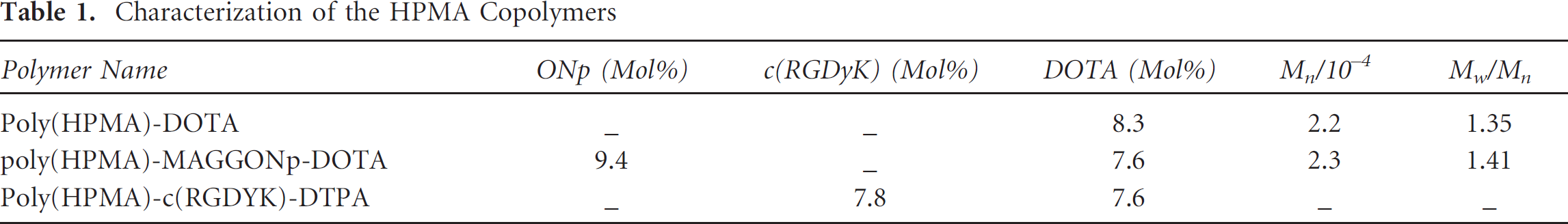
The purified poly(HPMA)-DOTA and poly(HPMA)-MAGGONp-DOTA were characterized by 1 H-NMR to determine the content of DOTA groups in the copolymers. We used the integral intensities of the -CH2-COOH protons of DOTA (< 3.45 ppm) and the integral intensity of the CH3-CH(OH)-CH2-NH-protons of the HPMA (≈ 3.66 ppm) to calculate the content of DOTA in the poly(HPMA)-DOTA and poly(HPMA)-MAGGONp-DOTA conjugates. The data show that the poly(HPMA)-DOTA and poly(HPMA)-MAGGONp-DOTA contained 8.3 and 7.6 mol% of the DOTA monomer units, respectively. Assuming Mn of poly(HPMA)-DOTA and poly(HPMA)-MAGGONp-DOTA to be 22 and 23 kDa, that translates into 10.9 and 9.3 DOTA groups per average polymer chain, respectively.
The purified poly(HPMA)-MAGGONp-DOTA was characterized by 1 H-NMR to determine the content of MAGGONp. We used the integral intensities of the -O-C6H4-NO2 protons of MAGGONp (≈ 7.2 and ≈ 8.1 ppm) and the integral intensity of the CH3-CH(OH)-CH2-NH-protons of HPMA (≈ 3.66 ppm) to calculate the content of MAGGONp, which was found to be 9.4 mol%, corresponding to 11.4 MAGGONp per average polymer chain. Purified poly(HPMA)-c(RGDyK)-DOTA copolymers were characterized by 1 H-NMR to determine the content of c(RGDyK) groups. We used the integral intensities of the -CH2-C6H4-OH protons of c(RGDyK) (≈ 6.9 and ≈ 6.5 ppm) and the integral intensity of the CH3-CH(OH)-CH2-NH-protons of HPMA (≈ 3.66 ppm) to calculate the content of c(RGDyK) in poly(HPMA)-c(RGDyK)-DOTA copolymers and found it to be 7.6 and 7.8 mol% of the monomer units modified with DOTA and c(RGDyK), respectively, corresponding to 9.2 DOTA and 9.5 c(RGDyK) groups per average polymer chain. The composition, molecular weight, and molecular weight distribution of the synthesized copolymers are shown in Table 1. Finally, the poly (HPMA)-c(RGDyK)-DOTA-64Cu copolymers were prepared after incubation of the conjugates with 64CuCl2 in 1 mL of acetate buffer (pH 5.5) at 50°C for 45 minutes, with a radiolabeling efficiency > 92% and specific activity of 12.2 to 17.4 MBq (0.33-0.47 mCi)/nmol. The purified poly(HPMA)-c(RGDyK)-DOTA-64Cu copolymers were dissolved in normal saline and sterilized by infiltration prior to use for PET. Poly(HPMA)-DOTA-64Cu copolymers without cyclic RGD ligand (specific activity of 15.2-19.2 MBq [0.41-0.52 mCi]/nmol) were prepared with the same method and used for tumor-bearing mice of the control group.
Characterization of the HPMA Copolymers
Tumor Localization of Poly(HPMA)-c(RGDyK)-DOTA-64Cu Copolymers Visualized by PET
Increased tumor 64Cu radioactivity was well visualized on the PET images of the tumor-bearing mice (n = 3) injected intravenously with poly(HPMA)-c(RGDyK)-DOTA-64Cu copolymers 1 and 3 hours postinjection of the copolymers (Figure 3A). In contrast, no significant increase in tumor radioactivity was visualized in the tumor-bearing mice (n = 3) injected intravenously with nontargeted poly(HPMA)-DOTA-64Cu copolymers 1 and 3 hours postinjection of the copolymers (Figure 3B). PET quantitative analysis demonstrated significantly higher tumor 64Cu radioactivity (2.75 ± 0.34 %ID/g) in the tumor-bearing mice 3 hours following intravenous injection of the poly(HPMA)-c(RGDyK)-DOTA-64Cu copolymers (Table 2), compared to that in the tumor-bearing mice injected with the nontargeted poly(HPMA)-DOTA-64Cu copolymers (1.29 ± 0.26 %ID/g, p = .004). There was a time-dependant increase in 64Cu radioactivity in the tumor region derived from tumor localization of the poly(HPMA)-c(RGDyK)-DOTA-64Cu copolymers (1.98 ± 0.18 %ID/g 1 hour postinjection versus 2.75 ± 0.34 %ID/g 3 hours postinjection), although this difference did not reach statistical significance (p = .12). In contrast, there was no significant time-dependant increase in tumor 64Cu radioactivity in the tumor-bearing mice injected with the nontargeted poly(HPMA)-DOTA-64Cu copolymers (p = .45; see Table 2). Abundant 64Cu radioactivity was detected in the kidneys (7.44 ± 0.85 % ID/g 1 hour postinjection and 8.83 ± 2.03 %ID/g 3 hours postinjection) of the tumor-bearing mice injected with the poly(HPMA)-c(RGDyK)-DOTA-64Cu copolymers, compared to much lower renal 64Cu radioactivity in the mice injected with the nontargeted poly(HPMA)-DOTA-64Cu copolymers (2.27 ± 0.19 %ID/g 1 hour postinjection and 2.17 ± 0.17 %ID/g 3 hours postinjection).

Representative PET images of the tumor-bearing mice injected with the poly(HPMA)-c(RGDyK)-DOTA-64Cu copolymers (A) or poly (HPMA)-DOTA-64Cu copolymers (B). Increased 64Cu radioactivity in the tumor region was visualized in the tumor-bearing mice injected with the poly(HPMA)-c(RGDyK)-DOTA-64Cu copolymers intravenously but not in the control tumor-bearing mice injected with the poly(HPMA)-DOTA-64Cu copolymers. %ID/g = percentage of inject dose per gram of tissue; p.i. = postinjection.
Biodistribution of 64Cu Radioactivity in Tumor-Bearing Mice Injected Intravenously with Poly(HPMA)-c(RGDyK)-DOTA-64Cu or Poly(HPMA)-DOTA-64Cu Copolymers by PET Quantitative Analysis

%ID/g data are expressed as mean ± SD; PET = positron emission tomography; PI = post-intravenous injection via the tail vein.
Discussion
Tumor angiogenesis is an attractive target for the development of radiolabeled theranostic agents for diagnostic cancer imaging and imaging-guided anticancer drug delivery. In the present study, poly(HPMA)-c(RGDyK)-DOTA-64Cu copolymers were synthesized, which consists of poly(HPMA) as a carrier for anticancer drug delivery, a DOTA metal chelator for 64Cu radiolabeling, and a c(RGDyK) peptide ligand for targeting tumor angiogenesis. High-affinity binding of c(RGDyK) peptide ligand or poly(HPMA)-RGD4C copolymer to αVβ3 integrin on vascular endothelial cells was previously demonstrated by cell binding or endothelial cell adhesion assays.11,22,23 In the current study, PET quantitative analysis demonstrated a time-dependent increase in tumor 64Cu radioactivity in the tumor-bearing mice injected with the poly(HPMA)-c(RGDyK)-DOTA-64Cu copolymers (see Figure 2 and Table 2) but not in the tumor-bearing mice injected with control poly(HPMA)-DOTA-64Cu copolymers. These findings suggested that incorporation of cyclic RGD peptide enhanced tumor localization of poly(HPMA)-c(RGDyK)-DOTA-64Cu copolymers, likely through αVβ3 integrin-mediated binding of the copolymers to tumor neovasculature. Conversely, a passive EPR effect might also play a partial role in tumor localization of poly(HPMA)-c(RGDyK)-DOTA-64Cu copolymers.
In the study by Line and colleagues, 99mTc-labeled HPMA copolymer with RGD4C peptide ligand was found to provide higher tumor uptake than that of 99mTc RGD4C-DPK in PC-3 prostate cancer xenografts 24 hours postinjection of the tracers. 23 In the study by Li and colleagues, increased uptake of 64Cu-DOTA-RGD tetramer and 64Cu-DOTA-RGD octamer in U87 malignant glioma tumors was demonstrated 20 hours postinjection of the tracers. 11 Additional studies are needed to compare tumor localization of poly(HPMA)-c(RGDyK)-DOTA-64Cu or poly(HPMA)-RGD4C-DOTA-64Cu copolymers to tumor localization of monomer, dimer, tetramer, or octamer of RGD4C or c(RGDyK) peptide ligand in a xenograft model of prostate cancer. This will provide additional evidence to support the development of poly-(HPMA)-c(RGDyK)-DOTA-64Cu copolymers as a theranostic scaffold for cancer imaging and targeted anticancer drug delivery, based on the EPR effect and multivalency of the targeting moiety on the polymer backbone.
In addition to β+, 64Cu also emits β− particles. 25 It was reported that 64Cu had a lethal effect in mammalian cells similar to that of 67Cu radionuclide, a pure β− emitter. 26 In addition to cancer imaging, the poly(HPMA)-c(RGDyK)-DOTA-64Cu copolymers may also be useful for systemic radiochemotherapy of cancer, combining targeted delivery of anticancer drugs conjugated with poly(HPMA) polymer and targeted delivery of 64Cu radionuclides conjugated with DOTA metal chelators. HPMA is a hydrophilic, biocompatible, and nonimmunogenic polymer with low toxicity.13,14 Indeed, no acute toxicity or death of mice was observed in the tumor-bearing mice injected intravenously with poly(HPMA)-c(RGDyK)-DOTA-64Cu copolymers. Based on the previous successful use of HPMA as a carrier for cancer drug delivery,15–21 demonstration of tumor localization of poly(HPMA)-c(RGDyK)-DOTA-64Cu by PET in the current study supports further testing of the radiochemotherapeutic effects of poly(HPMA)-c(RGDyK)-DOTA-64Cu copolymers loaded with anticancer drugs, such as docetaxel or aminohexyl-geldanamycin.20,21
Increased accumulation of poly(HPMA)-c(RGDyK)-DOTA-64Cu copolymers in the kidneys was noted after systemic administration of these copolymers. In contrast, there was much less accumulation of poly(HPMA)-DOTA-64Cu copolymers in the kidneys after systemic administration of this nontargeted copolymer without a c(RGDyK) peptide ligand. It is likely that increased accumulation of the poly(HPMA)-c(RGDyK)-DOTA-64Cu copolymers in the kidneys was caused by binding of cyclic RGD to integrin αvβ3 physiologically expressed in kidneys.31,32 Therefore, it is desirable to use a tumor-specific drug release linker for anticancer drug conjugation to the poly(HPMA)-c(RGDyK)-DOTA-64Cu copolymers to achieve tumor-specific drug release and minimize kidney toxicity.
Summary
Poly(HPMA)-c(RGDyK)-DOTA-64Cu copolymers were synthesized and characterized, which consists of HPMA as a carrier for anticancer drug delivery, DOTA as a metal chelator for radiolabeling with 64Cu radionuclide, and cyclic (RGDyK) as a peptide ligand targeting tumor angiogenesis. Tumors with increased 64Cu radioactivity were well visualized by PET in the tumor-bearing mice injected intravenously with the poly(HPMA)-c(RGDyK)-DOTA-64Cu copolymers but not in the tumor-bearing mice injected with the poly(HPMA)-DOTA-64Cu copolymers without the c(RGDyK) peptide ligand. Additionally, PET quantitative analysis demonstrated increased tumor localization of the poly(HPMA)-c(RGDyK)-DOTA-64Cu copolymers, compared to that of nontargeted poly(HPMA)-DOTA-64Cu copolymers. The findings from the current study provide evidence to support further study of the potential of poly(HPMA)-c(RGDyK)-DOTA-64Cu copolymers as a new theranostic scaffold for cancer imaging and radiochemotherapy of prostate cancer targeting tumor angiogenesis by noninvasive tracking with PET.
Footnotes
Acknowledgments
We thank Xin Lu for assistance with the microPET.
Financial disclosure of authors: This study was supported by a faculty research grant to F.P. from the Carman & Ann Adams Foundation through the Department of Pediatrics, School of Medicine, Wayne State University, and a faculty research startup grant to F.P. from the Harold C. Simmons Comprehensive Cancer Center, University of Texas Southwestern Medical Center at Dallas. This study was also partly supported by a grant to J.Y. from the National Natural Science Foundation of China (20964003). The production of 64Cu at Washington University School of Medicine is supported by National Cancer Institute grant R24 CA86307.
Financial disclosure of reviewers: None reported.