
Abstract
As cancer stem cells (CSCs) are postulated to play critical roles in cancer development, including metastasis and recurrence, CSC imaging would provide valuable information for cancer treatment and lead to CSC-targeted therapy. To assess the possibility of in vivo CSC targeting, we conducted basic studies on radioimmunotargeting of cancer cells positive for CD133, a CSC marker recognized in various cancers. Antibodies against CD133 were labeled with 125I, and their in vitro cell binding properties were tested. Using the same isotype IgG as a control, in vivo biodistribution of the labeled antibody retaining immunoreactivity was examined in mice bearing an HCT116 xenograft in which a population of the cancer cells expressed CD133. Intratumoral distribution of the labeled antibody was examined and compared to the CD133 expression pattern. The 125I-labeled anti-CD133 antibody showed a modest but significantly higher accumulation in the HCT116 xenograft compared to the control IgG. The intratumoral distribution of the labeled antibody mostly overlapped with the CD133 expression, whereas the control IgG was found in the area close to the necrotic tumor center. Our results indicate that noninvasive in vivo targeting of CSCs could be possible with radiolabeled antibodies against cell membrane markers.
IN RECENT YEARS, the cancer stem cell (CSC) hypothesis has become more widely accepted.1–3 In this hypothesis, CSCs, a subpopulation of cancer cells capable of self-renewal and differentiation in a manner similar to tissue stem cells, are postulated to give rise to cancers. As they play critical roles in metastasis and recurrence, in vivo detection of CSCs is expected to provide valuable information for cancer treatment and lead to CSC-targeted therapies.
In CSCs, signaling pathways associated with embryonic development, such as Wnt and Notch, are activated, which accounts for their stemness and cancer-initiating characteristics.4,5 CSCs can be distinguished by their capacity to exclude Hoechst dye.6,7 In addition, several membrane proteins have been identified as CSC markers, such as CD133 and CD44/CD24. 8 Although the functionality of these cell membrane markers is not yet fully understood, they are of great value for in vivo detection of CSCs as they can be targeted by molecular probes through systemic administration.
CD133-positive human colon carcinoma HCT116 cells were shown to possess CSC properties, 9 although it is still controversial if CD133 can serve as a CSC marker in colon cancer or in the HCT116 cell line.10,11 Recently, CD133-positive cells in xenografts were targeted using a fluorescent dye–labeled antibody and successfully imaged. 12 In the present study, we assessed the possibility of radioimmunotargeting of CSCs. Imaging methods that use radiolabeled molecular probes, such as positron emission tomography and single-photon emission computed tomography, are more quantitative than fluorescence imaging and can be more informative in a clinical setting. In addition, targeting with radiolabeled molecular probes can also be used in the design of internal radiotherapy strategies. In the present study, we tested several anti-CD133 antibodies that were reported to have their epitopes on the extracellular domain and/or to be adequate for fluorescence-activated cell sorting, which suggests that they can bind to intact cells expressing CD133 on the surface. The biodistribution and intratumoral distribution of the 125I-labeled anti-CD133 antibodies in HCT116 xenograft-bearing mice were examined.
Materials and Methods
Cell Culture
Human colorectal carcinoma HCT116, retinoblastoma WERI-Rb-1, and embryonic kidney HEK293 cells were obtained from the American Type Culture Collection (Manassas, VA). For in vitro cell binding and competitive inhibition assays, the HEK293 cells stably transfected, and overexpressing CD133 (HEK293-CD133) cells were established as described in the following section. The cells were cultured in Dulbecco's Modified Eagle's Medium (Sigma-Aldrich, St. Louis, MO) containing 10% fetal bovine serum (SAFC Biosciences, Lenexa, KS), 50 units/mL of penicillin and 50 mg/mL of streptomycin (Gibco Invitrogen Corp., Carlsbad, CA) in a humidified atmosphere of 5% CO2 in air at 37°C.
HEK293 Cells Stably Overexpressing CD133
CD133 (Prominin1) complementary deoxyribonucleic acid (cDNA) was obtained from the WERI-Rb-1 cells. The cells were lysed in Trizol reagent (Invitrogen, Grand Island, NY) according to the manufacturer's protocol, and total ribonucleic acid (RNA) was prepared. First-strand cDNA was synthesized using the Transcriptor First Strand cDNA Synthesis Kit (Roche Applied Science, Indianapolis, IN), and full-length human CD133 cDNA was amplified by polymerase chain reaction. After cleavage of the fragments and pcDNA3.1 vectors (Invitrogen) with the restriction enzymes, the fragments were inserted into the vectors using Ligation High (Toyobo Co., Ltd, Osaka, Japan) to obtain the pcDNA-CD133 construct, which was transformed into Escherichia coli DH5α (Toyobo Co., Ltd.) and subsequently screened by restriction analysis. The pcDNA-CD133 vectors were transfected into HEK293 cells using GeneJuice Transfection Reagent (Merck, Whitehouse Station, NJ) according to the instructions included in the manual. The transfected cells were replated and exposed to G418-containing medium. The positive clones were identified and selected. The expression of CD133 was confirmed by Western blot analysis.
Antibody Labeling with 125I
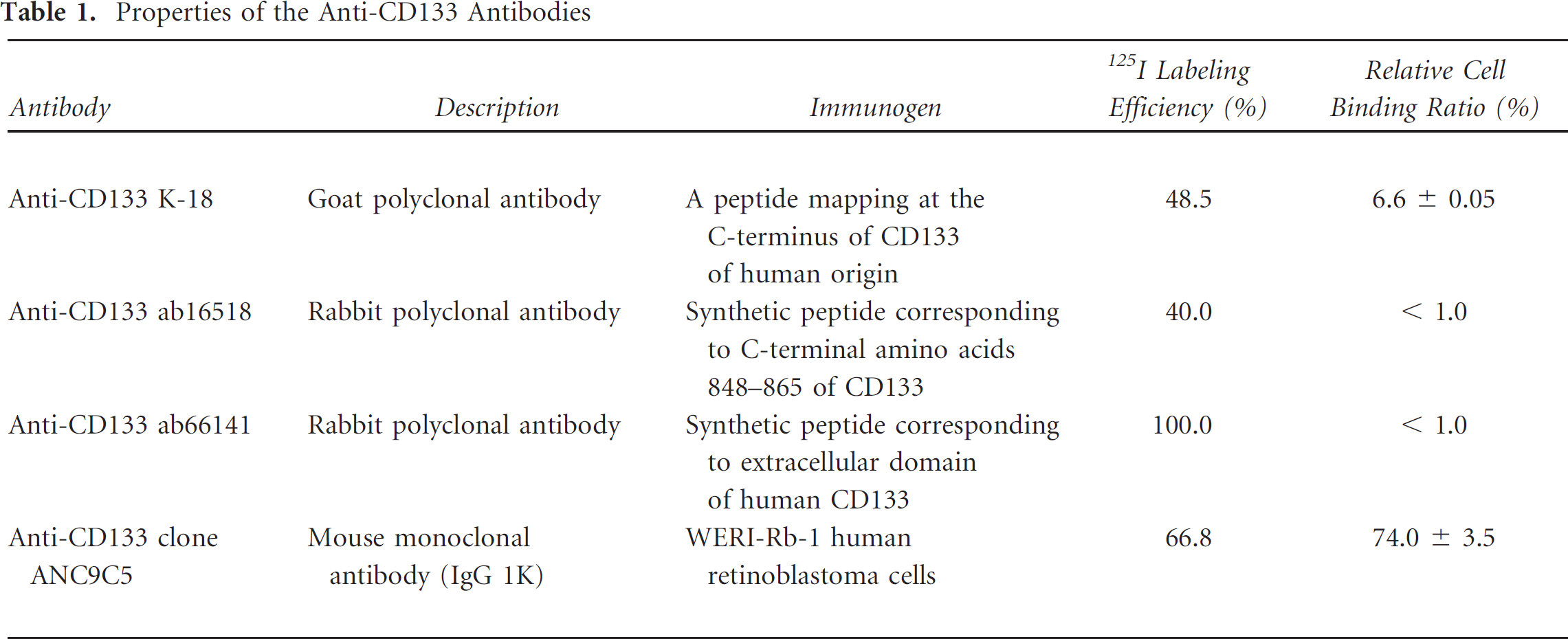
Four commercially available antihuman CD133 antibodies were labeled with 125I. The CD133 (K18) antibody was purchased from Santa Cruz Biotechnology Inc. (Santa Cruz, CA), ab16518 and ab66141 were from Abcam (Cambridge, MA), and ANC9C5 was from Ancell (Bayport, MN). The properties of the antibodies are summarized in Table 1. The antibodies and control IgG, mouse IgG1k (Alpha Diagnostic International Inc, San Antonio, TX), were radiolabeled with 125I using the chloramine-T method. In the case of ANC9C5, 20 μL of a reaction mixture containing 10 μL of antibody solution (1 mg/mL), 2 μL of Na125I (3.7 GBq/mL, PerkinElmer Japan Co., Ltd., Yokohama, Japan), and 2 μL of chloramine-T (2 mg/mL) in 0.3 M phosphate buffer (pH 7.5) was incubated at room temperature for 5 minutes. The reaction mixture was then loaded onto a Sephadex G- 50 spin column (GE Healthcare Life Sciences, Piscataway, NJ). The purified 125I-labeled antibody solution was assayed for the radioactivity bound to the antibody by a γ-counter (Aloka ARC370M, Tokyo, Japan) and for protein concentration by the DC Protein Assay Kit (Bio-Rad Laboratories, Hercules, CA). The radioiodination of the other antibodies was carried out in a similar way.
Properties of the Anti-CD133 Antibodies
Cell Binding Assay
HEK293-CD133 cells were suspended in 1 mL ice-cold phosphate-buffered saline containing 1% bovine serum albumin (1% BSA/PBS) at densities from 10 × 106 to 0.039 × 10⁶ cells/mL and incubated with 125I-labeled anti-CD133 antibodies (approximately 30,000 cpm) on ice for 1 hour. The parent HEK293 cells were used as a negative control. After incubation, the cells were centrifuged and washed twice with ice-cold 1% BSA/PBS, and the cell-bound radioactivity was measured. The relative cell binding ratio was defined as the percentage of the cell-bound radioactivity to the added radioactivity.
Competitive Inhibition Assay
HEK293-CD133 (1 × 10⁶) cells were incubated with 30,0 cpm of 125I-anti-CD133 antibodies in the presence or absence of increasing amounts of unlabeled anti-CD133 antibodies (0, 50–10,000 ng) on ice for 1 hour. After incubation, the cells were centrifuged and washed twice with ice-cold 1% BSA/PBS, and the cell-bound radioactivity was measured. The blocking efficiency was expressed as the percentage of cell-bound radioactivity to that of the control (without blocking). The Scatchard plot analysis was used for evaluation of the affinity constant and the binding capacity.
Biodistribution of 125I-Labeled Antibodies in Tumor-Bearing Mice
Five-week-old BALB/cAJ nu/nu mice (CLEA Japan, Inc. Tokyo, Japan) were subcutaneously injected in the thigh with 2.5 × 10 of HCT116 cells. Four weeks later, when the tumors were 5 to 10 mm in the diameter, tumor-bearing mice were intravenously injected with 37 kBq of 125I-labeled ANC9C5 (n = 5) or control IgG (n = 6). The injected protein dose was adjusted to 5 mg per mouse by adding unlabeled antibody. At 24 hours after the injection, the mice were killed, and blood and tissues of interest were collected and weighed. Their radioactivities were measured by a γ-counter and expressed as a percentage of the injected dose per gram of the tissue normalized to a mouse of 20 g body weight (%ID/g [20 g]). The animal experiments were approved by the Institutional Animal Care and Use Committee of the National Institutes of Radiological Sciences (NIRS) and performed in compliance with national and institutional rules.
Autoradiography and Immunohistochemistry
The HCT116 tumor-bearing mice were injected with 185 kBq of 125I-labeled ANC9C5 (n = 4) or control IgG (n = 4) antibody with a protein dose adjusted to 5 μg per mouse. At 24 hours after the injection, the tumors were resected and fixed overnight in 10% formalin neutral buffer solution (Wako Pure Chemical Industries, Ltd., Osaka, Japan). The specimens were dehydrated and embedded in paraffin. Sections of 4 μm thickness were prepared and exposed to an imaging plate (BAS-MS 2040, Fuji Photo Films, Tokyo, Japan). The imaging plate was analyzed by a bioimaging analyzer (FLA-7000, Fuji Photo Films). After deparaffinization and rehydration, the sections were treated with 3% hydrogen peroxide, followed by heating in citrate buffer, pH 6.0. Nonspecific binding was prevented using a protein blocking agent from Dako (Glostrup, Denmark), and the sections were then incubated for 1 hour at room temperature with rabbit anti-CD133 antibody (1:100 dilution; ab19898, Abcam). After washing the sections with Tris-buffered saline, secondary antibody (K400211, Dako) was applied for 30 minutes at room temperature. After another wash, the sections were incubated with 3,3'-diaminobenzidine tetrahydrochloride solution (K3465, Dako) for color development and counterstained with hematoxylin. Images were obtained using a microscope (Olympus, Tokyo, Japan) with a charge-coupled device camera, and whole-section images were reconstructed using an image-tiling system (e-Tiling, Mitani Corporation, Fukui, Japan).
Statistical Analysis
Statistical analysis was performed using the Aspin-Welch t-test or Student t-test, depending on the results of the F test.
Results and Discussion
Radioiodination and Binding Activities of the Radiolabeled Antibodies
The efficiency of radioiodination for clone ANC9C5 was 66.8%, the obtained specific radioactivity was 486.5 kBq/μg, and the molecular ratio of 125I to IgG was 0.9. The relative cell binding ratios of 125I-labeled ANC9C5 to CD133-overexpressing HEK293-CD133 cells increased with an increase in the cell number and reached a plateau when the cell density was higher than 5 × 10⁶ cells/mL (Figure 1A). The highest relative cell binding ratio was 74.0 ± 3.5%, which might be a good estimate of the immunoreactive fraction of the 125I-labeled ANC9C5. In contrast, negligible binding of 25I-labeled ANC9C5 was observed in the parent HEK293 cells (see Figure 1A). The results of the labeling and cell binding studies of the four anti-CD133 antibodies tested are shown in Table 1. As ANC9C5 was the only antibody retaining high binding activity after radioiodination, ANC9C5 was used for further experiments.

Cell binding studies of 125I-labeled anti-CD133 antibody. A, Binding of 125I-labeled ANC9C5 to HEK293-CD133 and HEC293 cells. Approximately 2 ng of the labeled antibodies (30,000 cpm) was incubated with different numbers of cells. B, The competitive binding assay of 125I-labeled ANC9C5 in HEK293-CD133 cells. The 125I-labeled antibodies were incubated with HEK293-CD133 cells in the presence of unlabeled antibodies (0–10,000 ng). The inset shows the Scatchard plot.
The binding of 125I-labeled ANC9C5 to HEK293-CD133 cells was inhibited by the unlabeled antibody in a dose-dependent manner, with approximately 99% inhibition achieved at 10 μM (Figure 1B). Scatchard plot analysis of the competitive inhibition revealed an affinity constant of 3.4 × 10⁸ M−1, and binding capacity was 3.1 × 10⁵ per cell.
Biodistribution of 125I-Labeled Antibodies
The biodistribution of 125I-labeled ANC9C5 and control IgG in HCT166 tumor–bearing mice 1 day after intravenous injection is shown in Figure 2. The 24-hour time point for in vivo studies was selected because in our earlier studies on other antibodies, at this time point and later, specific accumulation of radioiodinated antibodies to tumors was observed with whole IgG (data not shown), whereas an earlier time point was preferable if the expression of CD133 could change depending on the tumor microenvironment, 13 along with the possibility of the release of radioactivity from the target cells increasing with time, making it difficult to examine the relationship between the expression of CD133 and the distribution of the labeled antibody, especially at the intratumoral or histologic level. The radioactivity in the tumor was significantly higher for ANC9C5 compared to the control IgG in terms of %ID/g of tissue (4.54 ± 0.90 vs 2.51 ± 0.52, p < .01). The accumulation of 125I-labeled ANC9C5 in HCT116 tumors was not as high as previously reported with other tumor-targeting radioiodinated antibodies.14–16 However, considering that CD133 expression is limited to a population of cancer cells in the tumor (2.5 to 8.5% by a fluorescence-activated flow cytometry analysis; unpublished data from T.F., 2012), the value we obtained seemed reasonable. The radioactivity levels in the blood and stomach were also significantly higher with ANC9C5, whereas the radioactivity in the liver was significantly higher with the control IgG. Radioactivities in the other tissues were comparable between ANC9C5 and the control. The reasons for the differences in radioactivity between ANC9C5 and the control observed in blood, liver, and stomach are unknown, although internalization and degradation followed by the release of free 125I after the binding of radiolabeled ANC9C5 to the cell-surface antigen could be partly responsible, as well as intrinsic differences in the properties of specific immunoglobulins. Although the radioactivities in the blood and the tumor were both higher with ANC9C5, the tumor to blood ratio was significantly higher with ANC9C5 compared with the control IgG (0.26 ± 0.05 vs 0.18 ± 0.02 %ID/g, p < .01). The tumor to muscle ratio was also significantly higher with ANC9C5 (4.06 ± 0.56 vs 2.45 ± 0.39 %ID/g, p < .01). These ratios could be useful when evaluating the image data.

Biodistribution of 125I-labeled anti-CD133 antibody in the HCT166 tumor–bearing mice. The mice were intravenously injected with 37 kBq of 125I-labeled ANC9C5. At 24 hours after the injection, blood and tissues of interest were collected. Radioactivities were expressed as a percentage of the injected dose per gram of the tissue normalized to a mouse of 20 g body weight (%ID/g [20 g]). The asterisk indicates significant differences between the 125I-labeled ANC9C5 and the control IgG: *p < .05 and **p < .01, n = 5–6.
Autoradiography and Immunohistochemistry
Figure 3A compares the intratumoral distribution of 125I-labeled ANC9C5 captured by autoradiography of an HCT116 tumor section prepared from a tumor excised 1 day after injection of 125I-labeled ANC9C5 on the left panel with the expression of CD133 in the same section captured by immunohistochemical staining against CD133 on the right panel. Intratumoral distribution of 125I-labeled ANC9C5 and CD133 expression overlapped in many areas, although they did not show a complete match. Both accumulation of 125I-labeled ANC9C5 and expression of CD133 were found in the areas close to the tumor rim and, in some areas, toward the center that were viable. Figure 3B shows a comparison of the intratumoral distribution of 125I-labeled control IgG and CD133 expression. In contrast to 125I-labeled ANC9C5, 125I-labeled control IgG was mainly found in the area overlapping or peripheral to the necrotic regions located in the center of the tumor, and the distribution pattern was quite different from that observed with 125I-labeled ANC9C5. These findings indicated that the distribution of 125I-labeled ANC9C5 reflected the expression of CD133, although the presence of other factors influencing the distribution and causing some discrepancy between the intratumoral antibody distribution and CD133 expression cannot be ruled out.

Comparison of the intratumoral distribution of the 125I-labeled anti-CD133 antibody and the control IgG with CD133 expression in HCT116 xenografts. A, Autoradiograph of an HCT116 tumor section from a mouse treated with 125I-labeled ANC9C5 (left panel). Immunohistochemical staining of the section against a CD133 antibody (right panel). B, Autoradiograph of an HCT116 tumor section from a mouse treated with 125I-labeled control IgG (left panel) and immunohistochemical staining of the section against CD133 (right panel). Original magnification ×40.
Conclusion
In the present study, we showed that biodistribution and intratumoral distribution of 125I-labeled ANC9C5 could reflect the expression of CD133, a membrane protein recognized as a CSC marker in several types of cancers. Although more extensive studies are necessary, these results suggest that in vivo targeting of CSCs is possible with radiolabeled antibodies. Noninvasive imaging of CSCs would provide critical information for cancer treatment planning, and CSC targeted therapy could be ground-breaking in cancer treatment. Radioimmunotargeting techniques have made significant progress over the decades through the improvement of labeling methods and antibody engineering. Application of those methodologies would further facilitate the implementation of clinical CSC imaging and CSC-targeted therapy
Footnotes
Acknowledgments
We would like to thank the members of the Diagnostic Imaging Program, Molecular Imaging Center, NIRS, for helpful discussions and valuable suggestions.
Financial disclosure of authors: This work was partly supported by the Japan Advanced Molecular Imaging Program of the Ministry of Education, Culture, Sports, Science and Technology and the NIRS.
Financial disclosure of reviewers: None reported.