
Abstract
We hypothesized that imaging-based assessment of cellular proliferation in prostate cancer may improve tumor characterization. We therefore evaluated the biodistribution and effect of androgen on tumor uptake of the cellular proliferation imaging marker [18F]-2'-fluoro-5-methyl-1-beta-D-arabinofuranosyluracil (18F-FMAU) in xenograft mouse models of human prostate cancer. Castrated and noncastrated athymic male mice were implanted with androgen-independent PC3 and androgen-sensitive CWR22 human prostate cancer cells. Dynamic micro–positron emission tomography (PET)/computed tomography was performed for 1 hour followed by 10-minute static scans at 2 and 3 hours. Animals were sacrificed after imaging for biodistribution studies and immunohistochemical staining of tumors for androgen receptor and Ki-67/MIB expression. 18F-FMAU uptake was significantly higher in all major organs of the castrated animals in comparison with noncastrated mice, with the highest uptake in liver and the lowest uptake in muscle and bone. When compared to PC3 tumors, CWR22 xenografts showed significantly higher tumor to muscle (2.56 ± 0.30 vs 1.99 ± 0.30, p 5.008) and tumor to liver (1.72 ± 0.12 vs 1.26 ± 0.17, p = .0003) uptake ratios in the noncastrated animal at the 3-hour time point. Androgen receptor and Ki-67/MIB expressions were higher in CWR22 than in PC3 xenografts. Our initial preclinical observations suggest that there may be an association between androgen signaling and thymidine metabolism and that 18F-FMAU PET may be useful in prostate tumor characterization.
PROSTATE CANCER is the most common cancer and the second leading cause of cancer death affecting men in the United States. There is currently a lack of an accurate objective imaging-based method for the characterization (indolent vs lethal) of the primary prostate tumor. Image-guided detection, localization, and characterization of tumor will alleviate the current major problems of the conventional “blind” biopsy approach, which includes overdiagnosis (and overtreatment) of indolent tumors and underdiagnosis (and undertreatment) of aggressive prostate cancers.
There is increasing interest in the potential role of positron emission tomography (PET) in prostate cancer. Given the biologic and clinical heterogeneity of prostate cancer, PET would be an ideal imaging tool for noninvasive interrogation of the underlying tumor biology in different phases of the disease. The cumulative current experience with PET and the most studied radiotracers, namely 18F-fluorodeoxyglucose, 18F- or 11C –choline, and 18F- or 11C-acetate, suggests a generally limited role for these radiotracers in the imaging-based characterization of prostate tumor due to the overlap of uptake among normal, benign prostatic hyperplasia and prostate cancer tissues. 1 Several other promising radiotracers have been investigated in the imaging evaluation of prostate cancer, including 16β-18F-fluoro-5α-dihydrotestosterone (18F-FDHT), targeted to the androgen receptor anti-1-amino-3-18F-fluorocyclobutane-1-carboxylic acid (anti-18F-FACBC), which is a synthetic L-leucine analogue, and prostate-specific membrane antigen (PSMA)-based PET radiotracers. 2 However, the exact diagnostic roles of these radiotracers in prostate cancer remain undefined and will require continued investigations.
The goal of our study was to determine whether imaging assessment of cellular proliferation in prostate cancer might be useful in tumor characterization. Although 18F-fluorothymidine (18F-FLT) is the most studied PET radiotracer for imaging cellular proliferation in cancer, the high physiologic accumulation of this tracer in marrow and the high bladder urine activity may limit its utility in prostate cancer. On the other hand, [18F]-2'-fluoro-5-methyl-1-beta-D-arabinofuranosyluracil (18F-FMAU) has no or very little accumulation in bone (a common site for metastasis from prostate cancer) and in urinary bladder, which renders it a potentially ideal PET radiotracer for imaging deoxyribonucleic acid (DNA) synthesis in prostate cancer. 3 We therefore hypothesized that 18F-FMAU PET may be helpful in prostate tumor characterization for differentiating indolent from aggressive tumors with the supposition that tumors with higher tracer uptake are biologically more aggressive (eg, higher growth rate).
18F-FMAU is a thymidine analogue that is phosphorylated by thymidine kinase and incorporated into the DNA. 4 It was originally of clinical interest as an anticancer and an antiviral drug when used in pharmacologic doses. 5 In tracer doses, 18F-FMAU can be useful for imaging DNA synthesis and tumor proliferation.6–10 It has also been used for imaging reporter gene expression using the herpes simplex virus type 1 thymidine kinase (HSV-tk1) system.11–14 Pharmacokinetic studies have shown that radiolabeled 18F-FMAU behaves very similarly to the pyrimidine nucleoside thymidine with respect to cellular uptake velocity, saturability of cellular incorporation, and intracellular metabolite pools and is reflective of tumor cell division. 7 Initial imaging-based biodistribution of 18F-FMAU in normal dogs has shown that it is resistant to degradation and is selectively retained in DNA. 15
We set out to assess the biodistribution and tumor uptake of 18F-FMAU in androgen-sensitive and androgenindependent xenograft models of human prostate cancer. We also studied the potential effect of androgen on the implanted tumor uptake by using castrated and noncastrated mice to model, respectively, the absence and presence of physiologic levels of androgen.
Materials and Methods
18F-FMAU Radiosynthesis
The radiosynthesis of 18F-FMAU was performed using the newly developed one-pot labeling procedures. 16 Briefly, the radiosynthesis involves radiofluorination of 2-trifluoro-methanesulfonyl-1,3,5-tri-O-benzoyl ribofuranose to 2- [18F]-fluoro-1,3,5-tri-O-benzoyl arabinofuranose derivative followed by the conjugation with 2,4-bis-trimethylsilyluracil in the presence of hexamethyldisilazane (HMDS) and trimethylsilyl trifluoromethanesulfonate (TMSOTf). Finally, hydrolysis of the protecting groups from the sugar moiety and high-performance liquid chromatography (HPLC) purification produces the desired product. 18F-FMAU was obtained in 12 ± 3% radiochemical yield with 547 mCi/μmol specific activity. The overall radiosynthesis time was about 150 minutes, and the radiochemical purity was > 99%.
Tumor Cell Lines, Animals, and Experimental Protocol
We employed androgen-dependent CWR22 and androgen-independent PC3 human prostate cancer cell lines (American Type Culture Collection, Manassas, VA). The androgen-dependent CWR22 human prostate cancer cell line expresses androgen receptors and prostate-specific antigen and is stimulated by dihydroxytestosterone. The androgen-independent PC3 human prostate cancer cell line was initiated from a bone metastasis of a grade IV prostatic adenocarcinoma and displays low testosterone–5α-reductase activity.
Surgically castrated and noncastrated mice were purchased (Harlan Sprague-Dawley, Indianapolis, IN) and served as models for the absence and presence, respectively, of androgens. No testosterone pellets or blood sampling for androgen level was employed. Tumor volume was calculated using the formula (S 2 × L)/2, where S and L represent the small and large diameters of the lesion. 17 The lesion dimensions were measured using calipers at every 2- to 3-day interval.
We implanted either CWR22 or PC3 human prostate cancer cells at a concentration of 5 × 10⁶ cells per 0.1 mL in one thigh of 4- to 6-week-old, 20 to 30 g, castrated (n = 4) or noncastrated (n = 6) male athymic mice (BALB/c nu/nu), and the tumors were allowed to grow to 0.5 cm3. Separate control non–tumor-bearing athymic male mice were also included for imaging and biodistribution studies at 2 hours (three castrated, three noncastrated) and 3 hours (four castrated, four noncastrated) after tracer administration. Tumor cells were stained with trypan blue, and viable cells were counted using a hemocytometer under a light microscope.
All animal studies were approved by our Institutional Animal Care and Use Committee, the Biological Safety Committee, and the Radiation Safety Committee. Anesthesia was induced using 2% isoflurane in oxygen and maintained throughout all imaging studies. Euthanasia was performed by cervical dislocation while the animal was anesthetized. Animals were housed in the vivarium facility during the period of tumor growth and were fed regular rodent food and water ad libidum.
MicroPET Imaging and Biodistribution Studies
PET scans were performed on the microPET R4 (Concorde Microsystems, Knoxville, TN) and followed by microCT imaging (InveonCT, Siemens Medical Solutions USA, Knoxville, TN) for anatomic reference. Immediately following tail vein administration of 200 μCi of 18F-FMAU, dynamic PET scanning was performed for 1 hour (four castrated, six noncastrated) followed by additional 10-minute static scans at the 2- and 3-hour time points (four castrated, five noncastrated). PET data were all reconstructed using the 2D-OSEM algorithm supplied by MicroPET Manager (Siemens Medical Solutions USA) into 128 × 128 × 63 images with 0.084 mm × 0.084 mm × 1.21 mm resolution. Computed tomographic (CT) scans were acquired in two bed positions using the settings: 80 kVp, 500 uA, and 100 ms/180 steps covering 360° and reconstructed into 768 × 768 × 923 images with 0.105 mm isotropic resolution. The dynamic PET data were histogrammed into 11 frames, whereas static data were histogrammed into single frames using the MicroPET Manager software (Concorde Microsystems). PET and CT images were coregistered using rigid transformations as both scans were performed using warmed multimodality imaging chambers and animals were not moved between scans. Immediately following microPET imaging, animals were sacrificed for biodistribution studies. Blood, tumor, and organs were harvested, weighed, and counted for radioactivity on a Packard Cobra II Gamma Counter (Packard Instruments, Meriden, CT).
Immunohistochemical Assays
Xenograft tumor tissue samples were fixed in 4% freshly prepared buffered paraformaldehyde, embedded in paraffin according to standard histologic protocols, and sectioned at a thickness of 5 μm. Routine histologic staining with hematoxylin-eosin and immunohistochemical assays for cellular proliferation parameter, Ki-67/MIB, and androgen receptor were performed to obtain qualitative appraisal on the magnitudes of their expression in the implanted prostate tumors.
Data Analysis and Statistics
Tissue tracer uptake was calculated as standardized uptake value (SUV) from the PET data and as percent injected dose/gram of tissue (%ID/g) from the biodistribution data and expressed as ratios to the activities of muscle, blood, or liver. Two-group comparisons of data were performed between tumor types in a particular animal type and between animal types for a particular tumor type using a two-tailed Student t-test with unequal variance and with a significance probability level of less than .05.
Results
Biodistribution Studies
Control biodistribution studies in non-tumor-bearing mice demonstrated significantly higher 18F-FMAU uptake in all major organs of castrated animals in comparison with noncastrated animals, suggestive of generalized tracer retention in the castrated animal (Table 1). Similar observation was noted with the tumor-bearing mice biodistribution studies. Figure 1 shows biodistribution studies that were performed 3 hours after tracer administration in mice bearing either CWR22 or PC3 implanted xenografts. It is clear that there was consistently higher tracer uptake in all the major organs and in the implanted tumors in the castrated animals when compared to those in noncastrated mice. In both animal types, the organs with the highest tracer uptake were the liver, spleen, and kidneys. Bowel showed variable but generally high uptake. Bone uptake level was low and similar to muscle reference (noncastrated: 1.63 ± 0.86 vs 1.57 ± 0.95 %ID/g; castrated: 3.27 ± 1.19 vs 3.01 ± 1.38 %ID/g, respectively; all values at 3 hours).
Biodistribution of 18F-FMAU in Major Organs of Control Non–Tumor-Bearing Noncastrated and Castrated Mice at 2 and 3 Hours

The p values at 2 and 3 hours refer to comparison of the corresponding tissue mean uptake levels in the noncastrated and castrated animals. All comparisons were statistically significant except for the 2-hour time point renal and bowel uptake levels. Note the generalized tissue tracer retention in the castrated animal in comparison with the noncastrated animal.

Biodistribution of 18F-FMAU 3 hours after tail vein administration in athymic castrated and noncastrated male mice bearing either (A) CWR22 or (B) PC3 implanted human prostate cancer xenografts. In both animals, the organs with the highest tracer uptake were the liver, spleen, and kidneys. Note the generalized organ tracer retention in the castrated mice. The CWR22 tumor to liver and tumor to blood uptake ratios were significantly higher in the noncastrated animal.
MicroPET Studies
In the tumor-bearing animals, visually, there was high tracer uptake in both tumor types implanted in either animal type. The dynamic microPET demonstrated a representative uptake profile for the tumors that increased relatively rapidly to a plateau (within about 10 minutes after tracer administration), at which point, the uptake level increased slowly. Conversely, uptake in the reference tissues of muscle and liver showed a rapid decline in uptake, particularly at about 1 hour, such that by 3 hours post–tracer administration, the tumor uptake was substantially higher than the uptake levels in the liver (by an average factor of 1.45) and the muscle (by an average factor of 2.28) (Figure 2). The organs with the highest tracer accumulation on microPET were the same as those noted on the biodistribution studies (ie, liver, spleen, kidneys). Also, the excreted tracer progressively accumulated in the urinary bladder.

Representative 3-hour duration time-activity curves obtained from regions of interest placed over PC3 tumor (arrow on the mouse image in the top right corner), liver, and muscle on dynamic microPET images. Note the early rapid decline in the hepatic activity with a steady decline in both the liver and muscle uptake levels after 1 hour. The tumor uptake level rises rapidly, reaching a plateau with a continued slow rise. After 2 hours, the tumor uptake level is higher than the uptake levels in the liver and the muscle.
Effect of Tumor and Animal Type
For androgen-dependent CWR22 xenografts, when compared to the castrated animal, the noncastrated animal showed significantly higher tumor to liver (1.72 ± 0.12 vs 1.3 ± 0.17, p = .003) and tumor to blood (2.93 ± 0.21 vs 2.32 ± 0.13, p = .001) uptake ratios at 3 hours. Conversely for androgen-independent PC3 xenografts, no statistically significant difference was seen in the tumor to nonproliferating tissue reference (either muscle or liver) uptake ratios between the castrated and the noncastrated animals. Moreover, when compared to PC3 tumors, CWR22 xenografts showed significantly higher tumor to muscle (2.56 ± 0.30 vs 1.99 ± 0.30, p = .008) and tumor to liver (1.72 ± 0.12 vs 1.26 ± 0.17, p = .0003) uptake ratios, but this was observed only in the noncastrated animal at the 3-hour time point (Figure 3).
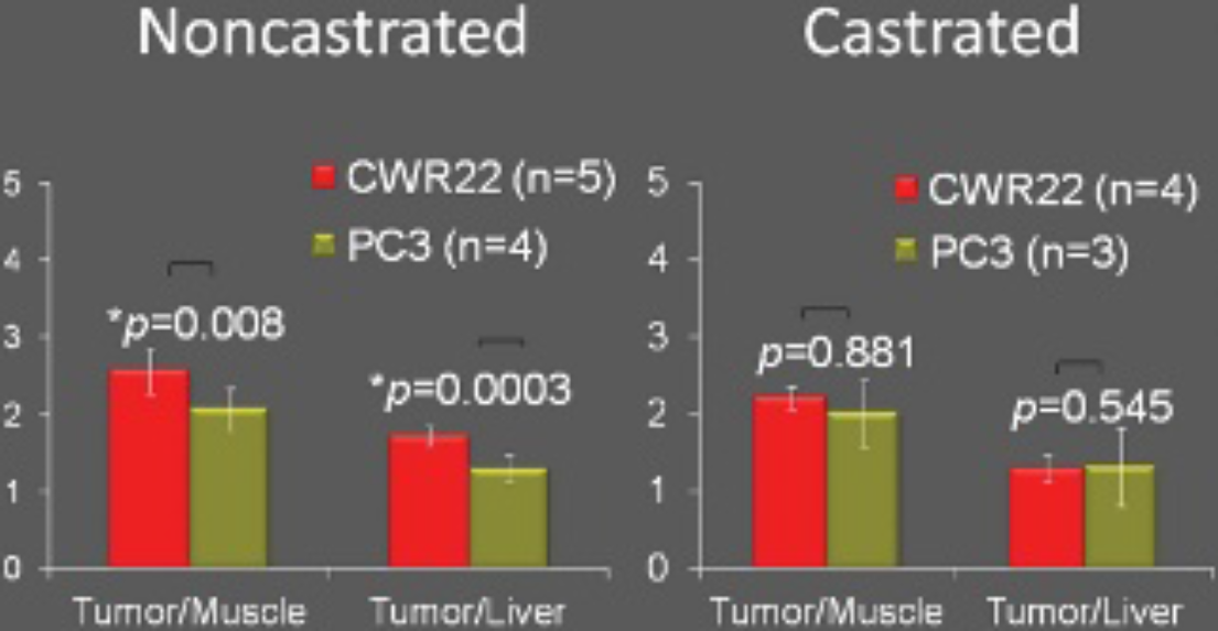
18F-FMAU uptake levels in the two tumor types were significantly different only in the noncastrated animal at the 3-hour time point.
Qualitative Assessment of Immunohistochemical Assays
Histologic assessment demonstrated few small areas of necrosis in the harvested tumors. Qualitative visual assessment of the immunohistochemical assays showed higher expressions of both androgen receptor and Ki-67/MIB in the CWR22 xenograft when compared to the implanted PC3 tumor (Figure 4).

Immunohistochemical stains: (androgen receptor [AR]; cellular proliferation index, Ki-67/MIB) for CWR22 (top panel) and PC3 (bottom panel) implanted tumors (×20 objective magnification). Note the relatively strong expression of AR and Ki-67/MIB in the CWR22 tumor tissue in comparison with those of the PC3 tumor tissue.
Discussion
The standard transrectal ultrasound-guided 10- to 12-core prostate biopsy misses about 38% of tumors due to multifocal distribution of prostate cancer. 18 Using this procedure, even when prostate cancer is diagnosed, the possible coexistent high-grade tumor may be missed leading to underdiagnosis of these tumors and subsequent therapy failures. Saturation (> 20) biopsy procedures cannot compensate for the shortcoming of the blind conventional biopsy procedure because it leads to overdiagnosis and overtreatment of clinically insignificant tumors at substantial morbidity to the individual patient and financial cost to the health care system. 19 Therefore, there is a critical need for image-guided biopsy that optimizes the probability of detection of biologically and clinically relevant tumors (eg, aggressive tumors) and reduces the biopsy rate of clinically insignificant tumors. Image-guided characterization will also pave the way for rational treatment decision making, including active surveillance for low-grade tumors and focal therapy (male lumpectomy) for localized aggressive tumors.
We therefore set out to assess the potential utility of the cellular proliferation imaging marker 18F-FMAU in a preclinical xenograft model of human prostate cancer. It is prudent to presume that biologically more aggressive prostate tumors would display higher cellular proliferative phenotype. A number of PET-based proliferation tracers have been explored, typically in conjunction with tracers for the thymidine salvage pathway of DNA synthesis. 20 The most studied thymidine analogue has been 18F-FLT. 18F−FLT is phosphorylated by thymidine kinase 1, incorporated by the normal proliferating marrow, and glucuronidated in the liver. Experience with 18F-FLT in prostate cancer is very limited, although, recently, in preclinical models of castrate-resistant prostate cancer, this tracer has been shown to be potentially useful for monitoring the response to docetaxel treatment. 21 However, the physiologic high marrow accumulation of 18F-FLT severely limits its clinical utility in the detection and assessment of treatment response in bone metastases.
On the other hand, 18F-FMAU is incorporated to DNA after phosphorylation and shows substantially less marrow uptake, which is a useful feature for imaging assessment of prostate cancer. Our biodistribution studies showed relatively high uptake in the normal heart, kidneys, and liver, similar to previous studies, which is thought to be related in part to the role of mitochondrial thymidine kinase 2. 4 We also observed high steady tracer accumulation in the xenograft human prostate cancer tumors. High activity was noted in the urinary bladder in relation to accumulating excreted tracer. Interestingly, previous studies have shown relatively low 18F-FMAU accumulation in the dog urinary bladder. 15 This may be due to species differences and will need further investigation in humans. Our results are also in line with those of previous studies in that bone uptake level is low, which renders 18F-FMAU useful for the imaging evaluation of osseous metastases of prostate cancer. 3
We found that despite some decay in radioactivity, delayed imaging at 3 hours post—tracer administration might be needed for improved tumor to reference tissue uptake ratio, which was essentially related to a steady general decline in the background activity in the setting of plateau or slowly increasing tumor uptake. In fact, at 3 hours, we observed a statistically significant difference in the tumor to reference tissue (either liver or muscle) uptake ratios between the two tumor types in the noncastrated mice. The higher 18F-FMAU uptake in the CWR22 tumor appeared to be associated with the relatively strong expression of the cellular proliferation index Ki-67/MIB. As expected, the androgen receptor expression level was higher in the CWR22 xenograft than in the PC3 tumor. When identical experiments were performed in the castrated mice, this difference between the tumor to reference uptake ratios for the two tumor types was not observed. Biodistribution studies revealed that this finding was largely due to the generally higher tracer retention in the castrated animal, thereby reducing the tumor to reference tissue uptake ratios even at 3 hours after tracer administration. Another contributing factor may be the reduction in cellular proliferation of the androgen-dependent tumor after androgen withdrawal.22,23
The exact biologic mechanism for the apparent generalized 18F-FMAU retention in the castrated mouse and the relationship of this observed phenomenon to the absence of androgen are unknown. We did not measure the plasma level of androgen in the noncastrated mice and relied on the intrinsic androgen level in the noncastrated mice to model and maintain the physiologic condition. The observation of generalized organ 18F-FMAU retention in the absence of androgen was an interesting finding. Although beyond the scope of the current report and our initial experimental design, we plan to perform follow-up longitudinal test–retest biodistribution and imaging studies using non–tumor-bearing castrated mice before and after subcutaneous implantation of a time-release testosterone pellet, which will enable us to assess the effect of androgen on 18F-FMAU biodistribution within the same animal. Additional imaging after systemic clearance of testosterone (≈ 2 to 4 weeks after pellet implantation) allows confirmation of our current preliminary observation of a possible association between androgen and physiologic 18F-FMAU biodistribution on which additional biochemical investigations may be necessary to elucidate the exact mechanistic association between androgen signaling and thymidine metabolism. One possibility may be the androgen control of mitochondrial function, which may include thymidine kinase 2 enzymatic activity. 24
Our study should be interpreted in the context of the preclinical subcutaneous xenograft model that was employed. Generally, factors such as tumor microvasculature, oxygenation distribution, and differences in the growth pattern and tumor microenvironment may limit extrapolation from tumor models in animals to the native spontaneous tumors in humans. 25 Despite this broad limitation in extending results from animal models to the human condition, xenograft tumor models are commonplace in preclinical studies.
Conclusion
Our preliminary observation with 18F-FMAU in prostate cancer xenografts suggests that there may be a role for 18F-FMAU in image-based characterization of tumor in the noncastrate state when men present at the time of initial evaluation for prostate cancer. Moreover, 18F-FMAU uptake in the androgen-dependent prostate tumor may be sensitive in responding to the differences in circulating androgen levels and as such may provide useful imaging-based information on the impending castrate-resistant state. Additional preclinical work in other prostate cancer cell lines and spontaneous tumors in mice as well as pilot human studies will be needed to decipher the exact role of 18F-FMAU in the imaging evaluation of prostate cancer.
Footnotes
Acknowledgments
We thank Fatemeh Abdollahi Mofakham for help with the animal procedures.
Financial disclosure of authors: This work was supported by the National Institutes of Health, National Cancer Institute grant number R21-CA142426 (to H.J.). The radiochemistry preparation and procedures for 18F-FMAU have been patented under US patent 7,273,600 B2 (inventors: P.S. Conti, M.M. Alauddin, J.D. Fissekis), and the patent has been assigned to the University of Southern California, Los Angeles, CA. None of the authors have any financial interest in this patent.
Financial disclosure of reviewers: None reported.