
Abstract
Multimodal molecular imaging can offer a synergistic improvement of diagnostic ability over a single imaging modality. Recent development of hybrid imaging systems has profoundly impacted the pool of available multimodal imaging probes. In particular, much interest has been focused on biocompatible, inorganic nanoparticle-based multimodal probes. Inorganic nanoparticles offer exceptional advantages to the field of multimodal imaging owing to their unique characteristics, such as nanometer dimensions, tunable imaging properties, and multifunctionality. Nanoparticles mainly based on iron oxide, quantum dots, gold, and silica have been applied to various imaging modalities to characterize and image specific biologic processes on a molecular level. A combination of nanoparticles and other materials such as biomolecules, polymers, and radiometals continue to increase functionality for in vivo multimodal imaging and therapeutic agents. In this review, we discuss the unique concepts, characteristics, and applications of the various multimodal imaging probes based on inorganic nanoparticles.
Molecular imaging characterizes the biologic functions of the body on the molecular level, allowing for a detailed understanding of the disease and an application of an individualized treatment to the patient. Standard molecular imaging systems commonly used in the clinic are positron emission tomography (PET), molecular MRI, and single-photon emission computed tomography (SPECT). Multimodal imaging combines the advantageous parameters of each individual modality into one system, allowing synergistic effects.13–15 For example, the bimodal PET-MRI system incorporates the high sensitivity of PET with the high resolution of MRI, thereby circumventing the inherent low resolution of PET and low sensitivity of MRI. 16 Various molecular imaging techniques require different properties from probes, such as para- or superparamagnetic materials for MRI, radionuclides for PET and SPECT, high atomic number atoms for computed tomography (CT), and various fluorescent particles for optical imaging. 14 The multifunctionality of inorganic nanoparticles allows these properties to be delivered to the body in one tunable nanoplatform system. In this review, the advantages of inorganic nanoparticles and their unique characteristics for various multimodality imaging are discussed.
IONP-Based Multifunctional Probes
IONPs are superparamagnetic iron oxide crystalline structures that have the formula Fe2O3MO, where M is a divalent metal ion such as manganese, nickel, cobalt, or magnesium. Superparamagnetic iron oxide particles are made up of iron alone and have been extensively used as commercial T2 MRI contrast agents such as Feridex, Resovist, and Combidex, with decreased toxicity compared to gadolinium (Gd)-based agents.17,18 IONPs, along with their intrinsic superparamagnetic properties and therefore their promising application as a MRI contrast agent, can serve as nanoscaffolds for additional imaging probes for bimodal imaging. 19 The most widely used method to multifunctionalize IONPs is surface modification by covalent conjugation. Such particles are called cross-linked iron oxide (CLIO). The surface of the IONP can be crosslinked with other imaging probes, such as fluorophores for magnetic resonance (MR)-optical imaging and radionuclides for MR-PET imaging, as well as with targeting groups for specific therapies.
Optical absorption, luminescence, and fluorescence have also been combined with IONPs for the development of optical and MR contrast agents. 13 Although numerous visible light fluorescent probes have been combined with IONPs, such as rhodamine, fluorescein, and others, these probes prove ineffective in vivo owing to their limited tissue penetration property. Near-infrared (NIR) imaging is particularly useful because of the “biologic window.” This window refers to the 700 to 1,300 nm spectral region where blood, water, and tissue have reduced absorption and autofluorescence, allowing for agents that absorb and emit in the NIR to be easily detected. NIR dyes have been combined with IONPs to produce MRI-near-infrared fluorescence (NIRF) probes. CLIOs are chemically crosslinked dextran-coated IONPs that are widely accepted to be biocompatible owing to their stable hydrophilic polymer coating. Josephson and colleagues synthesized CLIO conjugated to the NIR dye Cy5.5 for dual MR and optical imaging. 20 Cy5.5 was conjugated to CLIO by amine groups on the dextran-coated IONP to the succinimidyl ester on the Cy5.5. This system has been repeated in numerous animal models ranging from sentinel lymph node (SLN) to brain tissue imaging, showing low cytotoxicity and multimodal functionality as MRI-NIRF imaging probes.20,21
In another embodiment, Santra and colleagues coencapsulated an NIR dye, dialkylcarbocyanine, and an anticancer drug, paclitaxel, into the poly(acrylic acid) (PAA) matrix on the surface of IONPs. 22 Carbodiimide chemistry and click chemistry were used to functionalize the surface of the PAA-IONP. This allowed versatility in the types of functional groups that can be used on the surface of IONPs, enhancing targeting and delivery of these molecular imaging and drug delivery probes. The probe is able to be (1) monitored by MRI and NIRF, (2) targeted to cancerous tissue by surface functionality, and (3) used to deliver an anticancer drug. Cellular uptake of the targeted probes was monitored by MRI relaxivity and NIRF, and induced cancer cell death was observed when paclitaxel was encapsulated within the PAA matrix. Such molecular imaging probes require a long circulation time and low uptake into the reticuloendothelial system (RES) to allow for accurate imaging.
Chen and colleagues developed an MRI-NIRF probe demonstrating high targeting specificity and low in vivo clearance by coating IONPs with a polyethylene glycol (PEG)-conjugated amphiphilic triblock copolymer made up of polybutylacrylate, polyethylacrylate, polymethacrylic acid, and octylamine. 23 A NIRF dye, IRDye800, and cyclic Arg-Gly-Asp-D-Tyr-Lys (cRGDyK) were further conjugated for NIR imaging and targeting for integrin αvβ3 receptors. Cyclic RGD analogues are peptides commonly used in nanoparticle engineering to target tumor integrin αvβ3 and in this example were shown to significantly improve tumor uptake of the bimodal imaging probe over non-RGD functionalized probes. 24 Successful tumor homing, as seen in this study in a subcutaneous U87MG glioblastoma xenograft model, gives true molecular imaging capabilities as a small amount of probes is required to obtain strong contrast enhancement at the site of imaging. The 20 nm diameter engineered particles exhibit increased circulation time owing to their PEG coating and reduced uptake by the liver. 23 PEGylation has been shown to increase circulation time in other nanomaterials, such as QDs, and may be applied to other nanoparticles to improve the biodistribution.25,26 Recent works also demonstrated improved integrin αvβ3 receptor targeting with a copolymer coating between the targeting ligand and IONP. Chen and colleagues reported an amphiphilic diblock copolymer, poly(ethylene oxide)-block-poly(gamma-methacryloxypropyltrimethoxysilane) (PEO-b-PgammaMPS), which can be used to coat IONPs to reduce nonspecific cell and RES uptake because of its neutral charge. 27
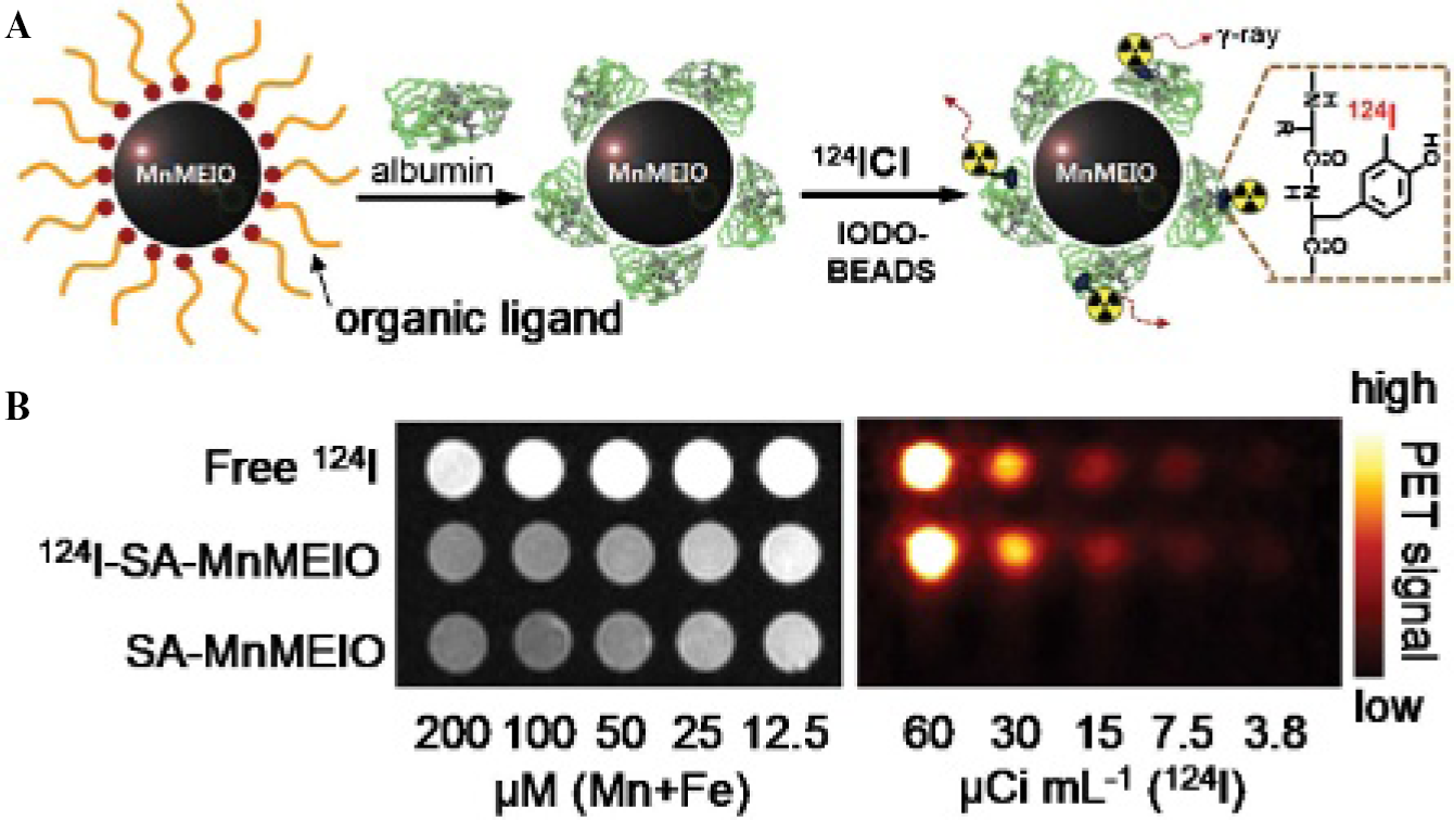
Beyond IONP conjugation to optical probes, numerous MRI-PET bimodal imaging agents have been developed with IONP as the main platform. An MRI-PET bimodal imaging system combines the sensitive molecular imaging of PET with the high-resolution imaging of MR. In this manner, the same molecular target can be imaged, evaluated, and analyzed with sensitivity and high resolution, giving anatomic and functional images. The design of an MRI-PET probe should involve proportionate amounts of each required material as MRI requires high doses of contrast agents, whereas PET uses only trace amounts. Radioisotopes for PET have been attached onto the IONP scaffold to provide dual-modality MR/PET imaging. Choi and colleagues designed a probe for MRI-PET made up of MnIONPs combined with the isotope 124I. 28 MnIONPs have been shown to have T2 relaxivity coefficients two to three times higher than IONPs without additional elements, therefore giving this hybrid nanoparticle a synergistic high MR contrast. 29 MnIONPs were first coated by serum albumin (SA) to prevent aggregation of the particles under physiologic conditions as well as to provide a binding point for the isotope. Second, 124I binds directly to the tyrosine residue of SA. The authors demonstrated that the contrast enhancement of the conjugated isotope to the MnIONP did not differ from the contrast enhancement of the probes separately. 124I-SA-MnIONP nanoparticles were verified in vivo to identify and distinguish the SLNs, giving both functional and anatomic information. SLN identification requires molecular imaging to determine if cancer has metastasized from its primary tumor site, thereby enlarging the SLN. 28 The MR and PET imaging capabilities of the 124I-SA-MnIONP are seen in Figure 1.
Another MRI-PET probe design incorporates DOTA chelated radionuclide 64Cu on the IONP scaffold. 30 IONPs were coated with polyaspartic acid (PASP) through the carboxyl group, whereas the remaining amine group was used as a binding site for another linker, NHS-PEG-MAL, to conjugate cRGD and DOTA chelators on the surface. PASP and PEG, besides allowing conjugation of the PET probes onto the MRI probes, serve to stabilize the nanoparticles in physiologic conditions and reduce uptake through the RES, which is useful for long-term imaging. As this example suggests, linkers used to join two imaging inorganic probes together can be designed to improve in vivo capabilities. In this case, PASP linked the IONPs and radionuclide together and gave the bimodal probe enhanced tumor uptake and improved molecular imaging capabilities.
Although chemical linkers of imaging probes can themselves serve as multifunctional molecules, the composition of heterostructured nanoparticles for multimodal imaging provides a simpler approach to probe development. The combination of iron oxide with high absorbance agents, such as gold, has been used for bimodal optical absorbance sensing and MRI. Choi and colleagues synthesized various heterostructured nanoparticles made up of IONPs or MnIONPs and gold by thermal decomposition of the metal in surfactant complexes. 31 In particular, Au-Fe3O4 nanoparticles coated with a PEG-phospholipid shell and conjugated with 12-base oligonucleotide sequences showed an increase in T2 contrast and red shift in the surface plasmon band. These probes were studied for both DNA sequence sensing and biomedical imaging. Complementary DNA binding self-assembly guided these particles to aggregate. During aggregation, the particles increase in size, as detected by dynamic light scattering, AuNP absorbance decreases and red shifts, and the T2-weighted MRI contrast from the IONP decreases. The design of this system exemplifies that imaging by MR (IONPs) and optical absorbance (AuNPs) can be used for biosensing of specific DNA sequences and multimodal imaging.
Recently, Wang and Irudayaraj synthesized Fe3O4-Au-rod-Fe3O4 nanodumbbells and Fe3O4-Au-rod necklace-like particles that show variation in plasmonic and magnetic properties, which can be used for bimodal imaging.
32
The necklace-like probes are used as therapeutic, separation, and imaging agents because they can thermally ablate multiple pathogens such as
IONPs, as seen by these previous examples, can serve as excellent scaffolds for additional imaging probes while maintaining the MRI capabilities. In this way, our laboratory has developed a multimodal imaging probe based on IONPs. 34 The nanoparticles were coated with human serum albumin (HSA), 64Cu-DOTA, and Cy5.5 to allow for MRI, PET, and NIRF imaging for both in vivo and in vitro applications. A schematic image of the particle and imaging capabilities is shown in Figure 2. A key approach to this design is the addition of dopamine to the surface of the IONP, allowing the particles to be encapsulated within an HSA matrix. Once the particles are encapsulated within a molecular matrix, various drugs can be loaded within the matrix, giving it optimal drug release properties compared to covalent linking of the drug onto the particles. These particles not only envelop three different imaging modalities but also have excellent nanoparticle properties for in vivo applications: a prolonged circulation half-life, high accumulation within lesion sites, low uptake by macrophages, and a high extravasation rate. 34

QD-Based Multifunctional Probes
QDs are semiconductors that are confined on the nanometer scale. At the reduced size, the oscillation strength is focused to a reduced number of transitions with electronic excitation shifts at higher energy. 35 The use of QDs for bioimaging arises from their unique optical properties, such as photostability, broad absorption sections, wide absorption spectra, and narrow emission spectra. These confined particles are used mainly as luminescence probes that have narrow, typically 20 to 30 nm full width at half maximum, and tunable emission spectra in the spectral range of 400 nm to 2 μm. The emission spectrum is tuned by the size of the confinement. For example, when the diameter of cadmium selenide (CdSe) is reduced from 20 nm to 2.0 nm, the band gap shifts from a deep red to a green color. 35 However, QDs can cause severe cytotoxicity when semiconductor leaching occurs, especially by cadmium (Cd), and the fluorescence emission has been reported to “blink.” 36 To overcome this limitation and improve multifunctionality of nanoparticles, a new class of biocompatible QDs has been reported. The recent development of various QD-based imaging probes has been described and reviewed elsewhere.37–39 Besides the optical properties, QDs can also serve as scaffolds for additional imaging agents for the design of bimodal molecular probes.
By blending luminescent QDs and MRI contrast agents, enhanced probes can be designed for noninvasive MRI. Paramagnetic quantum dots (pQDs) were developed by coating CdSe/ZnS core/shell QDs with a PEGylated phospholipid and a Gd lipid, making the particles biocompatible and MRI active, respectively, without affecting the absorption and emission spectra of the QDs. 40 The synergistic effects of combining paramagnetic Gd with the semiconductor QDs show relaxivities at 2,000 mM−1s−1 per QD. The relaxivity of the pQDs at 60 MHz is 12 mM−1s−1 per Gd molecule, which is three times higher than the clinical contrast agent Gd-DTPA that coats the pQD. The pQDs were also conjugated by maleimide to cRGD for targeting angiogenic vascular endothelium as demonstrated by in vitro experiments with human umbilical vein endothelial cells.
In another example, fluorescence MRI bimodal agents were developed by doping silica nanoparticles with a QD coated with paramagnetic and PEGylated lipids. 41 In this way, surface functionalization steps where steric hindrance and coupling reactivity reduce the coating efficacy and form large variations in the ratio of materials covering the particle were avoided. 41 First, CdSe QDs were coated with seven monolayers of inorganic shells using successive ion layer adhesion and reaction (SILAR) 42 and then incorporated in silica spheres by reverse microemulsion to form silica-coated QDs. After purification of the silica synthesis reactants, the hydrophobic particles were coated with a densely packed monolayer of PEG-DSPE, MAL-PEG-DSPE, and Gd-DTPA-DSA. Then these water-stable particles were conjugated with cRGD via the Mal-PEG-DSPE coated on the surface of particles. These particles can be detected by fluorescence with a 35% quantum yield and MRI with relaxivity of 14.4 mM−1s−1. Nontargeted particles had no significant MRI signal confirming that the target-specific molecular probes greatly improve molecular imaging. 41 A major disadvantage of using Cd-based QDs is cellular toxicity. But the doping of the QDs within silica particles may prevent or at least slow down the leaking of Cd in vivo. Current works with QDs stay away from the toxic Cd-based QDs and focus on silicon-based QDs. 43 Recently, paramagnetic silicon QDs doped with manganese (SiMn QDs) have been synthesized and are detectable by MRI and NIR excited two-photon imaging. 44 By doping Si QDs with a magnetic impurity, T1-weighted relaxivity is 25.5 mM−1s−1, significantly higher than commercially available Gd contrast agents, and fluorescence is emitted at 441 nm at 8.1% quantum yield in water. 44 However, the synthesis of water-soluble SiMn QDs is complicated, involving oxygen-sensitive materials, high-energy milling, and high reaction temperature. Also, the QD two-photon emission spectra were red-shifted with increasing excitation wavelength, requiring sensitive data correction. To perform cellular studies, these probes were coated with dextran sulfate to target macrophages, as seen in Figure 3A. Compared to uncoated QDs, these probes accumulated in macrophages by a receptor-mediated process with limited toxicity. Strong fluorescence within the cell was verified to arise from the probes, and MRIs show a strong contrast between cell lysate with and without the SiMn QD probes, as seen in Figure 3B.
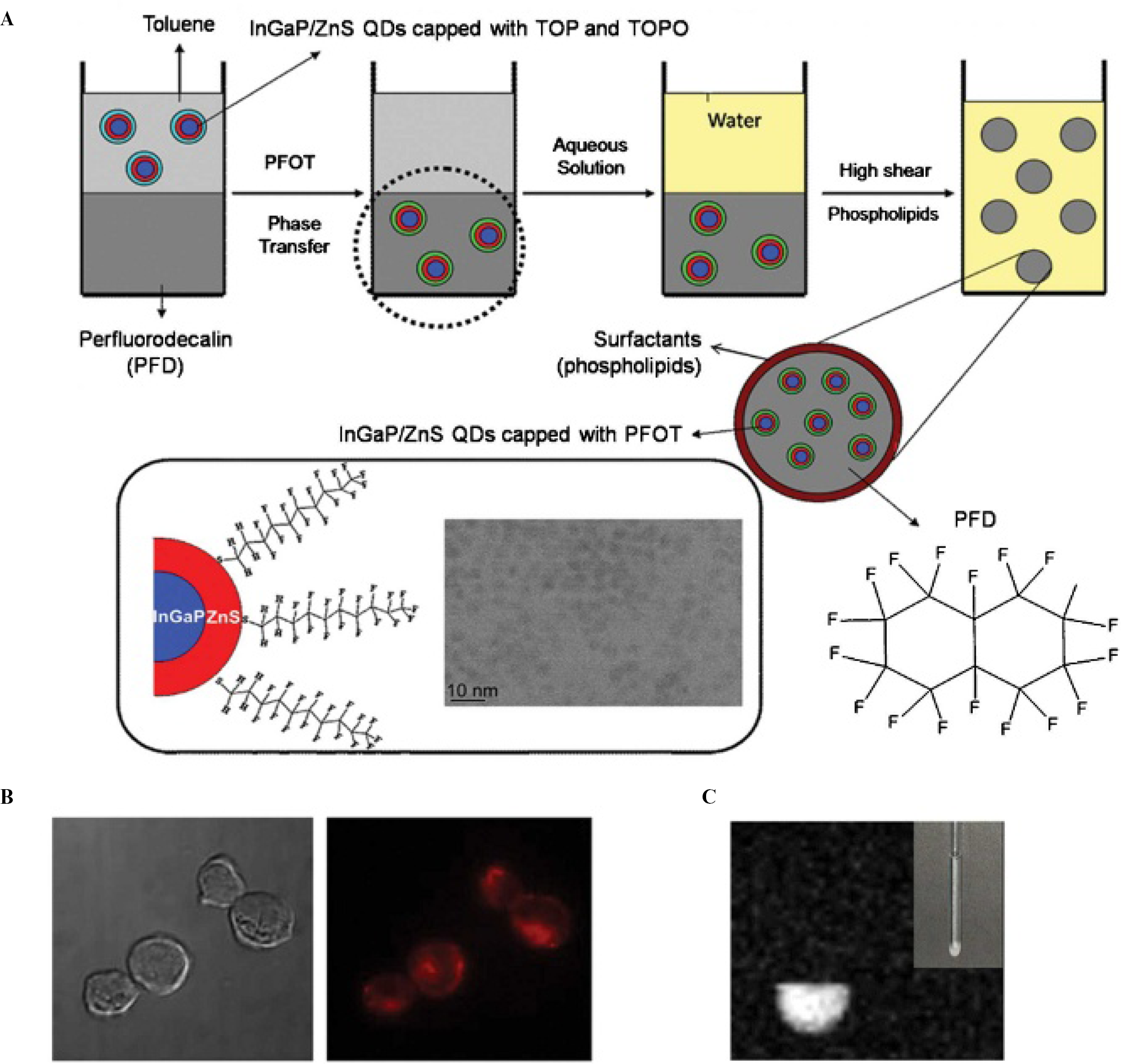
QDs have also been studied as MR and optical imaging contrast agents when combined with perfluorodecalin (PFD) synthesized into nanoemulsions. 45 InGaP/ZnS QDs dispersed in PFD (PDF/[InGaP/ZnS QDs]) were synthesized as nanoemulsions that contain 19F molecules, which serve as 19F-based MRI contrast agents. 45 This bimodal imaging agent can be delivered intracellularly into phagocytic and nonphagocytic immune cells without the use of transfecting agents and without affecting cell viability. By labeling such immune cells, which are involved in the early defense and elimination of tumor cells, immunotherapeutic strategies for preclinical or clinical cancer therapies can be applied. The important feature of these particles involves the exchange of the hydrophobic surface ligands of QDs with perfluorocarbons for proper mixing into the hydrophobic and lipophobic PFD to form nanoemulsions by phospholipid encapsulation. Figure 4A represents the synthesis process for these bimodal imaging agents. 45 The PDF/[InGaP/ZnS QDs] nanoemulsions exhibited bright red fluorescence (Figure 4B) and a 19F-based MR signal when excited at the resonance frequency doublet peak (Figure 4C). Efficiency of intracellular delivery into macrophages was 91% with localization in the lysosome, whereas the efficiency for nonphagocytic cells was about 38%. No significant cytotoxicity was seen at 2% doses of PFD/QD nanoemulsions.
These MR and fluorescence imaging agents incorporate all requirements for a successful bimodal molecular imaging agent with good intracellular delivery, low cyotoxicity, and multifunctional targeted and sensing. Fluorescent core-shell CdSe/ZnS QDs can also be used as photoacoustic and photothermal microscopy contrast agents, as demonstrated by Shaskov and colleagues. 46 The core-shell CdSe/ZnS QDs have an absorption cross section in the visible and NIR range of 10−15 cm2, about an order of magnitude better than other NIR fluorophores. 47 By adjusting the excitation laser pulse width, excited QDs can exhibit thermal energy, which can then be transformed into acoustic energy. Nanosecond pulse laser excitation at ranges between 420 and 900 nm is required to induce photoacoustic and photothermal effects. 46 QD photoacoustic and photothermal effects were not sensitive to scattering and autofluorescence background signals, but no in vitro images were taken. 46 A major problem with using high laser energies is the QD disintegration by core melting and explosion, leaving the potentially toxic semiconductors exposed in vitro and in vivo, as well as toxic bubble formation. Although numerous in vivo experiments have been performed with QDs for targeting and fluorescence imaging,36,48–52 few studies have been reported for bimodal imaging with QDs.

One example of bimodal in vivo work with QDs was shown by Cai and colleagues with the use of 64Cu-labeled DOTA-QD-RGD conjugates in a U87MG tumor mouse model. 53 In this work, CdSe/ZnS QDs were modified with RGD peptides and 64Cu-labeled DOTA for integrin αvβ3-targeted PET and NIRF imaging. Quantitative data by in vivo PET and ex vivo NIRF imaging were collected to determine the tumor targeting efficiency of these nanoparticles by both imaging modes with over 90% correlation, thereby overcoming tissue penetration limitations of optical imaging. About 4% injected dose per gram (% ID/g) was taken up by the tumor after 18 hours postinjection, an amount much higher compared to 1% ID/g by the nontargeted 64Cu-labeled DOTA QDs.
Both QD conjugates were shown to have high liver, spleen, lymph node, and bone marrow uptake. Over 50% ID/g liver uptake was seen for both DOTA-QD and DOTA-QD-RGD for all time points postinjection. A new direction following this study is to investigate the less toxic, non-Cd QDs for bimodal imaging.
Other Inorganic Nanoparticle-Based Multifunctional Probes
As seen by the silica nanoparticle QD doping example in the previous section, silica nanoparticles offer a variety of hybrid molecular imaging probes owing to their biologically inert surface and well-established loadable nanoplatform. 54 Silica particles can be used as shells for optically interesting core materials. For example, silica particles have been used to build luminescent and paramagnetic particles. 55 The core of the silica particle is filled with the luminescent, inner transition metal ruthenium, whereas a paramagnetic Gd monolayer coats the surface of the nanoparticle. The 37 nm diameter particles were synthesized using a well-controlled water-in-oil reverse microemulsion. A ligand-to-metal charge transfer is required for the ruthenium to show luminescence, with an absorption peak around 450 nm and emission at 595 nm. The nanoparticles were taken up by 98% of the monocyte cells with limited cytotoxicity. Laser scanning confocal microscopy was used to visualize the nanoparticles by the Ru luminescence, and T1-weighted images showed significant contrast owing to the Gd doping. T1 relaxivity is 19.7 mM−1s−1. Owing to the versatility of the platform, the multimodal imaging particle can be targeted to image specific diseases, such as rheumatoid arthritis in mice. 55
Owing to their high absorbance and heating abilities, AuNPs have been used for photothermal therapy agents in the form of gold nanoshells, gold nanocages, and gold nanorods.56–58 A 5 nm diameter AuNP has an absorption cross section of 3 nm2 at 514 nm, which is twice as high as for a common organic fluorophore.
59
Owing to this high absorbance, AuNPs exhibit a photothermal effect, where an increase in temperature is exhibited around the particle when illuminated by light. Photothermal imaging depends on the detection of the temperature changes or the effects of that temperature change. Recent advances in photothermal imaging allow for the detection of slight phase changes owing to the increase in heat when the high absorbance particles are excited with laser light energy using a standard optical microscope.
60
Photoacoustic imaging is based on the photoacoustic effect. Just like the photothermal effect, the photoacoustic effect depends on the absorption of energy that is converted to heat. However, the heat energy leads to a thermoelastic expansion and emits as ultrasonic waves.
61
Photoacoustic imaging depends on detecting those ultrasonic emissions by ultrasonic transducers.
61
An advanced design of a fluorescence MRI nanoparticle also exhibits photothermal heating to induce cell death.
62
This type of particle is based on a gold nanoshell, thereby exhibiting photothermal capabilities, coated with a silica epilayer doped with the T2 contrast agent iron oxide and organic fluorophore indocyanine green (ICG).
62
An interesting advantage to the design is a significant fluorescence enhancement (45-fold) owing to the synergistic combination of all components. As proposed by the authors, the increased quantum yield can be attributed to the gold plasmon properties and its enhanced absorption. The particles were functionalized with anti-
Recently, gold was incorporated onto carbon nanotubes for efficient photoacoustic and photothermal signals and demonstrated hundred-time NIR contrast enhancement when used to map the lymphatic vessels in mice. 63 These so-called golden carbon nanotubes (GNTs) incorporate the properties of gold to act as NIR absorption enhancers by depositing a thin layer of gold around the carbon nanotubes. Not only are these GNTs adjustable in length, size, and thickness, they also exhibit minimal cytotoxicity. To study the photoacoustic and photothermal mapping in vivo, GNTs were conjugated with an antibody specific for the lymphatic endothelial hyaluronan receptor 1 (LYVE-1) for lymphatic endothelial cell mapping. Low laser fluence levels can be used to image the endothelial cells with 300 nm spatial resolutions owing to the photothermal method and with high sensitivity in detecting GNT and deep tissue imaging owing to the photoacoustic method. GNTs have also been applied in a unique detection system for circulating tumor cells devised by Galanzha and colleagues. 64 This system involves using two separate nanoparticles. The tumor cells are first targeted by PEGylated magnetic nanoparticles (MNPs) and GNTs, captured noninvasively in the bloodstream under a magnet, and then verified by rapid photoacoustic detection. This unique multiplexed photoacoustic detection uses the unique properties of the nanomaterials not only for imaging but also for in vivo targeting, magnetic enrichment, signal amplification, and multicolor recognition. Both the MNP made up of a Fe2O3 core (commercially obtained) and the GNTs have photoacoustic spectra but at two different wavelengths, 639 nm and 900 nm, respectively. In tumor-bearing mice, this system was able to collect circulating tumor cells by a magnet placed on the skin to collect MNPs and to detect a strong photoacoustic signal coming from the MNPs and GNTs. 64
AuNPs also have unique molecular imaging capabilities for optical 65 and CT imaging, 66 but these individual capabilities have not yet been applied for multimodal imaging. Molecular-level fluorescent signals can be achieved by unique activation strategies that conjugate fluorophores, peptides, and AuNPs. 65 Owing to the surface plasmon resonance of AuNPs, these nanomaterials can quench fluorescence when in close contact with a fluorophore. In one example, 20 nm AuNPs were conjugated with a Cy5.5-labeled peptide substrate selective for matrix metalloproteinase 2 (MMP-2). This probe had strong quenching properties when the MMP-2 substrate was not digested. In the presence of MMPs, the substrate is digested and the dye is released from the AuNP, giving off a strong fluorescence signal. Such activation has been exemplified in vivo in MMP-2-positive tumor-bearing mice. 67 CT contrast has been achieved by AuNPs, allowing for longer imaging time and reduced toxicity over iodine-based compounds. These AuNPs were coated with PEG to increase circulation lifetime. Owing to the higher atomic number and therefore higher X-ray absorption coefficient than iodine, CT contrast was 5.7 times higher than current iodine-based contrast agents. In addition, in vivo results showed clear differences between cardiac ventricles and great vessels. 66 Such unique imaging capabilities of AuNPs can be applied for multimodality molecular imaging.
Single-walled carbon nanotubes (SWCNTs) are organic materials that can be conceptually seen as a single graphene sheet rolled up into a tubular structure with diameters on the nanoscale. Without the presence of any inorganic materials, and when these nanotubes are well dispersed, SWCNTs exhibit NIR photoluminescence within the 700 to 1,300 nm biologic window,68,69 strong Raman scattering, 70 and photoacoustic signals 71 that can be exploited for multimodal imaging with inorganic nanoparticles. Taking advantage of the NIRF emission, O'Connell and colleagues were able to distinguish SWCNTs from biologic systems, 68 and Welsher and colleagues demonstrated the use of SWCNTs stabilized with PEG and targeted to B cells and breast cancer cells as NIRF tags for cell surface probing and imaging. 72 Signature Raman bands for SWCNTs appear at 150 to 300 cm−1, the radial breathing modes, and can be related to the diameter of the nanotubes 73 and at 1,590 to 1,600 cm−1, the G band. 70 Liu and colleagues used Raman spectroscopy to determine the circulation time and biodistribution of SWCNTs functionalized with various lengths of branched and linear PEG by measuring the G-band Raman counts in blood. 74 The photoacoustic signal is also used for detecting and imaging SWNCTs, as seen by De la Zerda and colleagues. 71 Cyclic RGD and PEG conjugated SWCNTs were observed in vivo by the SWCNT intrinsic photoacoustic signal in U87MG tumor-bearing mice. 71 Biodistribution of SWCNT-RGD labeled with 64Cu could also be analyzed in vivo by PET and Raman spectroscopy. 75 Besides the intrinsic properties, Gd doped within SWCNTs can have enhanced effects to the MRI relaxivity. Sitharaman and colleagues loaded ultrashort SWCNTs with nanoscalar, superparamagnetic gadolinium ion (Gd3+) clusters, termed gadonanotubes. 76 These tubes were shown to have 100 times greater MRI T1-weighted contrast than current Gd3+ ion-based clinical contrast agents. This enhancement of paramagnetic relaxation rate of gadonanotubes is due to the nanoscale confinement of Gd3+ ions within the carbon capsule sheath. As these individual cases show, carbon nanotubes have potential as multimodal imaging probes by detecting multiple intrinsic and doped material properties.
Other nanoparticles have been combined with NIR photoluminescence probes to produce multimodal molecular imaging probes. IONPs and NIRF SWCNTs encapsulated within oligonucleotides, d(GT)15, have been used as MRI-NIRF probes. 77 The IONP used to grow the SWNCTs, Fe(CO)5, attaches to one end of the nanotube during the synthesis. In this MRI-NIRF probe, the SWCNTs act as the NIR fluorophore, the IONP on the end of the tube is the T2 MRI contrast agent, and the DNA that wraps around the nanotube acts as a stabilizing agent to prevent aggregation and improve biodistribution, as seen in the schematic of Figure 5.68,69 After SWCNT synthesis, magnetic separation is necessary to select for the IONP-bound SWCNTs. The attachment of the IONP to the SWNT does not reduce the NIR emission of the SWCNTs compared to Fe-enriched and Fe-depleted SWCNTs. Although this nanoparticle was not compared alongside commonly used MRI contrast agents, the combined functionality of SWCNT high-absorbance properties and NIRF emission and the IONP magnetic susceptibility show promise as a new multimodal biomedical imaging agent. 77 These particles were incubated with murine macrophage cells to study the capability of in vitro MR and NIR imaging. MR (see Figure 5B) and NIR (see Figure 5C) imaging were successful with the MRI contrast dependent on the IONP concentration, whereas the NIR emission within the cells was due to SWCNTs present, as verified by the SWCNT signature Raman peaks. No future works have been reported, but with the use of SWCNTs, many targeting groups can be attached for molecular imaging, along with the potential use in therapeutics for tumor ablation by hyperthermia.
SPECT-CT and MRI can provide high-sensitivity and high-resolution imaging if proper contrast agents are used. Lijowski and colleagues developed a targeted nanoparticle that can be detected by sensitive SPECT-CT along with high-resolution MRI. 78 The particle is targeted to integrin αvβ3 to characterize tumor angiogenesis with three-dimensional mapping capability. The core of the nanoparticle is radioactive technetium 99m (99mTc) with functional groups for targeting, and the outer phospholipid layer is made of Gd chelates for MRI. The synthesis of these nanoparticles is initialized by emulsified perfluorooctylbromide. The emulsification includes a surfactant with the integrin-targeted 99mTc nanoparticles complexed with high-molecular-weight PEG. The surfactant comixture of the nuclear nanoparticles includes a high molar concentration of Gd diethylenetriaminepentaacetic acid-bis-oleate. The final nanoparticles are emulsified, and these particles were intravenously injected into a Vx2 tumor model in male New Zealand white rabbits. With the use of 99mTc αvβ3 nanoparticles, SPECT-CT produced a neovasculature signal from the tumor that was not seen in the nontumor side. In addition, MR molecular imaging allowed for three-dimensional mapping of angiogenesis in the tumor after the 99mTc αvβ3 nanoparticles were conjugated with Gd nanoparticles. With the use of SPECT-CT and MRI, these particles were able to locate neovasculature around the tumor that was asymmetric and patchy. 78

The use of rare-earth oxide nanocrystals has been characterized for bimodal fluorescence and paramagnetic imaging of cancer cells. 79 Y2O3 nanocrystals doped with Eu3+ to show red visible light fluorescence and Gd3+ for paramagnetic functionality as MRI contrast agents were also targeted to folate receptor-positive cancer cells by conjugating the cancer targeting ligand folic acid. Y2O3 nanocrystals had significant fluorescence quantum efficiency of about 60% and five times greater relaxivity compared to clinically used Gd-based contrast agents when codoped with Eu and Gd, respectively. The development of these nanocrystals, although well developed for use in commercial displays and optical communication, was performed in a wet chemical method for biologic uses. The conjugation scheme of the nanocrystals is seen in Figure 6, where the folic acid is first activated for amine reactivity, then the Eu and Gd doped Y2O3 nanocrystals are aminized by polyethylenimine (PEI), and, finally, the amine-reactive folic acid reacts with the PEI nanocrystals to create the folic acid-conjugated nanocrystals. MRI contrast and fluorescence images of the nanocrystal incubated with the folate receptor-positive human nasopharyngeal carcinoma cells are shown in Figure 6. 79 Das and colleagues synthesized Tb-doped and Yb/Er-codoped Gd2O3 ultranarrow nanorods that exhibit fluorescence and paramagnetism, leading to good T1-weighted MRI contrast. 80 The nanorods have diameters at 2.5 nm and lengths at 18.8 nm, which can be attributed to the low cell viability after incubation (about 60%) because larger diameters in the range of 20 to 100 nm have shown more promising biocompatibility and cellular uptake. Although the particles were not used for further in vitro work, fluorescence and MRI signals were possible by the inclusion of rare-earth oxides in a nanorod shape using a single-phase synthesis.

Conclusion
Applications of inorganic nanoparticles continue to advance as more imaging modalities and therapeutic agents are added onto the nanoplatforms. Initial inorganic nanoparticle designs consisted of bare nanoparticles, but as nanotechnology progressed, particles with nuclear-magnetic-fluorescence properties were developed. As advancement continues, biocompatibility, biodistribution, and reproducibility are the main concerns in the development of inorganic nanoparticles. Aggregation and disintegration are the fate of inorganic nanoparticles, and the aggregate or disintegrated nanoparticles can be toxic. Effort is needed to keep their desirable properties intact in physiologic conditions. Not only do these newly developed hybrid nanoparticles need to be nontoxic, but they also must be biocompatible for potential clinical applications. Biocompatibility is defined as the ability of the material to perform biologic function with an appropriate immune response. Further review of the cytotoxicity of all nanomaterials discussed here is found in Lewinski and colleagues. 81 Biocompatibility needs to be understood and studied in a long-term manner before clinical applications are possible. Biodistribution is also an issue, and it is important to understand how these particles are excreted through the body and what organs are being targeted when the particles are delivered. Furthermore, as these nanomaterial imaging agents advance, sophisticated characterization techniques are required to understand if the particle that was intentionally made is still the same particle being injected. Contaminants that may not have affected previous inorganic material synthesis may now play a critical role in the reproducibility of the particle synthesis. Not only do biocompatibility and biodistribution need to be well understood, but the interactions of all the parts used to develop the materials also have to be studied. More importantly, chronic toxicity and metabolism mechanisms of inorganic nanoparticles in the body need to be revealed.
The recent examples of inorganic nanoparticles as multimodal imaging agents demonstrate the major advantages of engineered nanoparticles. Unique nanoparticle coating, biomolecules, fluorescent dyes, and doped metals can be applied to build bimodal or multimodal imaging probes. Synergistic effects can be achieved among the different molecular imaging agents when combined on nanoplatforms. As nanotechnology progresses and new materials are engineered, the development of multimodal molecular imaging probes will mature in the direction of molecular sensing and therapy, giving doctors and patients the understanding needed to diagnose and treat disease.
Footnotes
Acknowledgment
Financial disclosure authors and reviewers: None reported.