
Abstract

Thursday 17:30–19:30
Poster Session I: P01: Advances in Optical Imaging
Abstract ID: 200 Poster board space: 1
Despite the awareness that proteolysis is essential for cancer progression, and that proteases represent potential drug targets, clinical trials for cancer treatment with inhibitors of MMPs have failed. Moreover, a broad and comprehensive strategy to identify potential protease targets has not been employed. We hypothesize that proteases are valid therapeutic/prevention targets in cancer and that imaging of proteolysis and its inhibition will provide a means to confirm this hypothesis. We have developed functional optical imaging techniques to monitor tumor progression and tumor-host interactions based on proteolytic activity, both in vitro and in vivo.
In vitro, we have used a 3-dimensional assay system to study tumorstromal interactions, utilizing confocal microscopy. We have found that both pericellular and intracellular proteolysis occur during tumor invasion. Furthermore, there is significant interaction between tumor and stromal cells. Our results indicate that tumor cells actively recruit stromal cells and that these cells contribute significantly to tumor proteolysis. In addition, our most recent results show that the degree and the ratio of intracellular to pericellular proteolysis may be dependent on the density of the surrounding medium. It appears that in a low-density environment there is less overall proteolysis that is mostly intracellular.
In vivo, we have utilized quenched fluorescent probes that are activated by proteases. We have found that upon injection of these probes into tumor bearing mice, the probes are activated at the tumor site and the resulting fluorescence can be detected thereby revealing the position/size of the tumor in vivo. The probes are not specific to any particular tumor type. However, since there is increased expression of proteases in tumors, the resulting fluorescent products do accumulate at the tumor site. Our preliminary observations suggest that utilization of such probes will provide both a sensitive method for cancer diagnosis and a means to monitor therapeutic efficacy.
Abstract ID: 201 Poster board space: 2
Abstract ID: 202 Poster board space: 3

Abstract ID: 203 Poster board space: 4
Colorectal cancer is one of the leading causes of cancer death worldwide. Enhanced expression of Cathepsin B in colorectal cancer has been associated with advanced tumor stage and the potential for metastasis, highlighting the benefit of this protease as a disease biomarker. The aim of this study was to detect colon cancer in an orthotopic tumor model using a fluorescent protease-activated near-infrared (NIR) probe and multiple imaging modalities, in vivo and ex vivo.
To allow evaluation of lesions in the colon, human colon carcinoma CT-26 cells were implanted orthotopically into the mucosal and submucosal layers of the colons of nude mice, creating lesions of known age and location. Mice were injected intravenously with a Cathepsin-B-activated NIR probe (ProSense™750), imaged 24 hours later with a custom designed 2-channel (white light/NIR fluorescent) mouse colonoscope, and also with Fluorescence Molecular Tomography (FMT), a novel quantitative in vivo 3D imaging system. Colon imaging was corroborated by excision of the colons for both ex vivo conventional planar NIR imaging and assessment of tumor growth by histologic analysis.
We obtained real-time in situ images and tumor fluorescence data with both endoscopy and FMT (N=6) with a high tumor to background ratio and clear differentiation from the colons of normal control animals (TBR=3.43). Ex vivo planar imaging and histology confirmed the presence, localization, and size of tumors. Fluorescence microscopy further revealed a sub-endothelial distribution of NIRF signal within the tumor.
Colorectal cancer can be imaged in vivo non-invasively with protease-activatable NIRF agents via endoscopy and 3D fluorescence tomography, validating the benefits of these new imaging modalities in cancer research.

Abstract ID: 204 Poster board space: 5
The therapeutic potential of human embryonic stem cell (hESC) derivatives will rely on the establishment of protocols that maximize the long-term survival of suitably engrafted cell types in disease-specific models. The ability of hESC-derived skeletal myoblasts (hESC-SM) to survive and integrate into artificially-injured host muscle in vivo was investigated using optical reporter gene strategies and serial bioluminescence imaging (BLI). We utilized a lentiviral-mediated gene delivery system containing a triple reporter construct (TGL) to assess in situ graft viability, localization, and proliferation in mouse muscle injury models. Highly-enriched populations of skeletal myoblasts, directly derived from our published protocols, were transduced ex-vivo using a triple reporter lentiviral construct (TGL), expressing HSV-tk (thymidine kinase), eGFP, and luc (firefly luciferase). No alteration in cellular morphology, phenotype, or proliferative capacity was observed.
Lentiviral-transduced (luc+) hESC-SM (500,000 luc+ cells) were implanted into locally damaged right tibialis anterioris (TA) muscles of 8 week old SCID/Beige mice (n=6) using cardiotoxin, in order to assess whether human cells could survive and participate in muscle regeneration. Non-transduced (luc-) cells were administered into damaged left TA muscles as a control. Serial BLI was repetitively performed via retro-orbital injections of D-luciferin for up to 6 months. All animals demonstrated robust long-term survival of luc+ hESC derived myoblasts in the host TA muscle, with initial declines in graft signal (first post-transplantation week), followed by relative stabilization of the signal over the remainder of the study. Prior to histological analysis, ex-vivo BLI on luc+ and luc-engrafted TA muscles demonstrated luciferase expression in the right TA muscles, while no signal was detected in left TA muscles. Immunohistochemistry performed on frozen muscle sections demonstrated the presence of human-specific and skeletal muscle markers, confirmed further by RT-PCR analysis using human-specific skeletal muscle mRNA transcripts, and evidence of successful graft integration.
Abstract ID: 205 Poster board space: 6
The development and application of photonics-based molecular imaging modality, such as bioluminescence tomography (BLT) and fluorescence molecular imaging (FMT), depend on improvement of data acquisition techniques and appearance of novel reconstruction algorithms. In this work, the non-contact measurements method is adopted, which can remarkably improve the spatial sampling of the measured signal and facilitate the experimental equipment and operation. In the aspect of the reconstruction algorithms, an adaptive finite element based reconstruction algorithm is proposed, which can accurately localize the bioluminescent source and significantly decrease the computational cost with the automatically fromcoarse-to-fine mesh evolution. In addition, the permissible source region strategy is used to approximately infer the source position through the surface measured signal distribution and the anatomical information of the phantom, and establish a direct linear relationship between the measured data and the unknown source variable, which further reduces the ill-posedness of the BLT problem.
Figure 1 (A) shows the non-contact based data acquisition schematic illustration. The multi-view detection realized with the rotatable and movable stage and the single nitrogen-cooled CCD camera and the subtle design of camera calibration effectively improve the signal-to-noise ratio of the measured data. Figure 1 (B) depicts the initial finite-element mesh, which is generated by the microCT based images of the physical phantom. The optical parameters of each component are obtained from optical tomography techniques. Figure 2 illustrates the final reconstruction results through four mesh refinements. The preferable source reconstruction shows the availability and effectiveness of the adaptive finite element based reconstruction algorithm with non-contact measurements.

(A) Schematic illustration of data acquisition. (B) The initial mesh of physical phantom.

Reconstruction results
Abstract ID: 206 Poster board space: 7
This is the first demonstration of real-time fluorescent protein based optical imaging of metastatic progression at wavelengths above 600 nm in the Xenogen IVIS imager. Mammary fat pad xenografts of MDA-MB-231 cells that stably express an orange-red fluorescence protein, tdTomato1, defined the invasive breast cancer model. At two weeks post tumor cell injection just palpable tumor burdens were detected at the sites of injection (0.25 sec exposure). By 8 weeks a metastatic lesion at a contra-lateral mammary fat pad became visible (1 sec exposure) despite a small primary tumor burden (82 mm3). Metastases at axilliary lymph nodes were detected at 13 weeks. At 15 weeks large areas of bright fluorescence within the rib cage were resolved as clusters of smaller fluorescent masses (Figure). The nodular composition of these metastases was confirmed at necropsy. Using 3-D reconstruction it was estimated that fluorescence could be detected at 1 cm below the surface. Fluorescent microscopy facilitated the post-mortem evaluation of lung micro-metastases by eliminating the need to score for specific tumor markers. This study demonstrates the utility of non-invasive optical tracking of cancer cells during metastatic progression with endogenously expressed fluorescence proteins as probes.
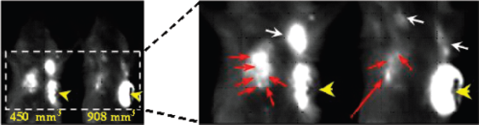
Fluorescent image (2.5 sec exposure) at 15 weeks post tumor cell injections. Caliper measured tumor volumes are given in the bottom left panel. Enlargement of the region of interest shows that the pleural/lung region fluorescence has been resolved as clusters of smaller fluorescent masses (short red arrows). A contra-lateral mammary fat pad metastasis (long red arrow), axilliary lymph node metastases (white arrows), and necrotic portions of the tumors (yellow arrow heads) are visible. 1. Shaner, N.C. et al. (2004) Nat. Biotechnol. 22, 1567-1572.
Abstract ID: 207 Poster board space: 8
Novel technologies are required for three-dimensional cell biology and biophysics. By three-dimensional we refer to experimental conditions that avoid hard and flat surfaces and favour unconstrained sample dynamics [1]. Light-sheet-based microscopes are particularly well suited for studies of sensitive three-dimensional biological systems. Their application can be illustrated with examples from the biophysics and three-dimensional cell cultures. Three-dimensional approaches reveal new aspects of a system and enable experiments in a more physiological, clinically more relevant context. A new implementation of the theta principle [2] takes advantage of parallel recording. This high-resolution light microscope [3] is designed to generate images of large samples (embryos, three-dimensional cell cultures) down to the sub-cellular level. The fundamental principle of EMBL's SPIM is the detection of fluorescence light perpendicular to the illumination axis. The illumination system provides the excitation light from the side of an object and hence excites fluorophores within a single plane. The illuminating light sheet overlaps with the focal plane of a regular fluorescence microscope. SPIMaging provides optical sectioning directly. Photo bleaching outside the thin volume of interest is completely avoided and photo toxicity is thus dramatically reduced. Millimetre-sized specimens can be observed in their entirety and as a function of time since a SPIM performs well with long working distance lenses. The specimen can be rotated to further increase the resolution and the information content of the data. Stacks recorded along different angles are combined in post-processing steps to yield high-resolution images of complete specimens [4]. The 3D resolution is then dominated by the lateral resolution and becomes isotropic. Since the SPIM provides an excellent signal to noise ratio image image processing procedures such as deconvolution work extremely well.
Abstract ID: 208 Poster board space: 9
We describe a novel design for a multi-view and multi-spectral bioluminescence tomography (BLT) system. The new BLT system acquires multiple bioluminescent views around a mouse in a number of spectral channels simultaneously. The multi-view feature is enabled by a multi-mirror signal collection module. The multi-spectral characteristic is implemented with a unique mouse holder or a dedicated cylindrical filter. The signal collection module consists of a mounting plate and a group of mirror stages which are right triangular blocks attached to the mounting plate such that the hypotenuse surfaces of the blocks all make 45° to the plate surface, and are symmetrically arranged. The semi-transparent cylindrical/polygonal mouse holder keeps the mouse at the center of the mirror array to create four bioluminescent views on the CCD camera. Rainbow band filters are on the side surface of the mouse holder for acquisition of spectrally resolved data. Computer simulation was performed to demonstrate the feasibility of this system. It has been shown in our study that bioluminescent signals collected using our system can produce a similar BLT reconstruction quality while greatly reducing the data acquisition time, as compared to a sequential BLT system. With the same data acquisition time, our system would give better signals, and may detect weaker bioluminescent sources. A prototype system was also built. Phantom experiments were conducted using the new system. Preliminary results are very promising.
Abstract ID: 209 Poster board space: 10
Fluorescence surgical resection has recently been shown to significantly improve survival time, as compared to conventional white-light guided surgical resection. Demonstrating its ability to enhance progression free survival and increase the number of full malignant glioma resections. The imaging approach adopted in this type of guidance uses the fluorescence provided by preoperative intravenous injection of patients with a prodrug, 5-aminole-vulinic acid, that the body biochemically synthesizes into Protoporphyrin IX (Pp-IX). The technology used for guidance in these studies simply maps the surface, and does not take advantage of the sub-surface light propagation to allow tomography. In this study, a fluorescence diffuse optical tomography (FDOT) system capable of producing B-scan-type images of localized fluorescence regions is demonstrated. The B-Scan mode is analogous to ultrasound where the excitation and remission signals are delivered from the same surface of the tissue, thereby providing depth information about the fluorescence. The system utilizes a 635 nm diode laser scanned with orthogonal galvanometers to raster the source across the tissue surface. Diffuse projections of the fluorescence intensity are detected by a filtered cooled video rate CCD camera. Excitation and remission intensity data are acquired for each source position and used as input data in a finite-element-based approach to reconstruction of where the emission came from. Experimental studies confirmed that the tomography-based system was more sensitive to smaller targets and lower contrasts as compared to a surface imaging system. The tomography approach could accurately recover the centroid location of fluorescent targets up to 15mm below the tissue surface. For these reasons, neurosurgical tumor resection under FDOT guidance has the potential to further improve the number of full resections, as a FDOT based system is capable of localizing residual tumors undetectable by broadbeam imaging.
Abstract ID: 210 Poster board space: 11
We have previously developed dual-color fluorescence imaging models of rodent and human tumor-host interaction based on a red fluorescent protein (RFP)-expressing tumor growing in either immunocompetent or immunodeficient green fluorescent protein (GFP)-expressing transgenic mice. This model system enabled visualization of the tumor-stroma interaction including tumor angiogenesis in fresh tissue by dual-color fluorescence [1,2]. In order to image tumor-host interaction and drug response in live animals, we have recently developed in vivo three-dimensional 3-color differential whole-body cellular imaging of tumor-stroma interaction using GFP-expressing mice, dual-color tumor cells labeled with GFP in the nucleus and RFP in the cytoplasm and the IV100 Laser Scanning Microscope (Olympus). This new model system images tumor-host interaction and in vivo cellular dynamics at the subcellular level in the intact live animal in real time with ultra-thin objectives (“stick objectives”) and scanning to a depth of 200 μm. Various in vivo phenomena of tumor-host interaction and cellular dynamics were imaged including mitotic and apoptotic tumor cells, tumor vasculature and blood flow. Whole-body subcellular imaging has shown that MMT mouse mammary cells with GFP in the nucleus and RFP in the cytoplasm lost their spindle-shape and showed morphological changes 24 hrs after i.v. injection of 10 mg/kg of doxorubicin. Many of the tumor cells lost their cytoplasm. In contrast, no dramatic morphological changes were observed in stromal cells with the exception of the tumor vasculature. This new model system enables the first true image of a live tumor at the subcellular level and demonstrates the highly complex juxtaposition of tumor and stromal cells. The model also offers a real-time imageable system of tumorstroma drug response at the subcellular level.
Abstract ID: 211 Poster board space: 12
Abstract ID: 212 Poster board space: 13
Bioluminescence imaging (BLI) of luciferase reporters in small animal models offers an attractive approach to monitor regulation of gene expression, signal transduction, and protein-protein interactions as well as following tumor progression, cell engraftment, infectious pathogens and target-specific drug action. Conventional BLI can be repeated within the same animal after bolus re-injections of a bioluminescent substrate. However, intervals between image acquisitions are governed by substrate pharmacokinetics and excretion, therefore restricting temporal resolution of re-injection protocols to the order of hours, limiting analyses of processes in vivo with short time constants. To eliminate these constraints, we examined use of implanted micro-osmotic pumps for continuous, long-term delivery of bioluminescent substrates. Pump-assisted D-luciferin delivery enabled BLI for ≥7 days from a variety of luciferase reporters. Pumps allowed direct repetitive imaging at < 5 minute intervals of the pharmacodynamics of proteasome- and IKK-inhibiting drugs in mice bearing tumors stably expressing ubiquitin-firefly luciferase or IκBα-firefly luciferase fusion reporters. Circadian oscillations in olfactory bulbs of transgenic rats expressing firefly luciferase under the control of the period1 promoter also were temporally resolved over the course of several days. We conclude that implanted pumps provide reliable, prolonged substrate delivery for high temporal resolution BLI, traversing complications of repetitive substrate injections.
Abstract ID: 213 Poster board space: 14
The overall goal of computational optical biopsy (COB) is to estimate key features of a bioluminescent source in vivo. Our recently developed COB system consists of an optical fiber based needle, a single photon counting module-16 (SPCM-16), an integrating sphere, a PC with in-house software, and a position tracking system. Bioluminescent data collected using the needle along one or multiple trajectories are processed for estimation of the bioluminescent source inside a living small animal. Because the COB needle is sensing a target locally and the mean free path is of submillimeter order, light migration can be characterized by the diffusion approximation in an infinite homogeneous medium. To evaluate the COB system, the prototype was applied to liposyn-10% liquid and an agar breast phantom with known optical properties. In our experiments, low energy light (1nw) was delivered by the integrating sphere into the specimen via the source fiber with a highly scattering sphere (2mm in diameter) on the top of the fiber tip. The small sphere helps form isotropic output from the source fiber to simulate a bioluminescent source. The optical signal was then collected using the COB needle. The distance between the tips of the source fiber and the needle was recorded with the position tracking system. The signal sensed by the needle was measured using the SPCM-16. Therefore, the location and power of the light source are estimated based on the diffusion approximation.
The errors of the proposed COB system were < 1.5mm in source localization, and < 30% in energy estimation in the phantom studies. Mouse studies also yielded promising results. The proposed COB system is instrumental to localize and quantify weak or deep light sources in vivo and is enabling techniques for optical molecular imaging and sensing, which is valuable for animal and patient studies in general.
Abstract ID: 214 Poster board space: 15
Fluorescence Molecular Tomography (FMT) is becoming a promising way for in-vivo non-invasive molecular-based imaging. However, limited surface detected data, as well as strong diffusion of near infrared light, make reconstruction of inside three-dimensional (3D) fluorescent yield distribution a typical underdetermine and ill-posed problem. Many reconstruction approaches to it utilize iterative methods for data inversion and they are always time-consuming.
In this paper we propose a fast pre-iteration reconstruction method for FMT, which pushes the iteration process off-line. Firstly the coupled diffusion equations are discretized by finite element analysis and a direct linear relationship between boundary measurements and inside unknown fluorescent yield is obtained. In order to solve the linear matrix equation, the fast algorithm divides the inversion into two steps of off-line pre-iteration process and on-line fast reconstruction. In pre-iteration step for the approximation of generalized inverse matrix a 2-order iteration expression is employed to exponentially accelerate the convergence rate. The on-line step consists of a single matrix vector multiplication and a post-processing of few steps Landweber iteration with range constraint of fluorescent yield value. Numerical experiments with a cylinder phantom of 25mm-diameter and 27mm-height are done to demonstrate the method, which contains 5125 unknowns and 980 measurements on the side surface. Fig. 1 shows the imaging geometry and the detailed computing time for linear equation obtaining, pre-iteration and on-line reconstruction. Results of computerized simulations (Fig. 2) and experiments using a self-made image system with Cy5.5 as probes prove that the method is efficient in estimating distributions and quantities of fluorescent yield.

Abstract ID: 215 Poster board space: 16
We analyze the successes and failures of translational research in bringing advanced optical imaging to the operating room, and present new results from our research aimed at establishing an improved path for these endeavors. Our approach is based on multimode optical imaging (combining spectral, lifetime, nonlinear and coherence-based methods), is designed not to necessitate contrast agents, and is optimized for an endoscopic implementation for minimally invasive surgery. This brings us closer to in vivo detection of molecular signatures and cellular-level abnormalities, and to the optimization and spatio-temporal guiding of surgical intervention by this type of molecular imaging.
We present examples of early cancer detection by spectral reflectance, autofluorescence and elastic scattering imaging, new ways of achieving optical coherence tomography and Raman imaging, and a complete description of an advanced molecular imaging endoscopic system and its uses. The design and implementation of a new “operating room of the future” setting for introducing these tools into surgical practice are also presented.
Abstract ID: 216 Poster board space: 17
Imaging of biological molecules provides cell and molecular biology with a powerful tool. The fluorescent probe has been widely used as a reporter of expression or to make biosensors. Using this technology and confocal microscopy, it is one of major challenges to spatiotemporally observe proteins tagged with the fluorescent probe in a living cell of a living animal. The technology once established will revolutionize the method of understanding in cell and molecular biology and will also contribute to molecular medicine and pharmacology. A technical difficulty of fluorescent confocal microscopy for in-vivo molecular imaging lies in that the microscopy is highly sensitive to change of relative distance and orientation, which naturally and necessary occurs for cells of living animals. One of the authors had developed visual stabilization technology for cardio-vascular surgery using robotic systems, which virtually reduces the motion of heart beat in endoscopic view and ease doctors' manipulation of surgical devices in beating-heart surgery. This paper describes our initial reports on the development of visual stabilization technology for molecular imaging. Our development consists of the high speed imaging system and the motion stage system. The high speed imaging system adopted a camera that captures approximately 1000 flames per second and measures the motion of targets. The motion stage has two degrees of freedom driven by piezoelectric actuators. The motion stage is controlled using target measurements to counteract the cells' natural motion. Cells of mice move due to breathing and more dominantly to heart-beat. The motion of illuminated small glass beads on the liver surface of living mouse was greatly reduced in the view of microscope. The experimental results clearly show that the visual stabilization is effective in cell imaging for living small animals and promising for fluorescent confocal microscopy for in-vivo molecular imaging.
Abstract ID: 217 Poster board space: 18
MRI imaging can be enhanced with inclusion of diffuse optical tomography to quantify hemoglobin levels, oxygen saturation, water fraction, lipid fraction and scatterer size estimation. This type of imaging is used to quantify the values in normal and diseased breast tissues in vivo, in a clinical trial to determine if this can be used to increase the specificity of Gd-MRI imaging of tumors. While a few different versions of this system have been tested, the current version includes fibers which encircle the breast and take data through a cross sectional plane. The regions are tagged from the T1 imaging, and spectroscopic reconstruction of these regions is completed. The accuracy in quantifying regions with broadband multispectral data is directly related to the number of wavelengths used in the fitting, and the ability to quantify phase as well as amplitude of the data transmitted. Adipose regions typically can be segmented out separately and quantified as a single region, whereas fibroglandular regions have similar T1 values to tumor and need to be quantified with an L-matrix type of regularization, thereby allowing the region some freedom to have internal heterogeneities, which could be tumors. The ability to eliminate false positive regions which enhance under MRI, but are not truly regions with a hemoglobin signature is significantly improved with large numbers of wavelengths included in the imaging system.
Abstract ID: 218 Poster board space: 19
A wavelength is light of 700–1500 nm, and in particular, in wavelength area of 700–900 nm, that absorption is low is known for aqua, fat, haemoglobin, and, as for the Near-Infrared, this wavelength area is called with NIR (Near-Infrared) Window by showing high organism tissue permeability. We developed the bioimaging system which used Near-Infrared fluorescence and reported it about imaging of coronary vessel, myocardium, brown adipose tissue, lymph node. The high efficiency CCD camera which has high quantum yield to Near-Infrared in order to catch weak Near-Infrared fluorescence is necessary for this system. We used ORCA-ER made in Hamamatsu Photonics Company by the previous system. ORCA-ER is high efficiency, however, it is heavy (about 25lb) and is expensive. In addition, the handling is hard because the cable is bold. It is lacking in mobility by clinical on-site use. We obtained ROLERA-XR made in Q Imaging which it had high quantum efficiency in a Near-Infrared area and could control with IEEE1394. We will present verification of our bioimaging system using ROLERA-XR.
Abstract ID: 219 Poster board space: 20
Up to now, in vivo molecular imaging has been limited to anaesthetized or restrained animals and, although it is needed for behavioral studies and dynamic signal transduction visualization, it has not taken motion into account.
Recently the use of new Ca2+ sensitive Bioluminescence Resonance Energy Transfer probes genetically encoded into transgenic animals has been demonstrated [1]. The millisecond time scale signaling pathways require high time resolution imaging.
Here we present an add-on to a photon counting system (Photon Imager) for bioluminescence, allowing to record simultaneously with a 40 ms time resolution the desired biological signal along with the video image of the animal. We use a near-infrared lighting and a beamsplitter to separate the bioluminescent signal from the one generated by the lighting. The video image is recorded by an additional CCD camera temporally synchronized with the intensified CCD camera dedicated to the bioluminescence.
We monitored local calcium transients in freely moving transgenic mice expressing the GFP-aequorin reporter protein and were able to correlate the bioluminescence signal taking place when calcium bursts occur to muscle contraction. The video movie and the bioluminescent sequence were co-registered at a video rate and a convenient exposure time was chosen after the acquisition to reveal even very low signals. Further biological validations and quantitative measurements are in progress to characterize and upgrade the system. Registration algorithms dedicated to long exposure time movies are developed to improve the spatial localization and the quantification of the signal.
The Video Imager system developed by Biospace Mesures enables bioluminescent signals in freely moving animals to be detected and spatially localized in the body. For the first time an in vivo molecular modality can perform non-invasive real time imaging in un-restrained or un-anaesthetized animals. This will greatly facilitate applications of bioluminescence in truly physiological conditions.
Abstract ID: 220 Poster board space: 21
The aim of the presentation is to provide technical specifications of a clinically relevant fluorescence endoscope designed to image at Cy5 wavelengths with nano molar concentrations of contrast agent.
Several clinical fluorescence endoscopes are commercially available for applications imaging native tissue auto fluorescence, and imaging with fluorescent contrast agents. Imaging is usually performed with blue/green excitation wavelengths (around 4-500nm) where auto fluorescence is relatively strong. There are several mechanisms that may be employed to enable optical molecular imaging with a contrast agent. Targeting molecules that are upregulated on the cell surface with a fluorescent contrast agent is an attractive approach, but suffers an inherent limitation to the concentration of contrast agent available at the imaging time point. An order of magnitude estimate suggests contrast agent concentrations in the region of nano moles / Kg tissue. The consequence for imaging is a fundamental limit on fluorescence light levels from the contrast agent (which should be significantly greater than native fluorescence for image formation without native fluorescence subtraction). Endoscopy is a real time imaging modality and real time imaging at video frame rates is considered mandatory for several applications. Funovics et al., (2004) have published details of a fluorescence mouse colonoscope system for Cy5.5 based imaging, but performance is inadequate for nano molar concentrations. We have constructed a system optimised for sensitivity by tailoring light delivery, collection, filtering and detection, in order to address the fundamental technical performance limits for endoscopic applications.
It is demonstrated through imaging system calibration, phantom based measurement and animal imaging data that low nano molar concentrations of Cy5 based fluorescent contrast agent in millimetre sized superficial lesions are adequately imaged with a clinically relevant endoscope system in real time. It is concluded that targeting is a technically viable approach for endoscopy applications.
Abstract ID: 221 Poster board space: 22
A new MR-coupled NIR diffuse optical tomography (DOT) imaging system based on parallel spectrometer detection is presented to image fluorescence and absorption contrast in deep tissue. The system is designed to compare imaging limits of MR-guided spectrally constrained absorption imaging of exogenous contrast, MR-guided fluorescence yield imaging, as well as standard clinical MR contrast agent imaging. A model-based technique relying on the diffuse nature of NIR photon propagation in tissue provides the foundation for recovering images of tissue chromophore concentration, scattering parameters, exogenous contrast concentration, and fluorescence yield of an administered fluorescing agent. The algorithm is guided by spatial information from MR images acquired simultaneously with the NIR measurements. A detection system of 16 spectrometers coupled to cooled CCD cameras provides spectral information of light emitted from the tissue. In absorption imaging mode, the absorption spectra from a white light source is combined with frequency domain PMT detection at selected laser wavelengths to recover chromophore concentration and scattering parameters directly using spectrally constrained reconstruction techniques. In fluorescence imaging mode, interference filtering, spectroscopic wavelength separation, and spectral fitting techniques are used to de-couple the fluorescence emission signal from excitation cross-talk.
MR images of breast tissue were used to generate realistic test domains for image reconstructions of Lutetium Texaphyrin, which provides strong absorption contrast at 733nm and a measurable fluorescence signal. It is shown that the background heterogeneity of chromophore concentration and scattering parameters has a dramatic impact on the ability to recover the concentration of LuTex, whether in absorption or fluorescence imaging mode. It is further demonstrated that spatial information from MR is critical to reconstructing accurate images of exogenous absorption contrast and fluorescence yield. The NIR image contrast of LuTex is compared to that obtained in MR contrast imaging using Gadolinium Texaphyrin, a contrast drug with similar pharmacokinetics to LuTex.
Abstract ID: 222 Poster board space: 23
Philips recently realized a novel imaging system for fluorescence optical mammography (Mammoscope) that can be used in combination with Schering AG's Omocianine dye [1]. Here, we report on a phantom study that demonstrates the feasibility of diffuse optical tomography of the female breast with fluorescent contrast agents for imaging of breast cancer.
The Mammoscope is a bed with a cup containing a coupling liquid in which the breast can be suspended. The breast is illuminated sequentially from 255 illumination positions and for every illumination position the fluorescent light distribution in the cup is measured in parallel at 255 detection positions [2]. Omocianine is a near-infrared fluorescent dye with favorable absorption and emission properties for in-vivo use in humans, including an up to five times higher quantum yield than ICG. Furthermore, the blood lifetime of Omocianine is much longer, favoring dye uptake in tumors [3].
In this study we have used a phantom that consists of a breast shaped shell and a hollow lesion that are both filled with a fluid with optical properties mimicking breast tissue and an appropriate amount of dye.
The results show that lesions with clinically relevant dye concentrations in the nM range can be detected. Based on these results we are currently preparing a clinical trial.
Abstract ID: 223 Poster board space: 24
The ability to image carcinogenesis in the lung using fluorescence opens exciting opportunities for facilitating longitudinal and multi-spectral observations of studying the disease evolution, molecular pathways and treatment effects. However planar (photographic) imaging approaches are generally limited in their ability to resolve in depth and quantify fluorescent signals non-invasively and in-vivo. In this work we developed and validated a 4th generation Fluorescence Molecular Tomography system for obtaining three-dimensional and quantitative information of lung-cancer related signatures in-vivo in small animals. The system utilizes free-space beam steering methods for scanning two laser beams at 665nm and 748nm onto the object imaged using a galvanometer-based set of mirrors. Photons propagating through tissue at the excitation and emission wavelengths are collected using a cooled CCD camera and appropriate band-pass interference filters. Tomographic reconstruction of fluorochrome distributions is based on the normalized Born approximation [1], adapted to 360°-projection geometries, or more advanced inversions schemes based on multimodality image priors as investigated by Soubret et al. in another submission to this meeting.
The technique developed is ideally suited for the three-dimensional study of deep-seated activity in animals. Herein we present results from mice injected in the lung with 1times106 LLC cells. Seven to ten days post tumor implantation the mice were injected with 2 nmols of Angioscence680 and Prosence750 (Visen Medical) and imaged 24h later. Preliminary results show well resolved areas of increased fluorescence concentration, that is congruent with the location of tumor implantation, confirmed also with non-invasive X-ray CT. We further demonstrate that planar imaging or 360° tomography is unable to resolve this activity. We conclude that FMT is a tool necessary for the accurate study of deep-seated activity.
Abstract ID: 224 Poster board space: 25
Targeted fluorescence optical imaging is an attractive method of detecting cancer because of its high sensitivity and specificity, its low cost, and the absence of ionizing radiation. However, fluorescent molecules are inherently limited by low target to background ratios because conventional fluorophores are “always on” regardless of whether they are bound to the target or not. To overcome this limitation, a target-specific activation strategy, in which the fluorophore is activated only in the targeted cells and not in background tissues, has been proposed. An example of the activatable fluorophore is a peptide backbone conjugated with self-quenched fluorophores, which can be digested and de-quenched by sequence-specific proteases. In this study, an alternate fluorescence activation strategy is proposed whereby self-quenched avidin-rhodamineX (Av-3ROX), which has affinity for lectins on cancer cell surface, is activated by degradation after endocytosis. Using the Av-3ROX, in vivo target-specific spectral fluorescence imaging of disseminated cancer microfoci was obtained in a mouse model of peritoneal ovarian cancer metastases with minimal contamination by background fluorescence signal. When dissociation of Av-3ROX was inhibited by crosslinking (CL) with a disuccinimidyl suberate, which covalently bound the avidin tetramer, non-crosslinked Av-3ROX emitted significantly higher fluorescence than crosslinked Av-3ROX on the gross tumor nodules (Fig. 1) as well as the submillimeter peritoneal implants (Fig. 2). Cellular internalization of receptor-ligand pairs with subsequent activation of fluorescence via “de-quenching” provides a generalizable and high S/N method for detecting cancer microfoci in vivo and has practical implications for assisting surgical and endoscopic procedures.

Abstract ID: 225 Poster board space: 26
Chemical biotinylation of cells can be a rapid method of biotinylating cells for conjugation to anti-biotin or anti-strept(avidin) antibodies. As the antibodies can be amenable to conjugation with contrast agents e.g., superparamagnetic iron oxide nanoparticles (SPIOs), the possibility of imaging such cells by MRI becomes feasible.
Human ovarian cancer cells were chemically biotinylated. Viability and cell death measurements, as assessed by trypan blue exclusion and annexin-V staining respectively, were comparable between biotinylated and mock biotinylated cells. Surface biotinylation was confirmed by incubation with anti-biotin-FITC and detection using flow cytometry. Significant biotinylation persisted 0–4 days after biotinylation but was negligible by day 6 due to the rapid proliferation (36h doubling rate) of this particular cell line. To determine the possibility of labeling biotinylated cells for MRI, cells were incubated with anti-biotin-SPIOs (Ab-SPIOs) or anti-H2K-SPIOs (control-SPIOs) after biotinylation. Following incubation with Ab-SPIOs or control-SPIOs, residual SPIO conjugates were removed and cells suspended in 1% agrarose for MRI. Significant negative enhancement of the anti-biotin-SPIO labeled cells were observed at TE values >10ms and is consistent with the significantly lower T2 values of the labeled cell pellets compared to the non-labeled pellets at 4.7T. To examine the feasibility of visualizing labeled cells in vivo, Ab-SPIO and non-labeled cells were injected into the contralateral legs of mice and MRI performed at 9.4T. Negative enhancement was observed in those regions in which Ab-SPIO labeled cells were injected supporting the in vitro visualization of Ab-SPIO labeling of biotinylated cells by MRI.
In conclusion, chemical biotinylation of cells provides a rapid, nondestructive and generic method of ‘tagging’ cells for imaging MRI in vivo.
Abstract ID: 226 Poster board space: 27
Despite the success of anti-angiogenesis drugs such as bevacizumab, incidences of therapeutic resistance necessitate further explanation, which may include the involvement of the Tie2 receptor and its angiopoietin ligands. While studies have validated the angiogenic function of vascular endothelial growth factor (VEGF) and the angiopoietins, it is unclear how combinations of these growth factors affect tumor susceptibility to anti-angiogenic therapy. Examination of tumors with differential expressions of growth factors would address the interaction between the growth factors, and could elucidate a more robust treatment mechanism. This study uses intravital fluorescent microscopy to perform serial characterization of in vivo tumors for examination of both tumor vasculature development and treatment response.
Wild-type K1735 melanoma tumor cells and clones engineered to over-express VEGF and angiopoietin-1 (Ang1) were implanted in dorsal window chambers of mice, allowing for serial imaging of in vivo tumor vasculature, as delineated by a fluorescent blood pool agent. Analysis showed 2.5-fold increase in vessel density, 37% increase in average diameter, and 17% increase in vessel complexity (fractal dimension) in VEGF-expressing tumors compared to wildtype tumors. In comparison, Ang1-expressing tumors showed 2.6-fold decrease in vessel density, 170% increase in average diameter, and 19% decrease in vessel complexity relative to wildtype. Comparison of immature vessel sprouting in VEGF, Ang1, and wildtype tumors—achieved through use of Tie2-GFP mice—highlighted an increased occurrence in VEGF tumors and a decreased occurrence in Ang1 tumors. To examine treatment response, mice with monoclonal and chimeric tumors were treated with either bevacizumab, anti-Tie2 therapy (using an inducible Tie2Ex construct), or both. Serial imaging of vasculature showed significant microvascular response to treatment, which was correlated positively with a decreased macroscopic tumor growth rate.

Abstract ID: 227 Poster board space: 28
In multi-step pre-targeting applications, avidin has been used as a mediator or a linker between a biotinylated targeting molecule (e.g. antibody) and a biotinylated probe. Our goal is to develop double- and singlestep targeting of metabolically biotinylated (avidin-binding) and avidin-fused (biotin-binding) engineered receptors, which will ultimately facilitate development of single-step targeted MRI of endogenous cell surface receptors.
Biotin- and avidin-binding receptors were produced by fusing an avidin-expressing sequence or a prokaryotic biotinylation epitope (GLN-DIFEAQKIEWHE) respectively, to the extracellular end of the PDGFR transmembrane domain. DsRed was fused to the intracellular end to monitor receptor expression and localization (Red; Figure a-d).
Confocal microscopy of transiently transfected live cells expressing biotin-binding receptor showed specific and selective binding of biotin-fluorescein (Green; 30min, 37°C; (a)) whereas binding was blocked by pre-incubation with free biotin (b). Bound biotinylated molecules colocalized (Yellow) with the DsRed-fused receptor, whereas binding was not detected in cells that do not express the receptor (and show only nuclear stain; Blue). Similar results were obtained with the dual optical-MRI probe, biotin-PAMAMdendrimer(generation 4)-GdDTPA-FAM.
Cells expressing avidin-binding receptor (biotinylated by biotin ligase (BirA; 1h, 37°C)) did not bind the biotinylated probe directly (c) but required an intermediate step mediated by avidin (Green; d).
Interestingly cellular distribution of biotin- and avidin-binding receptors was considerably different, with avidin probably inducing receptor multimerization and reducing the turnover rate of the receptor.
These models will enable assessment of a) tissue accessibility of various size imaging probes, b) binding specificity and c) minimal receptor concentration required for detection by MRI, thereby helping develop in-vivo targeted MRI, directed to endogenous cell surface receptor.

Abstract ID: 228 Poster board space: 29
Development of optical imaging biomarkers of tumor growth has been focused so far mostly on the use of cancer cells with genetically encoded optical probes. The potential need for optical imaging in transgenic animals and eventually in certain clinical applications has stimulated the design and development of new exogenous optical probes. Here we report on the evaluation of a new class of optical probes, termed “smart” probes, ProSense™ and AngioSense™ for the detection of tumor development in xenograft mouse tumor models. In order to determine the correlation between a probe reporting signal as a tumor growth biomarker and tumor growth we have used well characterized tumor models based on rat adenocarcinoma cells MTBIII with the genetically encoded optical probes. The correlations between tumor size determined either by caliper or by one of the anatomical imaging modalities (MRI, CT) and the probe volume distribution or the equivalent bioluminescent signal were measured in several subcutaneous xenograft models. The results using 2D and 3D tomographic systems are compared and limitations of individual instruments are discussed. The potential use of the probes in preclinical testing of cancer therapeutics will be demonstrated. The limitations of individual probes for detection of metastatic lesions were evaluated and will be discussed (Fig. 1). Results serve as a cross-validation between genetically encoded and exogenous optical probes.

Imaging of metastatic lesions developed in nu/nu mouse after tail vein implantation of 1times106 rat adenocarcinoma cells MTBIII-luc.
Abstract ID: 229 Poster board space: 30
We developed a mouse-sized cryomicrotome/imaging system that uniquely provides a large volume of view, high resolution, and multiple contrast mechanisms (color and exogenous/endogenous fluorescence). With this cryo-imaging system, we sectioned cryo-preserved tissues at 2–40 micron thickness and acquired block face images with micron in-plane resolution. Brightfield images of normal and pathological anatomy show exquisite detail, especially in the abdominal cavity (Figure 1). Multi-planar reformatting and 3D rendering allow one to interrogate tissue structures as small as 5 micron blood vessels. Because anatomy is preserved, we can easily register image data and correlate results with in vivo optical, MRI, and radionuclide imaging. We can find and image single fluorescently labeled stem and cancer cells and surrounding tissue structures. To date, we have imaged cancer, stem cells, kidney disease, and multiple transgenic mice. We will demonstrate several cryo-imaging capabilities relevant to small animal cellular and molecular imaging.

Abstract ID: 230 Poster board space: 31

Abstract ID: 231 Poster board space: 32
Green fluorescent protein (GFP) has been used for cell tracking and imaging gene expression in superficial or surgically exposed structures. However, in vivo murine imaging is often limited by several factors, including scatter and attenuation with depth and overlapping autofluorescence (AF). The AF signals have spectral profiles which are markedly different from the GFP emission spectral profile. The use of multi-spectral imaging (MSI) allows separation and quantitation of these contributions to the total fluorescence signal seen in vivo, by weighting known pure component profiles. Separation of relative GFP and AF signals is not readily possible using epifluorescent continuous wave single excitation and emission bandpass imaging (EFI).
Nude mice were subcutaneously injected with a series of 1times103 to 1times105 GFP expressing CT26 cells. Mice were imaged on MSI and EFI systems to compare detection limits. Initially, a pure GFP spectrum was produced for subsequent analysis by spectrally comparing known high GFP areas with adjacent GFP-devoid areas. For EFI imaging, optimized excitation and emission bandpass filters were used. Due to the ability to separate out AF contributions from the emission using MSI compared with the mixed contributions of GFP and AF emission in the single bandpass used to record emission in the EFI system, we achieved a 300 fold improvement in the cellular detection limit. The MSI detection limit was 3times103 cells vs. 1times106 cells for EFI. Integrating multiple frames on the MSI system improved its ability to discriminate low levels of GFP without saturating signal from nearby higher levels of GFP. Finally, MSI was able to separate signal interference of red fluorescent protein from GFP (subcutaneous detection limit of 1times104 GFP in 1times105 RFP; 1times104 RFP in 1times105 GFP).
These findings demonstrate the utility of MSI in detecting low levels of multiple fluorescent markers for whole animal in vivo applications.
Abstract ID: 232 Poster board space: 33
The main objectives of this research are to investigate the combination of reflectance and fluorescence confocal imaging and molecular-specific optical contrast agents (CA) in the detection of epithelial pre-cancers. A near-real time confocal microscope capable of both reflectance and fluorescence imaging of ex vivo human specimens and in vivo animal models was developed and molecular-specific CA targeting the epidermal growth factor receptor (EGFR) were designed. This presentation presents the confocal system and its capabilities, as well as the use of target-specific CA in the study of oral cavity cancer. The dual confocal microscope (DCM) has light sources at 488nm, 664nm, and 784nm, a frame rate of 15 fps, a maximum field of view of 350times250μm, and a resolution limit of 0.40μm laterally and 1.37μm axially. The DCM can image tissue architecture and cellular morphology, as well as molecular properties of tissue using reflective and fluorescent molecular-specific CA. In the hamster cheek pouch model of oral carcinogenesis, the DCM was used to image the epithelium and stroma of the cheek pouch, blood flow was visualized, and areas of dysplasia could be distinguished from normal epithelium using 6% acetic acid contrast. In human oral cavity tissue slices, DCM images show an increase in the nuclear-to-cytoplasm ratio and density of nuclei in neoplastic tissues as compared to normal tissue. After labeling the tissue slices with reflective and fluorescent CA targeting EGFR, an increase in the EGFR expression was detected in the cancerous tissue as compared to the normal tissue. Information obtained with the DCM will provide the knowledge necessary to further develop in vivo imaging tools and contrast agents for the realtime detection of dysplasia and identification of tumor margins, which will improve the early diagnosis and treatment of oral cavity cancer.
Abstract ID: 233 Poster board space: 34
We have developed an optical multichannel NIRF imaging system with a correction algorithm to account for fluorescence signal attenuation by opaque media, such as blood or superficial tissue. We demonstrate that this algorithm will allow for the quantitative in vivo assessment of intravascular or submucosal lesions without the strong tissue depth dependence that affect current epifluorescence imaging systems.
Our fiber optic catheter-based intravital imaging system allows for the simultaneous and real-time high resolution visualization of two near infrared fluorescence (NIRF) channels. For intravascular correction, we first calculated the auto-fluorescence of oxygenated murine blood by taking dark background and blood auto-fluorescence measurements over a range of exposure times. The difference was fit to a linear regression, allowing us to dynamically subtract the auto-fluorescence at any exposure time from subsequent images. Then, aqueous solutions of two fluorescent dyes (AF750 and Cy5.5) were prepared at different ratios of dye concentrations and covered with opaque media. Fluorescence intensity from the dye solution was imaged via a 0.8mm catheter through 0.5mm to 2mm of blood or tissue.
We found that whereas the raw NIRF signal, after accounting for the contributions of blood auto-fluorescence, demonstrated a ± 300% change from the mean over the measured distances, the corrected signal varied only ± 10%. The raw signals had an exponential decrease with distance; the corrected signal was essentially invariant. Moreover, the corrected signal was sensitive to the differences in the ratios of dye concentrations, while the uncorrected signals were not. These data demonstrate the ability to account for the fluorescence signal attenuating effects of blood and discriminate between relative concentration differences in two fluorescent dyes underneath a volume of blood. This result suggests the potential for the quantitative intravital assessment of vascular and intraoperative lesions.
Abstract ID: 234 Poster board space: 35
MicroRNAs (miRNAs) are 21-mer RNAs regulate the expression of genes by binding to the 3′-untranslated regions (3′-UTR) of specific mRNAs. Even though miRNAs are involved in multiple molecular pathways such like cell development and organ development, miRNA biogenesis is not enough to understand full processing of miRNA regulation. In this study, we developed dual luciferase reporter system that monitors functional flow of a specific miRNA, miR-23, during neuronal differentiation.
We constructed miR23P/Fluc (Firefly luciferase) plasmid carrying firefly luciferase under the control of miR-23 promoter to monitor the transcriptional level of miR-23. A 3xPT_mir23/Gluc (Gaussia luciferase) recombinant containing three copies of the target sequence of miR-23 was developed in order to monitor the targeting activity of mature miR-23. A 3xPT_mir23_reverse/Gluc including the reverse target sequence of miR-23 was designed as a negative control, 293 (Human kidney), HeLa (Human cervical adenocarcinoma) and P19 (Mouse embryonal carcinoma) were transfected with 3xPT_mir23/Glu and miR23P/Fluc, respectively. The expression of luciferase gene was measured by luminometer assay. The neural differentiation of P19 was induced by retinoic acid treatment.
The Fluc activity to see miR-23 transcriptional level indicated miR-23 activity in HeLa and P19 was higher than that in 293. The Gluc activity for miR-23 targeting showed significant decrease in HeLa and P19 but no difference in 293 compared with CMV_Gluc activity. There was a corresponsive change of Gluc and Fluc activities to P19 differentiation.
Recent studies of miRNA expression implicate miRNAs act as key regulators of brain development and neurodevelopment. Using both firefly and gaussia luciferase reporter system, we can evaluate the pri-miRNA (primary miRNA) and mature miRNA activity. Furthermore, this system can show the change of miRNA expression during neural differentiation. We expect that this system will be useful to understand the function and biogenesis of miRNA during neuronal differentiation.
Abstract ID: 235 Poster board space: 36
Abstract ID: 236 Poster board space: 37
As a popular numerical approach used in bioluminescent tomography (BT), the Monte Carlo (MC) method has the problem of cumbersome computation. To accelerate the simulation, we proposed a Table-Based Random sampling simulation (TBRS), which is a fast algorithm based on the conventional MC simulation of photon propagation. The key idea of TBRS is to simplify multi-steps of scattering to a single step process, through randomly table querying, thus greatly reduced the computing complexity of the conventional MC algorithm and expedite the computation. Here the theory of TBRS is given with discussions about the influential parameters involved. A linear mathematical model is set to evaluate the effectiveness of the TBRS simulation. To verify the feasibility of the TBRS algorithm, we compared its results with conventional MC method and finite element method (FEM) both in 3-D homogeneous and inhomogeneous mediums. Corresponding phantom experiments were also conducted. Good agreement is found among TBRS simulation, conventional MC simulation, FEM simulation, and the phantom experiments. And the computing time of TBRS decreases significantly compared to the conventional MC simulation. It retains flexibility and accuracy of MC method and adapted well to complex geometric media and various source shapes, but with much better efficiency. Besides, we present a reconstructing approach to estimate the position of the fluorescent source based on the Artificial Neural Network (ANN) theory, as a validation of the TBRS algorithm.

Abstract ID: 237 Poster board space: 38
Angiogenesis is the process of new vessel growth from existing blood vessels and is important in both physiological and pathological conditions. Despite many recent advances in angiogenesis research, little is understood about the molecular mechanisms and mechanics guiding vessel growth into the extracellular matrix. A major hurdle has been the lack of high resolution imaging techniques to measure angiogenesis in a living, three-dimensional model.
Here we present results of micro-vessels interacting with the extracellular matrix using multiphoton imaging. Our model is designed for in situ imaging of micro-vessels sprouting into a collagen matrix. We collect second harmonic generated (SHG) light from collagen fibrils and two photon excited fluorescence (2PEF) from vascular cells to provide endogenous contrast without staining or mechanical sectioning. Additionally, transmitted light in the NIR is collected to visualize the vessels.
Angiogenesis was induced in vessel constructs consisting of rat micro-vessel fragments reconstituted in a type-I collagen gel. Vessel constructs were maintained in a viable environment during imaging with a custom made incubator/imaging chamber.
Images from the vessels exhibited several striking features indicative of interactions with the collagen matrix resulting structural modification and re-modeling. Upon reconstitution with the collagen gel, the micro-vessel fragments attached to and condensed the collagen fibrils. As sprouting vessels migrated from the parent fragment, the leading end of the sprout attached the fibrils, pulling and anchoring to the collagen matrix (Figure, arrows; left: transmitted light, right: SHG + 2PEF), while collagen aligned and attached further down the sprouting vessel toward the parent vessel as the sprout grew over time.
These results indicate a dynamic relationship between growing vessels and collagen that may lead to new insights into angiogenesis.

Abstract ID: 238 Poster board space: 39
Transfection of human HT1080 fibrosarcoma cells with the plasminogen receptor gene in the sense and antisense orientation using retroviral system was performed to investigate the roles of plasmin in the tumor metastasis. The cell surface plasmin activation, plasmin-dependent in vitro transwell invasion, and cell surface plasmin-dependent matrix hydrolysis were studied in the transfected cells. In order to test the initial metastatic potential of the cells, the cells were stained with fluorescent cell tracker and injected to the liver portal vein of the nude mouse. The seeding procedure of the cells in the liver was monitored with intravital microscope. The titanium abdominal wall window was implanted over the liver and sutured between the skin and abdominal wall. The glass overslip was placed on the window and fixed with the snap ring. The initial metastatic potential was monitored until 48 hours. Transfection of human HT1080 cells with the human plasminogen receptor gene in the antisense orientation resulted in a loss of plasminogen receptor from the cell surface and concomitant decreases in cellular plasmin production, extracellular matrix degradation, and cellular invasiveness. Furthermore, the transfected cells demonstrated reduced liver metastasis in mice. In contrast, cells transfected with the plasminogen receptor gene in the sense orientation displayed increased cell surface plasminogen receptor and increases in cellular plasmin production, as well as enhanced extracellular matrix degradation and enhanced cellular invasiveness. The plasminogen receptor overexpressing cells also showed enhanced liver metastasis monitored with intravital microscope. These data establish that changes in the extracellular expression of the plasminogen receptor protein dramatically affect tumor cell-mediated pericellular proteolysis resulting in stimulation of its metastatic potential. In addition, intravital microscope appears a useful tool for longitudinal monitoring of initial liver metastasis.
Abstract ID: 239 Poster board space: 40
Tumor vessel imaging can be useful to identify angiogenic blood vessels as well as being a potential predictive marker of antiangiogenic treatment response. We recently reported the expression of the Neural Cell Adhesion Molecule (NCAM) in immature and tumor endothelial cells (TEC) lining vessels of human carcinomas. Exploiting an in vivo model of human tumor angiogenesis obtained by implantation of TEC in Matrigel in SCID mice, we aimed to image angiogenesis by detecting the expression of NCAM with magnetic resonance imaging. The imaging procedure consisted of 1) targeting NCAMs with a biotinylated derivative of C3d peptide that is known to have high affinity for these epitopes and 2) delivery of a streptavidin/Gd-loaded apoferritin 1:1 adduct at the biotinylated target sites. The remarkable relaxation enhancement ability of Gd-loaded apoferritin system allowed the visualization of TEC both in vitro and in vivo when organized in microvessels connected to the mouse vasculature. Gd-loaded apoferritin displayed a good in vivo stability and tolerability. The herein reported procedure may be easily extended to the magnetic resonance visualization of other epitopes suitably targeted by proper biotinylated vectors.

Abstract ID: 240 Poster board space: 41
We have developed a new technique for the analysis of image cubes that removes contaminating background emission from the emission of an in-vivo fluorescent probe. Because our technique operates on excitation-filtered light, our analysis does not suffer from optical distortions that degrade similar analyses of emission-filtered data cubes. Using Marquardt's method for finding a least squares solution of an over-determined system of non-linear equations, we simultaneously fit the excitation line spectra and auto-fluorescent emission of the data cube. In addition to removing the background emission, our method can easily be extended to identify multiple probes by adjusting the fit to account for the known differences in their excitation spectra. We present examples of the method in practice and discuss its application in the future.
Abstract ID: 241 Poster board space: 42
We have demonstrated simultaneous measurements of self-phase modulation (SPM) and two-photon absorption (TPA) in biological markers using shaped ultrafast laser pulses. Two-photon fluorescence microscopy overcomes many limitations of traditional fluorescence microscopy, most notably by providing increased penetration depth. However, still only molecules that fluoresce can be detected, excluding important endogenous molecular markers like melanin and hemoglobin. But even though these molecules show little two-photon fluorescence, they do exhibit two-photon absorption. In the long-wavelength water window tissue TPA is dominated by these two markers with an almost complete absence of endogenous two-photon fluorescence. Nonlinear phase contrast, such as self-phase modulation, can provide intrinsic signatures that depend on local tissue anisotropy, chemical environment, or other structural properties. TPA and SPM microscopy techniques offer novel contrast mechanisms that may for example aid in the diagnosis and treatment of melanoma and other skin disorders.
Our group has recently demonstrated that TPA permits deep-tissue imaging of melanocytes in human melanomas grown on nude mice [1]. Here we present simultaneous measurement of TPA and SPM with modest light power levels [2]. Cuvette samples of melanin (OD=0.5 at 600 nm) and oxyhemoglobin (2.3 mM) show strong SPM and TPA signals with only 400 μW average power, whereas the glass walls only show SPM. Self-phase modulation signatures of live tissue have also been observed and will be discussed.
Abstract ID: 242 Poster board space: 43
The biodistribution and integrity of nano-formulated contrast agent/drug carrier in vivo are critical in determining the imaging/therapeutic efficacy and the necessity of further design for controlled release. In the present study, the in vivo biodelivery of liposome-encapsulated carboxyfluorescein (CF) was monitored dynamically by home-made small animal optical imaging system and in situ microdialysis probe. The imaging shows that liposome containing 10 mM CF, with the size of 200–240 nm, accumulated in rat liver after the i.v. administration (Figure 1A). To validate the integrity of liposome in liver, the microdialysis probe with total diameter of 250 μm was in situ implanted for simultaneous fluorescence measurement of extracellular fluid. There was no significant increase of fluorescence intensity in extracellular fluid measured using microdialysis, comparing to the otherwise prominent increase of fluorescent intensity monitored by optical imaging (Figure 1B). It indicates that the CF-containing liposome was rather intact as it extravasated from the vasculature to the liver tissue, thus prevented its encapsulated CF from leaking out and collected through the size cut-off membrane of microdialysis probe. The integrity of liposome delivery to liver was also investigated by increasing the liposome-encapsulated CF concentration from 10 mM to 100 mM. No augmentation of fluorescence intensity in liver was measured by optical imaging due to the fluorescence self-quenching, and which manifested the liposome in liver still remained intact. Furthermore, the local delivery of Triton X-100 lysis buffer by microdialysis could result in an immediate increase of CF fluorescence intensity in the microdialysate. We herein demonstrated that the liposome biodelivery could be defined complementally with both optical imaging (macroscopic) and microdialysis probe (microscopic).

Abstract ID: 243 Poster board space: 44
In vivo fluorescence molecular imaging has shown great promise in expediting the drug discovery and development. Efforts have been made in the development of efficient fluorescent probes and quantitative imaging techniques to improve the sensitivity and specificity in tissue. In this work, we present our spectral unmixing and fluorescence tomography techniques for the newly developed imaging device which can acquire both reflectance images and raster-scanning transillumination images on the whole animal with a variety of narrow-band excitation and emission filter options.
A challenging problem is that signals from fluorescent reporters are often contested by tissue autofluorescence and instrument fluorescent background, especially for weak target or deep tissue in vivo applications. A spatially derived background elimination technique is developed to remove the instrument background within the collected images. A spectral unmixing technique based upon the multivariate curve resolution (MCR) algorithm can be used to separate the reporter of interest from tissue autofluorescence as well as other reporters. Structured light images are acquired to generate the animal surface topography which allows the modeling of light transportation through the animal. A simple and robust fitting tool is developed to estimate the depth and strength of the reporter quickly assuming the reporter can be treated approximately as a point source. A more general diffuse tomography reconstruction can provide three-dimensional localization and quantification of the reporter's distribution based upon multi-spectral images of planar reflectance measurements and transillumination measurements at multiple excitation locations.
Experimental results will be presented which validate our techniques. Data were collected on a mouse phantom as well as on real mice by varying the depth and concentration of fluorescent probes such as quantum dots and dyes.
Abstract ID: 244 Poster board space: 45
A wide variety of fluorescent dyes, proteins and nanoparticles are currently available, creating significant interest in the areas of in vivo molecular imaging. These reporters can be detected and monitored non-invasively to study the biological activity inside living animals. The sensitivity of detection of fluorescent probes in vivo is often limited by tissue absorption and tissue autofluorescence. Transillumination and spectral unmixing are two techniques that can help improve detection sensitivity.
In this work, a dual reporter optical imaging system that detects emission from fluorescent and bioluminescent probes is described. This instrument is fitted with ten excitation and eighteen emission filters in order to spectrally scan reporters in the range of 480–850 nm. For fluorescent imaging, the instrument can switch between reflectance and transillumination modes by means of a computer controlled optical switch. In both modes, filtered excitation light from a broadband lamp is used as the source. In the reflectance mode, light is delivered to four reflectors located on the ceiling of the imaging chamber and is projected down on the animal. In the transillumination mode, excitation light is delivered to an x-y translation assembly under the sample stage and focused to a 1.5 mm diameter beam that can be directed to any location on the underside of the animal. The instrument is also fitted with a structured light projector enabling the reconstruction of the surface topography. Using the combination of structured light and transillumination fluorescent images, 3D diffuse fluorescence tomography can be performed to determine source localization and concentration.
Experimental results using this system will be presented which compare reflectance versus transillumination fluorescent imaging modes as well as spectral techniques to reduce the effects of tissue autofluorescence. Various dyes and quantum dots have been evaluated at multiple locations inside phantoms and live mouse models.
Abstract ID: 245 Poster board space: 46
We have demonstrated that two-photon microscopy of non-fluorescent molecules in-vivo is possible by utilizing pulse shaping techniques. The nonlinear signal created by two-photon absorption can be revealed by measuring new frequency components generated in the amplitude modulated pulse trains. This is a novel method to probe biologically important non-fluorescent molecules such as melanin, hemoglobin, cytochrome c and quantum yield of fluorescent molecules. Previously we have presented micron resolution 3d images of melanin concentrations in human melanoma skin lesions by using two-photon absorption microscopy. From our current two photon absorption cross measurements of oxy-hemoglobin (HbO) and deoxy-hemoglobin (Hb), Figure 1, it is likely that micron resolution images of blood saturation can be obtained, by exploiting the substantial difference of Hb and HbO absorption cross sections around 760nm. Absorption cross-section measurements of cytochome c also show significant difference in reduced and oxidized state. Sensitivity of two-photon absorption process can be further enhanced by using two-different wavelengths instead of single color. We will present experimental results which show that eumelanin and pheomelanin can be distinguished by using two-color wavelength pairs for two-photon excitation, which is not possible by using one-photon events or single color two-photon absorption.

Two-photon absorption cross sections of HbO, Hb, Met-Hb and Rhodamine 6G.
Abstract ID: 246 Poster board space: 47
Targeted intracellular delivery of therapeutic and/or imaging agents is the ultimate goal of molecular therapy/image. Although monoclonal antibodies can provide selective targeting, they do not always cross the cellular membrane to reach the cytoplasm. Trastuzumab, an FDA approved humanized monoclonal antibody against the HER2 breast cancer antigen is one example. Trastuzumab binds to BT-474 breast tumor cells that express the Her-2/neu receptor and remains predominately on the cell membrane (Fig. 1A, Fig. 2A). We are reporting here that when the membrane bound Trastuzumab is cross-linked with a biotin avidin/streptavidin system, Trastuzumab is internalized efficiently. We demonstrated that fast internalization is achieved with cross-linking both in vitro (Fig. 1B) and in vivo (Fig. 2B) in HER-2/neu expressing tumor models. Furthermore, Her-2/neu receptor recovered via recycling on the cell membrane within four hours after being internalized (Fig. 1C, Fig. 1D). Going through this “internalization-recycling” cycle, we can amplify the loading of a cargo molecule to target cells by a repetitive multi-step labeling process. This strategy was demonstrated for MRI and optical imaging of BT-474 cells using several cycles of loading with biotinylated Trastuzumab, followed by T1 MR contrast agent, avidin-(Gd-DTPA), and fluorescent streptavidin conjugates respectively. We also followed the internalization pathway of receptors/antibody/cross-linker reagent to endosomal and lysosomal compartments using fluorescent microscopy with fluorescent markers. It is feasible to expand this intracellular delivery approach through internalization induced by cross-linking to other systems.

Abstract ID: 247 Poster board space: 48
The serine/threonine kinase Akt functions as a signaling hub wherein many upstream signaling pathways involving stimulation of receptor tyrosine kinases such as IGF-1R, HER2/Neu, VEGF-R and PDGF-R converge. The integration of these intracellular signals at the level of Akt and its kinase activity, regulates the phosphorylation of several downstream effectors, such as NF-κB, mTOR, Forkhead, Bad, GSK-3 and MDM-2. These phosphorylation events in turn mediate the effects of Akt on cell growth, proliferation, protection from pro-apoptotic stimuli, and stimulation of neo-angiogenesis. The Akt pathway is considered a key determinant of biologic aggressiveness of tumors, and a major target for novel anti-cancer therapies. To facilitate in vitro as well as in vivo studies of novel modulators of the Akt signaling pathway we have developed a genetically engineered reporter molecule that consists of the amino- and carboxyl- domains of luciferase connected by a phosphor-aminoacid-binding domain and a consensus Akt substrate sequence. Phosphorylation of the Akt substrate peptide causes intramolecular interaction with the phosphoraminoacid-binding domain. In this conformation (Akt-ON), the reporter molecule lacks luciferase activity. Upon treatment of cells or live animals with an Akt inhibitor (Akt-OFF), dephosphorylation of the Akt substrate peptide results in a conformation that restores luciferase activity. The use of this reporter molecule to interrogate Akt status dynamically, quantitatively and non-invasively by optical imaging in cells as well as in live animals is proving to be an invaluable tool for pharmacological studies of Akt inhibitors as well as inhibitors of upstream signaling molecules such as PI3-kinase and receptor tyrosine kinase.
Abstract ID: 248 Poster board space: 49
The development and validation of non-invasive or minimally invasive imaging techniques is critical for the clinical progress of vascular targeted therapies. In this study, we utilized two such advanced imaging techniques - contrast-enhanced magnetic resonance (MR) imaging and intravital microscopy (IVM) to visualize the response of Colon 26 murine carcinomas to the vascular targeting agent 5,6-dimethylxanthenone-4-acetic acid (DMXAA). Tumors were grown within dorsal skin window chambers for IVM and subcutaneously for MR studies. Vascular function was assessed at an early timepoint of 4h and a later timepoint of 24h after treatment following a single i.p. injection of DMXAA (30 mg/kg). Intravital imaging revealed a significant increase in vascular permeability 4h after DMXAA treatment. The enhanced permeability change resulted in vascular leakage that was directly visualized within the window chamber. Twenty-four hours after DMXAA treatment, complete disruption of the vascular architecture was visible. MR imaging also revealed leakage of the contrast agent at 4h after treatment followed by loss of vascular perfusion at 24h, consistent with IVM observations. Immunohistochemistry data showed good correlation with the imaging studies. Tumor sections showed faint staining for the endothelial cell adhesion molecule (CD31) with a virtual absence of endothelial cell clusters at the 24h timepoint suggestive of significant vascular damage. Histological sections (H&E) showed extensive ischemic tumor necrosis consistent with the vascular damage. These results demonstrate the usefulness of multiple imaging methodologies for morphological and functional evaluation of tumor vasculature and response to therapy.

Abstract ID: 249 Poster board space: 50
Surface Plasmon Resonance (SPR) has emerged as a leading technology for real-time label-free studies of biomolecular interactions [1]. Commercially available units (Biacore) [2] use angular and spectral characteristics of reflected light as sensing parameters, which conditions the detection limit of the method of the order of about 1 pg mm^-2 of biomaterial accumulating at the biosensor surface. Although this sensitivity is sufficient to detect many antibody-antigen, protein-DNA, or DNA-DNA interactions, it needs to be improved for detecting low molecular weight analytes, which is crucial for applications in pharmaceutical companies that are involved in drug discovery. We examine physical methods to improve the detection limit of SPR-based biosensing and imaging. Our approach is based on the employment of phase characteristics of light reflected under SPR. As we showed in [3], this approach enables a 100-fold increase of sensor sensitivity compared to conventional SPR. In particular, the imaging modification of phase-sensitive SPR technology, Interferometric SPR imaging [4,5], makes possible the real-time detection and imaging of bioobjects, non-resolvable by any other optical method. The presentation will review various configurations based on Interferometric SPR imaging and their applications for tasks of biomolecular multi-sensing (multi-channel arrays) and imaging.
Abstract ID: 250 Poster board space: 51
Bioluminescence tomography (BLT), which was invented in 2002, is a rapidly developing optical molecular imaging mode for small animal imaging. The state-of-the art results indicate that BLT can indeed produce valuable tomographic information in favorable cases or with appropriate prior knowledge. However, the primary challenge in this area remains that it is difficult to stabilize and improve the current BLT performance. It has been recently reported that bioluminescent spectra are significantly affected by temperature. This presents us a major opportunity to transform the ill-posed BLT problem into a well-posed setting. Specifically, with focused ultrasound heating, a bioluminescent source distribution in a mouse can be precisely perturbed in a local and controllable fashion. The resultant difference in the externally captured optical signals is solely related to the source parameters in the heated region. Here we propose to develop temperature-modulated bioluminescence tomography (TBT) based on the temperature dependence of bioluminescence. Our TBT system will produce one small hot spot (or a differently shaped volumetric region) each time in a living mouse by a focused ultrasound array. Also, our system uses a traditional BLT method to reconstruct an underlying bioluminescent source distribution from datasets measured before and after heating. This new approach promises major gains in BLT. Preliminary numerical results are encouraging. In all the simulation runs so far, it has been found that the source localization and power estimation can be done with on average < 1mm and < 25% errors, respectively.
Abstract ID: 251 Poster board space: 52
Optical approaches to small animal in vivo molecular imaging provide high sensitivity, stable non-radioactive probes, and an extensive array of functional reporting strategies. However, quantitative whole body assays remain illusive. While bioluminescence and fluorescence reflectance planar imagers provide quick assessment of probe concentrations, quantitative localization is lacking due to strong depth dependence, masking of buried targets by superficial tissues, and poor resolution. The potential benefits of whole body optical tomography are significant, and particularly relevant to cancer models involving internal organs that require assays of deep tissues (at depths >2 mm), taken longitudinally over weeks and months.
Recently, feasibility of in vivo continuous-wave fluorescence tomography (CWFT) has been demonstrated in small animals. CWFT approaches address some of the limitations of planar reflectance techniques and can provide even sensitivity versus depth throughout live, intact mice. Generally, in vivo CWFT involves differential measurements and homogeneous tissue models for light propagation calculations. However, light levels across a mouse vary by 2 to 3 orders of magnitude, indicating that the optical properties vary by an order of magnitude or more. Quantitative imaging requires obtaining the heterogeneous optical property map of each mouse.
We present here a time-resolved, tomography system for obtaining quantitative absorption and scattering optical property maps in small animals. To take advantage of CCD spatial sampling and achieve good temporal resolution we use an ultrafast gated image intensifier (ICCD). For small animal measurements with short source-detector separations, the measured pulse delays are small, suggesting that the use of higher modulation frequencies might be beneficial. Using Fourier-transformed time-domain data, we show that measurements with useful contrast-to-noise ratios can be obtained with modulation frequencies up to 1 GHz.
Abstract ID: 252 Poster board space: 53
Abstract ID: 253 Poster board space: 54
Detection of dim fluorescent signals near the surface of a mouse and small targets in deep tissue is an inherent problem in non-invasive molecular imaging. Signals are often masked by autofluorescence, i.e., unwanted emission from unlabeled tissues and/or ingested food. Although optical means are available for reducing autofluorescence considerably, physical reduction has potential to increase signal-to-noise ratios significantly. To determine whether different mouse diets contribute to the reduction of autofluorescence, we tested 5 different mouse chows obtained from Harlan (Madison, WI) and Research Diets (New Brunswick, NJ) in three different mouse strains. The C57/BL6 (“black”) mouse strain is the preferred background for genetically manipulated disease models, CD-1 (“white”) are the most commonly studied outbred mice, and the HSD Athymic Nude nu/nu (“nude”) mouse strain is primarily used in models for cancer research. Groups of mice were initially fed with standard chow (Pico Rodent Diet 20 from Purina, Framingham, MA,) and whole-body autofluorescence spectra were captured with a CRi Maestro System using multiple excitation wavelengths. After switching mice onto test diets, whole-body autofluorescent spectra were captured again at 3, 7, and 11 days. We found that chlorophyll-related fluorescence from food and skin could be reduced by more than 80% and 90% respectively, suggesting that changes in mouse diet may help to increase signal-to-noise ratio significantly. Quantifying food intake through recording temporal changes in food autofluorescence spectra after switching mice to non-fluorescent food may also allow tracking of metabolism and indicate the rate of drug uptake.

Abstract ID: 254 Poster board space: 55
Improved quantification of in vivo bioluminescent images is made possible by estimating three-dimensional locations and intensities of luminescent sources from the image data. In the past, we have presented results of our diffuse tomography algorithm, analyzing in vivo multi-spectral data of images acquired 360° around the transaxial axis. In this work, we present improvements to the reconstruction method, including better surface rendering, co-registration with other imaging technologies, and more robust solutions to the tomographic inverse problem.
Our goal for rendering the animal surface topography is to improve the detail of the limbs and the robustness to surface variations such as fur. This is accomplished by analyzing structured light images and back-projecting photographic binary images of views acquired around the animal. As the animal is lying in a prone position on the imaging platform much like on the beds for other small animal imaging systems, registration of bioluminescent reconstructions with these other modalities is possible. As bioluminescent imaging is a functional imaging modality much like PET or SPECT, registration with a structural imaging modality such as CT or MRI would enhance interpretation of the data in terms of identification of light source locations within a relevant anatomical framework, and the functional aspect can reveal presence of disease at the cellular level, which presently may not be resolved in structural modalities. Algorithms for registering 3D bioluminescent reconstructions with volumetric CT and MRI reconstructions will be demonstrated.
The inverse problem for light source intensities in a diffusive medium can be ill-conditioned. Regularization methods have been used to help stabilize ill-conditioned problems. As negative luminescent source intensity values are unphysical, it is necessary to constrain the solution to be nonnegative. We will present results of incorporating constrained regularization into the diffuse luminescent tomography inverse problem.
Abstract ID: 255 Poster board space: 56
Bioluminescence imaging has proven to be a valuable tool for monitoring physiological and pathological activities at cellular and molecular levels. This molecular imaging modality can be applied to study many diseases in mouse models. Hence, it becomes increasingly popular. Bioluminescence tomography (BLT) promises to localize and quantify bioluminescent molecular probes in a living mouse. This approach has a potential to accelerate biomedical research and generate healthcare benefits.
In bioluminescence imaging of a mouse, the photon fluence rate detected on the mouse body surface depends on not only the bioluminescence source distribution but also the geometrical and optical properties of the mouse. However, the in vivo optical parameters can be significantly different from in vitro counterparts. Also, the anatomical structures of the mouse are quite complex and highly heterogeneous.
In this work, we integrate the determination of optical parameters and the reconstruction of an underlying bioluminescence source distribution in a unified modeling framework. Based on the same geometrical and photon transport models, we work in the two-stages involving a surface excitation light source and an internal bioluminescent source to determine the optical parameters and the bioluminescent source distribution. First, we determine the optical parameters using diffuse optical tomography. Then, the bioluminescent source is reconstructed using the FEM method based on the estimated optical parameters and the first stage models. The methodology helps enhance the solution accuracy and numerical stability, because it compensates for the model mismatches in a systematic fashion. The numerical and physical phantom experiments demonstrate the feasibility and merits of the proposed techniques.
Abstract ID: 256 Poster board space: 57
As an important aspect of our bioluminescent tomography (BLT) project, an optical simulation software platform named MOSE (Molecular Optical Simulation Environment) was developed to simulate bioluminescent phenomena in small living animals, specifically to predict bioluminescent signals in and out of the body. Here, we report the major functions in our platform.
Compared to other popular programs, MOSE has several unique features. First, a key improvement lies in that the input is not only the analytical geometrical data but also the real micro-CT slices of small animal. After preprocessing, 3D reconstruction and mesh simplification, a biological model is generated in terms of a series of triangular meshes, which is more accurate and flexible to simulate the real tissues. Moreover, several interactive graphic editing tools are used to generate the accurate bioluminescent sources. These advantages are illustrated by Figure 1. Thirdly, the algorithm of bioluminescent simulation using Monte Carlo method is optimized, which is significantly less time-consuming than other software.
After simulation, bioluminescent signals and other physical quantities can be conveniently obtained, which is shown in Figure 2. Presented in Figure 3, the accuracy of the system is verified by comparing the MOSE results with independent data from analytic solutions, commercial software and phantom study. With gradual refinements and enhancements, it is hoped that the MOSE will become a standard platform for BLT.

Biological model of triangle meshes. From left to right are the lung, the bone and the surface of a mouse.

bioluminescent source model generated by interactive graphic editing tool.

Bioluminescent simulation result of MOSE

Comparison experiments (i) MOSE result (a) with the phantom data (b) (ii) MOSE result with the analytic solutions of diffusion equation (iii) MOSE result with the simulation of TracePro
Abstract ID: 257 Poster board space: 58
The in-vivo planar fluorescence imaging of murine brain pathologies easily leads to an inconsistent dataset due to the complex geometry of the head. The reading of the results often shows a dependency on the acquisition particulars.
To appropriately resolve fluorescent compounds in-vivo in small animals, a light propagation model which accounts for the tissue optical heterogeneity and overall geometrical form of the animal should be adopted. Fluorescence tomography deals with these features and has proven a significant novel technique capable of providing molecular level information on biological processes in small animals. The sparse contributions of tomographic murine head imaging relate to the challenges such measurements still represent. The complex anatomy and small distances involved challenge the widespread diffusion approximation to the light propagation. We have developed and benchmarked a tomography system which can overcome these issues.
The laser excitation light is coupled to the lying animal around the head using optical fibres connected to a switch. The fluorescence, which originates from a deep-seated locus, is detected through an interchangeable set of filters by a back-illuminated CCD camera situated above the animal. The experimental dataset feeds a RTE model where the inner inhomogeneities of the tissue are taken into account. The 3D quantitative fluorophore distribution is obtained using the sensitivities delivered by the Monte Carlo solution of the RTE.
The amplitude and position errors, as well as the expected resolution have been thoroughly investigated for reconstructed maps using near-infrared fluorophores. We used solid phantoms mimicking tissue properties and simulated noisy measurements using MRT anatomical data. We show the application to a C57Bl6 mouse having a fluorescent implant in the brain, thus simulating the effect of a targeted molecular probe.
Research supported by the European Regional Development Fund (EFRE, Germany) and the H. & L. Schilling Stiftung.
Abstract ID: 258 Poster board space: 59
The development of novel Indocyanine derivatives, targeted dyes and smart molecular beacon-like agents have opened exciting pathways for detecting function and disease using fluorescent imaging methods. Fluorescence Molecular Tomography (FMT) is a promising technology, developed to overcome the limitations of planar (photographic) imaging method and produce quantitative and three-dimensional reconstruction of fluorescence bio-distribution inside small-animals and other tissues.
Previously, we demonstrated that CCD camera-based planar geometry systems yield large data-sets that can be used to accurately reconstruct fluorescence bio-distribution in small animals in-vivo. In this work we show how the use of complete-projection (3600) tomography coupled to a-priori information, derived from anatomical modalities such as X-ray CT and Magnetic Resonance Imaging (MRI), offer an optimal implementation for achieving the next level in the quality, accuracy and quantification with FMT.
Experimental measurements on phantoms and animals were acquired using a 4th generation complete-angle (3600) projection FMT scanner which enables conventional 3600 photographic imaging (topography) as well as fluorescence tomography. MRI-guided anatomical information obtained under identical placement conditions of the phantoms and animals examined was used as image priors to calculate appropriate sensitivity functions based on a finite element solver, by using the high MRI spatial resolution to construct absorption and scattering maps. Furthermore, MRI information was also used in the FMT reconstructions for constraining the inversion using appropriate penalty functions. Finally, a background subtraction scheme, based on the diffusion approximation, was used to enhance image contrast and minimize artifacts. The key methodological steps and representative results and image quality improvement are showcased herein with phantoms and animals.

Abstract ID: 259 Poster board space: 60
Gold nanoshells represent a novel class of particles which can provide molecular targeting and contrast enhancement during optical coherence tomography (OCT) imaging. These particles consist of a 100–300 nm diameter silica core and a 5–25 nm thick gold shell. Nanoshells have backscattering and extinction coefficients which are a function of wavelength, core diameter and shell thickness. Furthermore, they exhibit a high level of backscattering and relatively little attenuation by scattering and absorption. While this approach holds significant potential for improving disease detection, a quantitative understanding of nanoshell optical properties and the influence of nanoshell parameters on detected signals is needed to facilitate optimization. In this study, measurements were performed by spectrophotometry and 1300 nm OCT and compared to Mie theory calculations. Samples included nanoshells in water and turbid phantoms. The effects of nanoshell concentration, core diameter, shell thickness and surface-attached polyethylene glycol (PEG) on signal characteristics were investigated. Experimental results indicated trends that were consistent with Mie theory predictions. Threshold concentrations for a 2 dB OCT signal intensity gain were determined for several nanoshell geometries. For the most highly backscattering nanoshells tested −291 nm core diameter, 25 nm shell thickness - the threshold concentration was 109 nanoshells/mL. Although quantitative agreement between experimental and theoretical data was excellent when extinction was low, theory tended to under-predict experimental values by up to 30% when extinction values were at their highest. Backscattering coefficients measured directly from the OCT signal showed reasonable agreement with theoretical values. Limited data also indicate that the addition of PEG causes a relatively small (10%) increase in extinction, likely due to absorption. The experimental data presented here help to elucidate the optical behavior of nanoshells and should facilitate optimization of molecular-targeted optical imaging.
Abstract ID: 261 Poster board space: 61
Abstract ID: 262 Poster board space: 62
Abstract ID: 263 Poster board space: 63
Abstract ID: 264 Poster board space: 64
The purpose of this study was to improve signal-to-noise ratio (SNR) and spatial resolution of magnetic resonance spectroscopic imaging (MRSI). Although MRSI is a powerful tool for measuring both indigenous metabolic molecules and exogenous molecules of contrast agents, its low SNR and low spatial resolution sometimes hamper detecting small lesions. With this drawback in mind, we have developed a new spatio-spectral edge-preserving filter for post-processing the MRSI data to improve SNR while preserving spatial resolution. The developed filter uses a weighted smoothing function in the spatial direction, while its weight is calculated according to the similarity of local spectrum. The weight is calculated as follows. For each spatio-spectral coordinate (v0,c0), the weight w(v1,c0) of the spatial neighboring coordinate (v1,c0) is calculated by the following equation: w(v1,c0) = exp(-a||f(v0,⋅)h(c0,⋅)-f(v1,⋅)h(c0,⋅)||), where f(v,⋅) denotes the spectrum at v and h(c0,⋅) denotes the cosine-shaped window function with a center at c0.
This approach was validated experimentally using a rat model of acute focal cerebral ischemia induced by a right middle cerebral artery occlusion. The data were obtained using a 4.7-T MRI system and 1H echo-planar spectroscopic imaging. To compare the resultant spectroscopic images (Fig. 1), post-processing of non-filter (a), a standard smoothing filter (b) and the developed spatio-spectral edge-preserving filter (c) were applied. The figure shows that the developed filter improves SNR compared to the non-filter (a), preserves spatial resolution compared to the standard smoothing filter (b) and also preserves spectral resolution. The developed filter (c) clearly shows that the N-acetylaspartate (NAA) in the right hemisphere is lower than the left one. These results demonstrate that the developed filter is effective in detecting small changes in metabolites concentration.

Post-processed MRSI data of a rat ischemia model: non-filter (a), a standard smoothing filter (b), and the developed spatio-spectral edge-preserving filter (c).
Abstract ID: 265 Poster board space: 65

Abstract ID: 266 Poster board space: 66
This work demonstrates an alternative approach, off-resonance saturation (ORS), for generating contrast with superparamagnetic particles, and that this contrast can be used to estimate nanoparticle concentration.
Iron oxide particles with core size of 6.6nm and overall diameter of 12-14nm were prepared in solution and agarose phantoms. Imaging was on a 3T Siemens TRIO using FLASH sequences, and analysis performed on MATLAB. Off-resonance irradiation consisted of a 6ms Gaussian pulse per TR prior to imaging pulses.
Experimental data demonstrate that the ORS effect highlights particles, due to a contrast different from standard T2- or T2*-contrast. By constructing an ORS ratio (Figure b), particles can be detected with signal increasing with particle concentration. pH studies demonstrate ORS being distinct from magnetization transfer (MT), and experiment with agarose phantoms, simulating tissue, demonstrates ORS can be seen even in the presence of MT. The present method leads to images whose intensity is correlated with nanoparticle concentration, especially at low concentrations (Figure c). This is important for applications in which contrast concentration can reveal not only the presence of a disease, but also its severity, as in the case of tumor grading. Data suggest a useful frequency offset range of 500Hz<|Δω|< 1500Hz at 3T. Given that this pulse is several folds longer than an off-resonance excitation pulse, the ORS effect is expected to be proportionally larger. Additional data show that this effect depends highly on diffusion which may prove useful in diseases where diffusion may be altered by pathology. As opposed to T2- and T2*- weighted imaging where signal voids can be due to both particles and tissue susceptibility differences, this method can offer more specificity in detecting iron-oxide contrast agent.

Abstract ID: 267 Poster board space: 67
Abstract ID: 268 Poster board space: 68
Abstract ID: 269 Poster board space: 69
The detection accuracy of primary malignancies in non-enlarged (occult) lymph nodes is typically low in current modalities. The development of lymph node targeted magnetic nanomaterials (LNMRI) has been shown to significantly improve diagnostic accuracies of MR imaging for nodal staging in prostate cancer. These techniques have been routinely utilized at 1.5T and have been previously shown to increase the accuracy of detecting small sized metastasis. It is known that at 3T there is an improvement in SNR with concomitant reduction in scan time. We wish to explore if these qualities translate into benefits when optimized at 3T for LNMRI.
A 12 echo a monopolar multi echo GRE (MEGRE) sequence was optimized on phantoms before translating to a human volunteer. The monopolar sequence was chosen because it maintains the chemical shift and phase errors between the echoes by using a rewinder gradient to maintain the direction of k-space traversal. Imaging was performed on Siemens 1.5T Avanto and 3T Tim Trio systems. FOV and slice positions were closely matched in both scanners. TR was varied in such a way as to reduce any T1 weighting and TEs were chosen such that the echoes always were in-phase with the optimized sequence.
The parametric sensitivity of the T2* maps increased at 3T, however B0 and B1 homogeneities had to be compensated. The SNR improved by a factor of 2 for 3T. While it was possible to obtain a minimum slice thickness of 3mm at 3T without any concatenations, the number of concatenations at 1.5T had to be increased by a factor of two. Smaller echo spacing was possible at 3T as opposed to 1.5T as the time between in phase echoes is halved. In both cases the bandwidth was ensured to be the same for all echoes in order to maintain the SNR.
Abstract ID: 270 Poster board space: 70
Estimation of T2* maps is essential for easy detection of lymph nodes targeted by magnetic nanomaterials (LNMRI). The detection accuracy of primary malignancies in non-enlarged (occult) lymph nodes is a crucial aspect of LNMRI, however practical sequencing restrictions can limit the extent of detection. Here we propose methods to enhance the detection accuracy.
We have previously demonstrated that a 12 echo mono-polar multi echo GRE (MEGRE) sequence when optimized can pick up nodes less than 4mm in diameter with reduced noise over conventional dual echo bipolar sequence, however there is still considerable noise in the images that can destroy the T2* fits in metastasis that are only a few millimeters in diameter. The noise increases even more when using parallel imaging techniques in order to reduce the scan time, hence any benefits that are obtained by the optimization are offset by the practical considerations. In order to achieve noise reduction we first optimized the sequence with respect to flip angle, bandwidth and maximum echo time acquired. It was necessary to limit the maximum echo time as the susceptibility effects are higher at higher echo times thereby inducing measurement noise. Next, statistical techniques were applied for estimation of noise in the images and then compensated in the estimation process. Also the standard assumption of mono-exponential fit was extended to a multi-exponential fit, typically a bi-exponential in most cases and the first mode used to compute fits.
The parametric sensitivity of the T2* maps increased considerably with these techniques due to the reduction of noise.
Abstract ID: 271 Poster board space: 71

Abstract ID: 272 Poster board space: 72

Images obtained using 3D FIESTA on a 1.5T clinical MR scanner.
Abstract ID: 273 Poster board space: 73
T2* relaxometry is usually performed by multiple gradient echo imaging. However, in tissues containing highly concentrated iron labeled cells, T2* can be below 1 ms and therefore the signal decay is too rapid for regular gradient echo times. Taking advantage of the relative long T2 decay of cell bounded SPIO, we have developed a new method to measure fast decaying T2* relaxation using a series of spin echo images (Figure 1). The first image was obtained as a regular spin echo image. The next images were acquired by shifting the readout towards the T2* decay by steps below 1 ms. In vivo experiment was performed with Feridex labeled tumor model in rats. Five sets of spin echo images were obtained with the readout echo shifted 0 ms, 0.4 ms, 0.8 ms, 1.2 ms and 2.3 ms for ultrashort T2* relaxometry. The signal void in the labeled tumor (Figure 2A) was induced by highly concentrated SPIO labeled cells. The T2* map measured using regular gradient echo failed to detect any signal due to the fast T2* decay in the center of the tumor (Figure 2B). The ultrashort T2* map (Figure 2C) revealed T2* values in the center of the tumor of below 1 ms. The overlaid image illustrated a clear distribution of T2* of the whole tumor. Combined with the regular T2* map, the new technique is expected to improve the in vivo quantification and monitoring of tissues containing highly concentrated SPIO labeled cells.

Superparamagnetic iron oxide particles (SPIOs) are commonly used in molecular and cellular imaging. However, signal voids arising from changes in susceptibility generated by SPIOs are indistinguishable from changes in bulk susceptibility by conventional (gradient-based) MRI methods. Thus, we have developed an ultra-short TE imaging method, diagonal-SPRITE, to distinguish susceptibility induced by SPIOs from bulk susceptibility. Cells were labelled in vitro with SPIOs and diagonal-SPRITE performed with different values of Tp (Tp is comparable to TE). At ultra-short Tp values uniform signal intensity was observed in all cell pellets, but with increasing Tp values negative enhancement was only observed for the SPIO-labelled cells and not the non-SPIO labelled cells. Subtraction of Tp=3.5ms images from those obtained at Tp=0.25ms (ultrashort TE) led to a difference image with positive contrast from the SPIO labelled cells. Similarly, this was observed in vivo following injection of SPIO- and non-SPIO labelled cells in different legs of a mouse (see Figure). We were also able to distinguish air cavities from SPIOs in vivo because at ultra-short Tp values the air produces signal voids whereas the SPIOs do not. Thus, we have shown diagonal-SPRITE can be used to provide positive contrast for SPIO labelled cells in vitro and in vivo, and to be able to distinguish signal voids arising from changes in bulk susceptibility from those arising from SPIOs.

Transverse diagonal-SPRITE MRI images of a mouse injected with (a) non-SPIO labelled cells and (b) SPIO-labelled cells in different legs at Tp values of 0.25ms and 3.50ms, and the corresponding subtraction image.
Abstract ID: 274 Poster board space: 74
Abstract ID: 275 Poster board space: 75
We evaluated the effects of NPC treatment of stroke on white matter reorganization (WMR) using MRI. We demonstrate that MRI fractional anisotropy (FA) identifies tissue with WMR in the ischemic boundary region after stroke. However, the distribution of labeled cells was not colocalized in the regions of WMR.
Male Wistar rats were subjected to 3 h of middle cerebral artery occlusion without (n=7) and with labeled NPC treatment (n=11) at 48 h after ischemia. MRI measurements of T1, T1sat, T2, 3D, and FA were performed one day, and weekly for 5–7 consecutive weeks after stroke. Prussian blue, Bielshowsky, and Luxol fast blue staining were performed to detect labeled NPCs, axons, and myelination.
MRI measurements revealed that grafted NPCs selectively migrated towards the ischemic boundary and the overall distribution of labeled cells was not localized in the WMR regions. WMR was coincident with increases of FA (p < 0.05) and coincident with decreases of relative T1, T2, T1sat (p < 0.05) in the ischemic recovery regions compared to that in the ischemic core region in both groups. The treated group appears early and large increase in FA (p< 0.01) in the ischemic recovery regions compared with control group at 5 weeks after stroke.
Our data suggest that MRI can be used to investigate the relationships between WMR and labeled cell distribution after NPC treatment of stroke. FA differentiated white matter reorganized brain tissue from other ischemic damaged tissues. T1, T2, and T1sat provide complementary information to characterize status of ischemic tissue with and without brain remodeling. Of these MRI methods, DTI related parameters appear to be the most useful MR measurements which identify the location and area of WMR.
Abstract ID: 276 Poster board space: 76
Tissue oxygenation is a critical parameter of many diseases, e.g. ischemia accompanying stroke and myocardial infarction. It is also critically related to tumor growth, development and therapy and hence the ability to non-invasively measure pO2 and dynamic changes in it is important. The linear dependence of the spin lattice relaxation rate, R1, of the 19F NMR resonances of fluorocarbons on pO2 is well known and has been studied extensively [1]. Recently, hexamethyldisiloxane (HMDSO) was presented as a potential analogous 1H NMR pO2 reporter molecule and bulk pO2 measurements were obtained by 1H spectroscopy [2]. We have now extended application to present an imaging based method, PISTOL (Proton Imaging of Silanes to map Tissue Oxygenation Levels), and use HMDSO to map tissue oxygenation in rat thigh muscle and rat breast and prostate tumors in response to oxygen challenge. Dynamic changes in pO2 were assessed with respect to respiratory challenge. Given the lack of toxicity and ready availability of HMDSO, we believe it has great potential as a pO2 reporter molecule in the clinic.

Mapping p02 dynamics with PISTOL in response to oxygen challenge in A) rat thigh and B) MAT-Lu prostate tumor.
Abstract ID: 277 Poster board space: 77
To attempt to investigate the potential utility of 1H chemical shift Imaging (CSI) technique in the body. Several fatty-containing lesions (4 angiolipoleiomyomas in the abdomen and 3 teratomas in the pelvis) were examined by a single breathhold multivoxel proton MR spectroscopy (1H MRS) with an individual or multiple coil elements of the body and spine array coil at a 1.5T MRI/MRS scanner. After the localizer images for MRS were selected from the breathhold anatomical images, the single breathhold (22sec) 2D chemical shift imaging PRESS sequence of 1H MRS were carried out with a single element of the phase array body and spine coil respectively; or the four coil elements consisting of two body array elements and two spine array elements. The same spectra parameter were: TR/TE: 700/30 msec, 1 average, FOV: 160mm*160mm, VOI:80mm*80mm, Resolution 8mm*8mm, Delta frequency: −3.4ppm, Non water suppressed. The lipid compartment of the lesions was successfully detected by the technique, but the signals were influenced by the sensitivity of receiver coil elements. The main resonances of the central voxel of CSI map in the lesions were at 1.13 to 1.50ppm (the methylene (-CH2) of FA, TG) and 4.30 to 4.9ppm (water). The signals in the same central voxel of each lesion varied by selecting different coil elements. The CSI map of each fatty lesion showed that the signals close to the phase array body coil elements or the spine coil elements were stronger than that farther away from them. The results of this study suggest a practical strategy for implementation of a single breathhold multivoxel 1H MRS in the body fatty lesions, but the quantification of lipid is complicated by using the different coil elements of the body or spine array coil.
Abstract ID: 278 Poster board space: 78
13C magnetic resonance spectroscopy (MRS) and 18F-FDG positron emission tomography (PET) have both been used to elucidate biochemical abnormalities in neurological diseases. Radioactive PET isotopes do not permit in vivo tracking, while stable isotope 13C MRS reagents do. However, 13C MRS suffers from inherently low signal-to-noise ratio (SNR) and is therefore inferior to PET in spatial resolution. Hyperpolarization of 13C reagents, using techniques such as PASADENA (Bower and Weitekamp, 1987), offers a solution to this problem by increasing SNR by a factor of over 10,000x. The goal of this study was to confirm that hyperpolarized 13C PASADENA reagents can be imaged in the brain.

Rats were cannulated in the carotid artery and placed on a dual-tuned 1H-13C surface coil at isocenter of a 1.5T clinical MR scanner (GE LX 9.1). Two PASADENA reagents: hydroxyl-ethylpropionate and sodium maleate were hyperpolarized individually or simultaneously at 25 mM 2.5 ml volumes and injected through a catheter into the rat. For individual reagent infusions, optimized 13C 3D steady-state free precession images were acquired with a resolution of 7times7×7 mm3. For simultaneous infusions, 13C fast chemical shift images were acquired.
Results demonstrated that both PASADENA reagents readily cross the blood-brain barrier and can be visualized over a time course. These in vivo results confirm that hyperpolarized PASADENA reagents are viable for the elucidation of cerebral metabolic processes at the nanomolecular level.
Abstract ID: 279 Poster board space: 79
The zebrafish is a current model organism of vertebrate development and organogenesis. MRI offers a non invasive means of acquiring three dimensional images that can map structure and function in vivo and could constitute a useful and relevant tool for the observation of biological processes in zebrafish. Using a routine contrast agent, we were able to drive high-throughput screens of zebrafish embryos by MRI at 7T. Developing an adapted probe for the small size of the zebrafish embryos, we obtained high spatial resolution images with a resolution of 31times31times62μm3 voxel size in about 2 hours. These developments offer significant whole-organism value in the experimental analysis of gene function, the detailed characterization of disease processes, the assessment of the action of gene targeted contrast agents or of pharmaceutical compounds.
This work was supported by ANR, Génopole Rhône Alpes and Fondation Rhône Alpes Futur.

MRI detection of DOTAREM injected zebrafish embryos. MRI was carried out 24 or 48h after the administration of the contrast agent using a 1.5 cm surface coil. A. A representative T2-weighted image of living embryos (hyposignal); C, non-injected control embryo; I, injected embryo. B. A representative T1-weighted image. Only the injected embryo appears in hypersignal.

High spatial resolution MR T1-weigted image of injected zebrafish embryo. MRI is performed 48h after injection at 7T using a 0.5 cm dedicated single loop coil. The high-intensity regions of the embryo indicate the presence of the contrast agent.
Abstract ID: 280 Poster board space: 80
Chemical shift-selective (CHESS) MRI was used to separately measure water and lipid signals. Percent-lipid images were calculated from the ratio of water-suppressed (i.e., fat only) images to unsuppressed T1W images. Animals were scanned on 1.5T and 7T MR scanners with solenoidal or surface coils. A calibration phantom gave a linear relationship (slope = 1.04, R^2=0.98) between lipid concentration in solution and MR ratios, and the fit was even better when a non-linear correction was applied to correct for the spin density as measured in a phantom comparing pure saturated lipid (mineral oil) to pure water (deionized). Transmitter and receive coil heterogeneities were inherently corrected by taking the ratio image. A SHROB shows constant ratios were typically obtained within subcutaneous fat (Figure 1, red, 83±2%) and visceral fat (green, 85±1%). Left-right, scan-rescan values were within 3 percent, indicating excellent reproducibility, at least over a short time frame. Image analysis methods (i.e. registration, segmentation, visualization) applied to ratio and anatomical images allow one to rapidly and conveniently measure lipids in various adipose tissue depots, muscle, and liver. We are using this method to phenotype genetic variants such as mouse models of obesity, a rat model of Metabolic Syndrome X, and a transgenic mouse that overexpresses phosphoenolpyruvate carboxykinase (PEPCK) in skeletal muscle. Example results in Table 1 show significant differences between spontaneously hypertensive rats (SHR) and a genetically obese variant (SHROB). We believe this technique to be suitable for rapid phenotyping and assessment of exercise, drug, and diet therapies.


Abstract ID: 281 Poster board space: 81
There is a demand for novel analgesics to provide relief for different types of pain. The development of new analgesics depends on traditional pain tests in animals. They measure behavioural reactions of awake animals suffering from pain. A solution could come from measuring responses to painful stimuli in the anesthetized animal non-invasively by functional magnetic resonance imaging (fMRI). This method provides highly resolved objective functional information on the processing of nociceptive stimuli throughout the whole brain as has been demonstrated in human pain studies. Consequently, this method could also improve the objective measurement of modulatory effects of analgesics.
We established such a fMRI testing system in anesthetized rats using a mild noxious heat stimulation applied to the rat hindpaw. Because the testing is applied to animals under anaesthesia and the stimulation is mild (temperature: 34–45°C, suitable for humans too) we are minimizing the stress for the animal. Moreover, we obtain reliable objective information of different pain competent structures along the pain pathway throughout the whole brain. Having established an optimized analytical framework, a rat “Talairach system”, with which we could define the degree of pain suppression of different conventional analgesics. Moreover, such a model can be applied for investigating (chronic) pain processes. This would open a new avenue for research on pain chronification and it may contribute to the evaluation of novel analgesics intended to inhibit or even reverse chronic pain.
Abstract ID: 282 Poster board space: 82
A Swiss-Webster mouse was examined with several morphologic and functional pulse sequences, including T1-weighted MRI with isotropic spatial resolution of 0.3times0.3times0.3mm? pre and post administration of a Gd contrast agent as well as proton-density- and T2-weighted MRI with and without parallel imaging.

Abstract ID: 283 Poster board space: 83
Fast, noninvasive means of assessing intrathoracic tumors are needed for pre-clinical models of lung tumors. Despite its excellent soft tissue contrast and resolution, MR has not traditionally been used for visualization of lung disease. Cardiac and respiratory motion and long acquisition times restrict MR acquisition strategies, particularly for T2-weighted imaging. We tested whether respiratory- and cardiac-gated gated MR imaging using a T2-weighted echo-planar (EPI) sequence can delineate intrathoracic lung tumors in mice. Nude mice bearing intrathoracic N417 tumors (human small cell lung carcinoma) were scanned using respiratory- and cardiac-gated, fat-suppressed EPI and respiratory- and a cardiac-gated, T1-weighted spoiled gradient echo (SPGR) sequence with and without intravenous contrast. They then underwent CT imaging followed by ex vivo whole thorax sectioning. Tumor visualization was assessed qualitatively and tumor volumes, based on tracing of tumor boundaries and slice thickness, were correlated. Another set of nude mice bearing intrathoracic H460 tumors (human non-small cell lung carcinoma) underwent respiratory- and cardiac-gated gated EPI with fat suppression and tumor volumes were correlated with the weight of excised tumors. The EPI acquisition required approximately 2 minutes per animal. In both models, all tumors abutted the pleura. Tumor borders were well delineated by EPI and contrast enhanced MR imaging, but not unenhanced T1-weighted MR or CT imaging. N417 tumor volumes derived from EPI or SPGR acquisitions and from whole-mouse sections correlated (r2 ≥ 0.97, P < 0.01, n=4). H460 tumor volumes and ex-vivo tumor mass correlated (r2 = 0.98, P < 0.0001, n=8). Thus, T2-weighted respiratory- and cardiac-gated EPI allows rapid, non-invasive evaluation of intrathoracic lung tumors in mice.
Abstract ID: 284 Poster board space: 84
Abstract ID: 285 Poster board space: 85

Abstract ID: 286 Poster board space: 86
MRI provides an effective method for the evaluation of experimental therapies in small animal models of cancer because it is non-invasive and provides excellent soft-tissue contrast and resolution. Relatively long acquisition times can be cumbersome to investigators by restricting the number of animals that can be evaluated proximal to a specific time of interest, and in some cases, instrument access charges that increase with the number of groups and animals required to achieve statistical significance. Recently, arrays of independent volume resonators have been introduced to improve the efficiency of MRI-based characterization of murine models of human disease by allowing multiple animals to be simultaneously scanned with a duration and image quality approaching that of a similar, single-coil configuration.
The purpose of our study was to investigate the use of multiple-mouse MRI to accelerate throughput in studies requiring dynamic contrast-enhanced (DCE-) MRI for the evaluation of vascular-targeted therapies. DCE-MRI is a long, technically demanding protocol that is often employed at our institution to evaluate tumor response to anti-angiogenic therapies.
Several arrays of independent volume resonators were designed, built, and tested on a four-channel, 4.7T Bruker Biospec MRI/MRS system. Array performance was compared, based on metrics such as minimum achievable echo and repetition times, RF and main field homogeneity, gradient linearity, and signal-to-noise ratio (SNR) demonstrated in phantom measurements. DCE-MRI experiments were performed on four animals simultaneously without compromising SNR or image resolution. Quantitative and semi-quantitative analysis of tumor microvascular function was performed on each animal (see Figure), illustrating that this approach can significantly increase throughput for the evaluation of novel therapeutic agents.

Abstract ID: 287 Poster board space: 87
Single invading cells are partly responsible for the poor prognosis presented by gliomas. The assessment of cell invasion has been limited by the static and destructive nature of histological methods and would benefit from a technique capable of yielding longitudinal information, ideally with sufficient sensitivity to detect single cells.
We have previously shown that single cells labelled with superparamagnetic iron oxide (SPIO) contrast agents can be detected both in vitro and in vivo with our optimized cellular MRI protocol. This includes a highperformance gradient insert coil and custom-built RF coils, and employs the 3D-FIESTA pulse sequence to achieve very high spatial resolution (100μm isotropic voxels) while retaining high SNR efficiency and iron sensitivity.
In the present work, 105 GL261 glioma cells, either unlabelled or loaded with micron-sized SPIO (20pg Fe/cell), were stereotactically injected into the caudate/putamen of C57/BL6 mice. Mice were sacrificed one or two weeks post-surgery and the heads were imaged on a 3T MRI scanner (GE) with the protocol described above. Contrast on 3D-FIESTA images was sufficient for visualization of unlabelled tumours (Figure 1A). The iron in labelled tumours gave rise to large areas of signal loss, preventing assessment of tumour progression, but leading to a preliminary conclusion that we should be able to reduce the number of labelled cells or the iron per cell, while retaining tumour cell detectability (Figure 1B). Through optimization of tumour cell labelling and sequence parameters, we aim to demonstrate sensitivity to single invading glioma cells, while maintaining the ability to assess bulk disease, with the inclusion of T1-weighted contrast-enhanced imaging.

MRI of GL261 tumours one week post surgery.
Abstract ID: 288 Poster board space: 88

Abstract ID: 289 Poster board space: 89
We have developed specialized tools for conducting cellular and molecular MRI at very high spatial resolution and cellular detection sensitivity, at the clinically relevant field strengths of 1.5T and 3T. Our technology is based around custom-designed ultra-high-performance gradient coils, adapted for use within whole-body clinical MRI scanners, but enabling an order-of-magnitude increase in peak gradient strength (500 mT/m) and peak slew rate (3000 T/m/s). We have also developed a range of highly efficient solenoidal radio frequency coils, customized for each model system (eg. mouse head, mouse body, rat head, etc). This combination of hardware has enabled the use of the thermally demanding but signal-efficient pulse sequence known as 3D SSFP (3D FIESTA on GE MR scanners), to achieve very high spatial resolution (< 100 micron isotropic voxels) in modest scan times (less than 2 hours) while retaining high sensitivity to iron-loaded cells. We have developed a theoretical model of contrast around single iron-loaded cells for this pulse sequence, and have used this theory (supported by experiment), to find optimal scan parameters for cellular MRI in various murine models.
We have used this methodology to follow brain metastases from the single cell level to the macroscopic tumor level. We have also been able to detect and track over time < 1000 iron-loaded cells in inguinal lymph nodes, and < 200 iron-loaded pancreatic islets implanted under the kidney capsule. Our unique 3T cellular MRI technology has been compared on the basis of SNR and CNR per unit scan time to recently published results from much higher field dedicated animal scanners, and found to be competitive or superior under many circumstances. These results provide intriguing evidence that ultra-high-sensitivity cellular and molecular MRI is possible at clinical field strengths, and lay the technological groundwork for performing such studies in human subjects in the future.
Abstract ID: 290 Poster board space: 90
Gene therapy shows promise for treating cancer and has been successfully exploited in several clinical trials. A major hurdle is establishing a method of verifying transgene activity in situ. The lacZ gene encoding β-galactosidase (β-gal) has historically been the most popular reporter gene for molecular biology and we are designing non-invasive NMR approaches to reveal β-gal activity in vivo. 2-Fluoro-4-nitrophenol-β-D-galactopyranoside (OFPNPG) and 4-Fluoro-2-nitrophenol-β-D-galactopyranoside (PFONPG) belong to a novel class of NMR active molecules (fluorophenyl-β-D-galactopyranosides), which are highly responsive to the action of β-gal. We recently reported chemical shift MRI to detect β-gal activity based on substrate and product development in tumor cells. We now have extended the work by exploiting separate agents: OFPNPG and PFONPG both have a single 19F peak at 55 ppm and 43 ppm, respectively, relative to aqueous sodium trifluoroacetate (NaTFA). Upon cleavage by β-gal, the pH sensitive aglycones OFPNP and PFONP are observed at a chemical shift of 59–61 ppm and 46.5–56 ppm, respectively. This difference in chemical shifts of the two substrates (OFPNPG and PFONPG) and their aglycones combined with a lack of overlap in the signals allowed us to simultaneously administer the two agents in different tumors (PFONPG in MCF-7 and OFPNPG in MCF-7-lacZ) on the same animals and compare the two tumor types with respect to β-gal activity. Supported by DOD Breast Cancer Initiative DAMD 17-03-1-0343.
Abstract ID: 291 Poster board space: 91
Abstract ID: 292 Poster board space: 92

Abstract ID: 293 Poster board space: 93
The growing increasing use of SPIO nanoparticles as MRI contrast agents has resulted in the development of newer formulations with different coatings and particle size. Recent work from our lab has focused on the preparation of SPIO encapsulated in micelles prepared from block copolymers. The efficacy of such agents in shortening water T2 depends upon several parameters including the magnetic susceptibility of the SPIO particles and the number and the distribution of water molecules residing near the hydrophilic polymeric layer which wraps the SPIO core. We demonstrate here that application of an off-resonance presaturation pulse (using a B1 50-200Hz applied 0.2 1 kHz off resonance) can substantially enhance the T2 effectiveness of these agents. This has been demonstrated in phantoms containing variable amounts of MFe2O4 (M = Fe, Co, and Mn) SPIO particles. It has also applied to detect cRGD labeled SPIO micelles for targeting cancer cells in vitro, and to detect trace amounts of SPIO-micelles in mouse brain. In addition to enhancing the detectability of SPIO particles in tissues, the effect can be turned on and off thereby allowing differentiationting a targeted SPIO particle from a tissue void.

Abstract ID: 294 Poster board space: 94
High resolution cellular and molecular MRI has been shown to be possible at clinical field strengths in small animals through the use of insertable gradient and custom RF coil hardware and an optimized pulse sequence [1]. This technology has been used to visualize inflammation in rodent models [2,3], and most recently, was shown to be capable of detecting single cells in a mouse brain in vivo [4]. The 100–200μm voxel sizes used in these studies were made possible by a custom-built MRI gradient coil for small animal imaging, capable of achieving continuous magnetic field gradient strengths of 200-300mT/m.
We introduce here a novel MRI gradient coil concept capable of the same 200-300mT/m gradient strengths, but physically large enough to allow imaging of the human head. Established electromagnetic coil design laws dictate that gradient strengths scale inversely with coil size. The concept we describe maintains peak gradient strengths at this larger size with an intelligent configuration of multiple concentric winding layers driven independently by user-selectable currents to enable dynamic manipulation of imaging volume and resolution. In high resolution mode the coil is expected to produce a gradient strength of up to 220mT/m at a reduced imaging volume of approximately 15times15times15cm3. Conversely, in low resolution mode the coil would be capable of 80mT/m at an imaging region volume of up to 40times40times40cm3. At peak gradient strengths this coil would enable the 100–200μm voxel sizes needed for local cell detection and tracking, while at lower gradient strengths it could be used for bulk localization and more general-purpose anatomical imaging. Accordingly, we expect this new coil concept to bridge the gap between preclinical and clinical cellular and molecular MRI.
Abstract ID: 295 Poster board space: 95
Liquid-phase hyperpolarized contrast agents based on 13C dramatically enhance the sensitivity of MRI [1] and may enable many new applications of MRI [2]. A number of questions must be addressed in determining the most promising applications of these agents. In particular, the spectrum of biomolecules that can be polarized using these methods must be determined. In addition, because existing agents have relatively short (1 minute) in vivo lifetimes [3], it is important to determine which metabolic processes can be usefully studied on short time scales, or alternatively whether there might be techniques that can extend the useful lifetimes of these agents.
Researchers interested in studying these questions are often hampered by the complexity of hyperpolarization technology. Here we present a simple and inexpensive apparatus for conducting experiments using parahydrogen-induced polarization (PHIP) [4]. The apparatus is based upon similar principles to existing, more sophisticated polarizers, and can be constructed using commercially available components. It can generate significant levels of polarization in both protons and 13C.
We illustrate the application of this apparatus to in vitro hyperpolarization of both protons and 13C in a variety of sample compounds, and quantify the dependence of the achievable polarization on structural and NMR-related features of the compound. In addition, we discuss the dependence of the signal lifetime on the magnetic field strength, the chemical environment of the polarized nucleus, and certain quantum-mechanical features of the polarized molecule [5].
Abstract ID: 296 Poster board space: 96
University of Missouri-Columbia, Columbia, MO, USA. Contact e-mail:
Magnetic Resonance Imaging (MRI) has emerged as a powerful diagnostic Molecular Imaging technique over the past decade. It has exhibited unlimited potential for obtaining reliable diagnostic information relatively quick and thus represents an important milestone in the field of molecular medicine. The most appealing aspect of this technique is that patient exposure to radiation which is common in other techniques such as CAT - X-ray or SPECT can be entirely avoided. The rapid developments in this field have resulted in a pressing demand for new generation of pharmaceuticals known as “MRI Contrast Agents”. The basic function of these agents is to enhance the image contrast between normal and diseased tissue. This makes it easier to locate a tumor and provide information on the extent of its development. These agents enhance contrast by altering the relaxation times of protons (mainly water protons) in biological tissues. As part of our overall research in molecular imaging and therapeutic medicine, we have developed trifluoro acetate functionalized gadolinium(III) paramagnetic contrast agents. Trifluoro alky functionalities are non toxic and provide excellent in vivo stability to Gd(III) ions. This presentation will discuss results of new and novel synthetic routes, in vitro and in vivo MRI imaging properties of this new generation of MRI contrast agents (see Figure).
This work has been funded by a NIH grant under the ‘Clinical Biodetectives Training Program(Grant Number: R 90 DK 70105).

(A) T1 and (B) T2 measurements for Gd(CF3COO)3 carried out using 7T MRI at RT.
Abstract ID: 297 Poster board space: 97

Examples of short-axis rodent heart images. (a) T1-weighted pre-Mn2+ infusion image; (b) T1-weighted post-Mn2+ infusion image; (c) pre-Mn2+ T1 map; (d) post-Mn2+ T1 map.
Abstract ID: 298 Poster board space: 98
Progression of diabetes mellitus and the ambiguity of β-cell mass continues to be an issue. It is very challenging to assess the dynamics as well as prognosis of the disease accurately. A novel MR imaging method, previously used for in-vitro islet imaging can provide in-vivo real time assessment of pancreatic function. Pancreatic β-cells are stimulated as a result of increased blood glucose levels which induce Ca+2 uptake through voltage-gated channels and followed by insulin release. Mn+2, a Ca+2 analog and a MR contrast agent, can enter β-cells and alter T1 relaxation time of the target tissue. The use of high field magnets and novel RF coils hardware have enabled us to observe cellular contrast change in isolated human islets Figure 1.

Anatomical μMRI of stimulated and non-stimulated isolated human islets. TR = 500 ms, TE = 8 ms, MTX = 256 × 256, FOV = 4.0 × 4.0 mm, slice thickness = 0.3 mm, Nex = 8.
Based on these data, we hypothesized that a correlation can be established between insulin concentration and a change in MR signal intensity. As a proof of concept, we performed interventional in-vivo MR imaging of a rat pancreas by surgically catheterizing the bile duct and infusing it with MnCl2 followed by an IP glucose injection. Anatomical difficulties localizing the rat pancreas were overcome by introducing small particle iron oxide (SPIO) into the pancreas. Blood glucose and serum analyses were analyzed at frequent intervals. Preliminary results in both in-vitro and in-vivo analysis indicate a spike in insulin concentration after glucose challenge. In-vivo magnetic resonance imaging of the rat pancreas showed an increase of one fold in signal intensity after an IP glucose injection, which was correlated with 50–70% elevated insulin concentration.

In vivo Pancreatic MRI. a. Section of rat pancreas surgically labeled. b: Mn infusion. c: After Mn infusion and IP glucose injection. d: Subtracted image of rat pancreas with glucose and manganese chloride.
Abstract ID: 299 Poster board space: 99
The research field of molecular imaging leads to an increasing demand of small animal MR Imaging. However, a major constraint is the limited access to dedicated animal MR scanners. Therefore, we developed procedures and techniques that allow in vivo mouse investigations using widely available clinical MRI scanners in a routine way.
Anaesthesia was applied using Rompun/Ketamin injection. Monitoring was performed with the built-in clinical respiration monitoring system with a pediatric breathing sensor. The animal's body temperature was kept constant by applying warm air.
A syringe was used as animal holder fitting perfectly into the standard 4cm loop coil of a Siemens Avanto System. The syringe was mounted in a box allowing positioning the mouse in the iso-center. The head of the mouse was fixed by a tooth bar.
It is possible to add an additional 4cm-coil for a larger imaging area and optionally Isoflouran narcosis can be applied through the tooth bar.
T1-Measurement protocols were performed using a slightly modified 3D VIBE - Sequence (TR/TE/@:20ms/10ms/25° 256?, typically 100Hz/pixel), with which voxel dimensions smaller than 100 μm were possible. The measurement times for the in vivo varied from 2 minutes (resolution: 0.25times0.25times0.5mm?) to 60 minutes (0.15times0.15times0×0.2 mm?).
T2-Measurement protocols were performed as multi-slab 3D-TSE with a slab thickness of 5 mm and 10–20 partitions creating a slice thickness between 0.5 and 0.25 mm. The measurement times varied from 12 to 30 minutes resulting in 150–180μm? with sufficient SNR.
Anatomic imaging of living mice can be done in clinical scanners without substantial investments. Current limitations of imaging are dominated by the lower SNR compared to dedicated high-field systems which wouldn't allow performing faster scan techniques like perfusion EPI. Other restrictions are related to the low gradient performance of clinical systems.
Abstract ID: 300 Poster board space: 100
Stem cells play important role in tissue engineering and the ability to visualize stem cells at single cell level allows us to evaluate cell-cell interaction. Magnetic Resonance (MR) imaging is a non-invasive imaging method which has advantages in stem cell trafficking. Micrometer sized paramagnetic iron oxide (MPIO) is an iron oxide particle coupled with fluorescent dye at one micrometer range. Human mesenchymal stem cells labeled with micrometer sized paramagnetic iron oxide (MPIO) were fixed in agarose gel. The test tube was scanned by using clinical 1.5 T MR System. Three dimensional gradient echo pulse sequence was selected. The scanning parameters are TR=71, TE=29, FA=15, and slice thickness was 0.7 mm. Punctate low signal intensity dots were found inside the gel by MR and microscopic identification proved these punctate dots are signals originate from stem cells at separate position. We mapped the MR images with fluorescent microscope and these punctate dots correlated with single cells in the microscope. The cells labeled with MPIO were evaluated for its viability and it showed no difference between MPIO labeled cells and non-labeled cells. The evidence of MPIO uptake was done by fluorescent microscope which showed fluorescent particles in the cytoplasm of the stem cells. We conclude that MPIO labeled stem cell is an advantageous method for MR stem cell trafficking and is potentially applicable in the clinical 1.5 T MR system.
Abstract ID: 301 Poster board space: 101
The microenvironment within tumors is significantly different from that in normal tissues. A major difference is the chaotic vasculature of tumors, which results in unbalanced blood supply and significant perfusion heterogeneities. As a consequence, many regions within tumors are transiently or chronically hypoxic. This exacerbates tumor cells' natural tendency to overproduce acids, resulting in very acidic pHe values. The hypoxia and acidity of tumors have important consequences for anti-tumor therapy and can contribute to the progression of tumors to a more aggressive metastatic phenotype. Methodologies to image the spatial distribution of tissue pHe would have considerable biomedical and clinical relevance in such cases because they would enable the noninvasive assessment of disease extent, progression, and response to therapy. Our approach to determine tumor pHe is to sequentially administer two contrast agents whose relaxivities (r1 or r2) are sensitive (GdDOTA-4AmP5-) or insensitive (GdDOTP5-) to pHe. The distribution of the pH-insensitive agent can be used to predict the concentration of the pH-sensitive agent and the r1 of the pH-sensitive agent is used to estimate the pHe. This has been performed in intracranial C6 gliomas in rats, and in mouse kidneys. A pixel-by-pixel analysis showed robust correlations between the pharmacokinetics of two reagents which allow improvements in spatial and temporal resolution (Figure 1). A goal of this research is to generate and analyze high resolution pHe maps of tumors by contrast-enhanced MRI that can be used in clinic. To this end we are developing single injection imaging methods for pH measurement.

Abstract ID: 302 Poster board space: 102
The prospect of noninvasive diagnostics by MRI/MRS with hyperpolarized nuclear spins motivates examination of the differential metabolism of normal and diseased tissue by NMR. We find that 25 mM maleate when injected in carotid artery of 9L brain tumor bearing rat undergoes much different metabolic transport, pathways and/or kinetics in tumor and normal brain tissues. Specifically, we find by ex vivo 13C spectroscopy at 11.7T that glutamine and glutamate C1 and C5 carbons are enriched with 13C label from C1 of maleate in tumors, most likely via the Krebs cycle. We recently demonstrated that small 13C hyperpolarized molecules can be injected in animals images with high contrast and high SNR can be readily obtained using spin polarization of the same chemical species. Among molecules efficiently hyperpolarized and imaged in vivo was 1-13C-maleate. The PASADENA (parahydrogen and synthesis allow dramatically enhanced nuclear alignment) method used for polarization enhancement requires a double or triple chemical bond for molecular hydrogen addition. Succinate hyperpolarization has also been achieved in this manner and may play a similar role. Because C1 and C5 glutamine and glutamate resonances are now seen to be specific markers for brain tumor, the question arises of which steps in this metabolic process will be significantly enhanced when hyperpolarized agents are used. The relatively long T1 of the carbonyl carbons (48 s in 1-13C-maleate) and the degradation of the blood-brain barrier in tumors suggest that these species may also provide discrimination between normal and diseased tissue on the time scale over which hyperpolarization is maintained.

1H decoupled 13C ex vivo spectra of brain and brain tumor tissues of 9L in rats obtained at 11.7T.
Abstract ID: 303 Poster board space: 103
Smart 13C PASADENA hyper-polarized agents have advantages over conventional 1H spectroscopy by providing real-time metabolic images and 13C chemical shift for resolution enhancement. Detecting metabolic profile and kinetics of biomarkers other than major brain chemicals has the potential to non-invasively distinguish the subtle differences which determine tumor aggressiveness. One such property is angiogenesis. Design of hyperpolarized 13C contrast agents capable of imaging angiogenesis requires a better understanding of the differences in biomarkers between ‘angiogenic’ and ‘non-angiogenic’ tumors.
We performed in vivo 1H spectroscopy on two groups of common rat brain tumors, 9L and 9L VEGF+ (courtesy of BDR). The latter tumor had been genetically modified to promote angiogenesis. As expected from earlier published work, VEGF+ was more aggressive, grew faster and had significantly higher blood flow than native 9L. 9L and 9L VEGF+ tumors were indistinguishable using MRI alone, but showed major metabolic differences in proton MRS. ‘Tumor’ marker Cho was equally elevated in both. 9L (Fig 1) showed reduced or absent neuronal marker NAA and excess of the anaerobic marker, lactate. In contrast, 9L VEGF+ tumors (representative spectrum Fig. 2) lacked lactate while the neuronal marker NAA was conserved.
We hypothesize that angiogenesis, by providing more aerobic metabolism results in an unique metabolic profile offering a number of possible biomarkers of tumor aggressivity. We are currently designing 13C PASADENA imaging reagents which will permit subsecond in vivo imaging of these differences. Initial results of hyperpolarized 13C imaging and spectroscopy of each tumor type will be presented.

1H localized spectra from brain tumors in 9L (top) and VegF+ (bottom) genetically modified rats obtained at 4.7T using PRESS from 64 μL voxel with 256 averages.
Abstract ID: 304 Poster board space: 104

T2 weighted Coronal image (left) and axial image (center top) showing the primary tumor of the TRAMP mouse and the corresponding spectra in the axial slice. The right spectrum demonstrates C-13 lactate in the region of tumor.
Abstract ID: 305 Poster board space: 105

Abstract ID: 306 Poster board space: 106
The potential of stem cell therapy in myocardial injuries has recently been demonstrated in animal models. Significant increase in cardiac function has been detected in stem cell treated animals. To better understand cell therapeutic effects and to further develop this therapy method, non-invasive techniques to both image the cardiac function and monitor stem cells in vivo will be of great importance, and MR imaging is the modality of choice. A few recent studies have shown the utility of MRI to track iron-labeled adult stem cells in pigs in the setting of myocardial infarction (MI). The purpose of this present study was to develop a cell labeling and sensitive imaging technique to track the stem cells that are administered in the mouse hearts with myocardial injury models.
Mouse amniotic fluid derived stem (AFS) cells were transfected with micrometer-size paramagnetic iron oxide (MPIO) particles (Bangs Laboratories) using a technique called nucleofection (Amaxa Biosystems). MPIO-labeled cells were then collected and directly injected into the left ventricle of the mouse heart.
MR imaging were performed at 24 hours, 3 days, 7 days and 14 days after injections, using a 7T horizontal bore small animal scanner (Bruker Biospin, Billerica, MA). ECG-gated trueFISP sequence was used for data collection.
In vivo results showed that MPIO particles at micrometer sizes can be incorporated with stem cells for cellular MRI. Effects of MPIO particles loading in relation to resolution on MR contrasts were examined experimentally with histological and optical image correlation. Significant signal effects due to MPIOs could be detected at resolutions on the order of several hundreds microns in the mouse heart. These results demonstrated that successful stem cell labeling can be achieved with MPIOs and labeled cells can be monitored noninvasively in the mouse heart.
Abstract ID: 307 Poster board space: 107
Poster Session I: P03: Advances in Multimodality Imaging
Abstract ID: 310 Poster board space: 133
Abstract ID: 311 Poster board space: 134
One of the most important mechanisms of the initial spreading of cancer cells is through regional lymph nodes. Evaluation for the presence of metastases to regional lymph nodes is currently being done via lymphoscintigraphy with sentinel node biopsy. While the technique has reduced morbidity of regional staging, this approach still obligates patients to invasive surgical procedures and up to two weeks of waiting for diagnosis. To overcome these limitations, magnetic resonance (MR) and near-infrared (NIR) optical imaging techniques have been proposed for sentinel lymph node (SLN) mapping. Here we have developed a dual modality MR/optical contrast agent that was used to probe regional lymph nodes. Poly(L-glutamic acid) (PG) was conjugated with paramagnetic metal chelate Gd-DTPA and a fluorescence dye NIR813 to obtain PG-DTPA-Gd-NIR813 conjugate. To determine its localization in the SLN, PG-DTPA-Gd-NIR813 was co-injected with isosulfan blue, the gold standard for SLN mapping. Pre- and post-contrast images were taken using 4.7T Bruker MRI scanner and Xenogen optical imaging system. PG-DTPA-Gd-NIR813 was injected subcutaneously into the front paw of nude mice at doses ranging from 0.002 mmol Gd/kg (4.8 nmol eq. NIR813) to 0.02 mmol Gd/kg (48 nmol eq. NIR813). When injected together with isosulfan blue, PG-DTPA-Gd-NIR813 co-localized with it, indicating drainage to the SLN. Axiliary and branchial lymph nodes did not have sufficient contrast with neighboring tissue without the agent in T-1 weighted acquisitions. However, these nodes were clearly visualized with both MR and optical imaging within 6 min of contrast injection, even at the lowest dose tested (0.002 mmol Gd/kg). Enhancement remained persistent beyond 24 hr after injection. The superior spatial resolution of MRI combined with high detection sensitivity with NIR optical imaging enabled visualization of lymphatic flow and SLN using a minimally invasive imaging procedure requiring no ionizing radiation, and may provide a powerful method for SLN mapping.
Abstract ID: 312 Poster board space: 135
LacZ and Luc have been used widely as reporter genes, to determine the location, the degree of activity and change in magnitude over time of the expression of therapeutic genes. We now report a novel double gene approach to detecting transgenic activity in tumors based on 1H MRI of the “black stain”, S-Gal™ (3, 4-cyclohexenoesculetin-β-D-galactopyranoside) and BLI. MCF7 cells were transfected with phCMV/lacZ to generate MCF7/lacZ cells, these cells were further transfected by adenovirus system with Luc gene and the highest expressing clones from each isolated and implanted in flanks of nude female mice. Luciferin-D (450 mg/kg) was administered S.C. in the neck region and BLI acquired 10 mins later over 2 mins with a CCD camera. MRI was performed using a 4.7 T Varian Inova system with T2* weighted images obtained before, and 2 min after the intratumoral injection of 50 mg/kg S-gal-Na and 25 mg/kg ferric ammonium citrate (TR=500ms, TE=15ms, Flip angle=20, matrix=128times128). BLI showed a good linear relationship between light intensity and tumor growth curve in MCF7/lacZ/Luc, while no signal was found in the MCF7/LacZ tumor (left). Black paramagnetic precipitate was detected rapidly in both MCF7/lacZ/Luc and MCF7/lacZ tumors by 1H MRI following S-gal + FAC injection, and confirmed by histology, Western blots and enzyme assay. This study demonstrates combined MRI and BLI to detect LacZ and Luc double gene expression in breast xenograft. BLI is relatively cheap and facilitates high throughput interrogation, while MRI provides high spatial resolution. We believe this combined approach can become a valuable tool for assessing tumor growth (e.g., constitutive Luc) together with in situ transfection.

Supported by DOD Breast Cancer Initiative IDEA Awards DAMD 170310363
Abstract ID: 313 Poster board space: 136
Research on the development of APD-PET imaging inserts for MRI compatibility has been reported in the literature in recent years. SPECT-MRI development has not been reported largely due to the perceived need for detector motion and photomultiplier tubes in SPECT instruments, both of which cannot be used in magnetic fields. We are developing CZT semiconductor-based SPECT for use in high magnetic fields as part of a multimodality, pre-clinical imaging system. The design and construction of a CZT SPECT prototype that is capable of operation within the 120 mm gradient coil diameter of a 7.0 T small animal MRI system is described. The field of view of the system is optimized for mice, being 2.5 cm × 12.7 cm with a parallel hole collimator. Materials for cabling, support, shielding, and cooling are selected to have minimal impact on the MRI functionality within the magnet core. A manually-positioned gear mechanism has been fabricated to change projection angles by up to 90° while minimizing the potential generation of eddy currents. Preliminary measurements of both PMT-based and CZT-based imagers have demonstrated that already at 100 Gauss the PMT fails to function as an imaging detector, but CZT images remain unchanged. We will report on the influence of a high magnetic field on the CZT system as well as the perturbations of the magnetic field by the CZT system. In future work we plan to build a complete semiconductor ring fully surrounding the subject, viewed with an annular, converging collimator or a cylindrical multiple pinhole or slit/slat collimators.

Abstract ID: 314 Poster board space: 137
Abstract ID: 315 Poster board space: 138
In Electron Paramagnetic Resonance Imaging (EPRI), the accumulation of contrast agent in the bladder can create a very large source of signal, often far greater than that of the organ of interest. This creates imaging artifacts, obscuring details from nearby anatomy and physiology. Mouse imaging models have become increasingly important in preclinical testing. To minimize bladder accumulation on mouse images, we developed a novel, minimally-invasive, MRI/EPRI friendly procedure for flushing a female mouse bladder. It is also applicable to other imaging techniques e.g., PET, SPECT, etc. where contrast agent accumulation in the bladder is also undesirable. A double lumen urethral catheter was developed, using a standard i.v. catheter with a silicone tube extension, having a polyethylene tube threaded into the i.v. catheter. Prior to the development of the double lumen catheter, 42% of our EPR images were spoiled by image artifacts directly attributed to bladder signal. Another 28% had enough contrast agent in the bladder to make it visible in the image but not cause noticeable artifacts. After implementing the double lumen bladder catheter with flushing, 7% of these images contained significant bladder induced image artifacts and 74% of these images had no bladder signal. The remaining 19% of the images taken using bladder flushing had a small bladder signal but no artifacts. The only limitation thus far is the confinement to the use of only female mice. Where a female mouse model is appropriate, flushing of the bladder provides a substantial reduction in artifacts.
Abstract ID: 316 Poster board space: 139
Image registration is crucial to fully describe changes induced by therapy assessed by multiple anatomic and functional images. Radiation induced gene therapy is a novel cancer therapy offering the potential of both improved efficacy and improved targeting. A genetically modified adenoviral vector (Ad.EGR-TNFα) which causes infected cells to express a gene for the production of tumor necrosis factor alpha (TNFα) is upregulated by a radiation inducible promoter, only when those cells have been given a significant dose of radiation. TNFα has an antivascular effect and potentiates the effect of radiation. Also, the cytotoxic effects of TNFα are confined to the irradiated region, limiting the systemic effects of TNFα.
For image guidance, a signature for a well treated area and deficient areas needed to be defined. Since vascular destructive effects are expected as a direct result of this antivascular therapy, oxygen imaging using EPR is one of the methods being used to define success of treatment. MRI complements EPRI by providing anatomical reference and changes in perfusion/permeability using dynamic contrast enhanced MRI.
Briefly, our protocol used athymic nude mice with a human prostate cancer (PC-3) xenograft on the right hind leg. On the initial treatment day, both EPR and NMR imaging were performed, followed by combined treatment. Three days later, the mice were again imaged with EPRI and MRI. All animal procedures were performed under IACUC approved protocols. To identify where the treatment requires another fraction, the images (both serial and multi-modality) need to be registered. Using dental material to make a cast of the tumor bearing leg provided a means to reliably translate one imaging coordinate system to the other. We present our progress in using these methods to register EPRI and MRI in an effort to define a signature for well treated and deficient tumors.
Abstract ID: 317 Poster board space: 140
Approximately 70 million Americans suffer from one or more types of cardiovascular disease, which are the major, primary or contributing causes of death in the United States. Current methods for imaging disease are geared towards indirectly imaging the lumen, rather than the lesion. We describe development of targeted contrast agents that will enable in vivo, combined Positron Emission Tomography (PET) and Magnetic Resonance (MR) imaging of early atherosclerosis. We propose to use the high sensitivity of PET to first locate regions of potential blockages of the blood vessel, which then can be probed at higher resolution by using MRI. Interest in multimodality imaging has surged in recent years and multimodality probes will play a pivotal role in clinical molecular imaging of the future.
This agent is targeted to macrophages, an early cellular component of developing plaques. Macrophages are labeled through the macrophage scavenger receptor A using an MRI contrast agent derived from scavenger receptor ligands. The agent is based on a maleylated bovine serum albumin with a conjugated 1,4,7,10-tetraazacyclododecane-1,4,7,10-tetraacetic acid (DOTA) ligand system, that tethers both paramagnetic Gd3+ and a positron emitter, e.g. 64Cu2+. Conjugation of 18 Gd-DOTA groups per molecule resulting in relaxivity of 31 mM−1s−1, comparable to literature values for similarly substituted proteins. Incorporation of ≥0.1 μCi/ml 64Cu2+ shows sufficient activity for imaging by PET.
Experiments in cultured cells demonstrate receptor-mediated uptake of the probes by macrophages, and show measurable contrast in MR image. Biodistribution and imaging of vessels in ApoE−/− knock-out mice are reported. Rats with balloon angioplasty injured vessel walls will give further information of the in vivo PET and MR imaging capabilities as well.
This novel MRI contrast agent has been combined with features for plaque lesion detection using PET, allowing the application of only one contrast agent for imaging using both these non-invasive modalities.
Abstract ID: 318 Poster board space: 141
Reconstructing 3D bioluminescence images of small animals is notoriously difficult because light undergoes scattering and absorption in living tissue, but the difficulties can be alleviated by prior knowledge of the anatomy. Here, we report the combined magnetic-resonance (MRI) and bioluminescence imaging (BLI) of live mice. To simulate bioluminescence, small tritium-powered light sources (Trasers from MB-Microtec) were implanted in nude mice under the left kidney. BLI images were acquired with a Xenogen IVIS-3D system from 8 viewing angles, and combined to produce a 3D map of the optical flux at the skin. The imaging was performed on a custom-made stage that could be moved directly from the IVIS-3D to a 7 T Varian small-animal MRI scanner. Whole-body MRI images (0.2 mm isotropic resolution) were acquired, nonlinearly registered to the BLI images to remove residual posture differences, and manually segmented for all major abdominal organs to produce the anatomical model. A Monte Carlo algorithm was used to simulate the propagation of the light from the Traser source to the mouse skin. The optical properties of the different tissues were obtained from Xenogen and were originally measured in vivo with a multi-fiber probe. With a global normalization factor as the only adjustable parameter, we found an overall good match between the simulated and measured intensities (see Figure).

Abstract ID: 319 Poster board space: 142
Several molecular therapies include the implantation of native or genetically modified cells that secrete biotherapeutic molecules. The curative cells must interact with the host while being immunoprotected. Microelectromechanical systems (MEMS) based strategies can be used to encapsulate cells in a nanoporous capsule, thereby immunoprotecting the encapsulated cells from the host while allowing the free exchange of nutrients, waste products, hormones and growth factors between the encapsulated cells and the host. These advances in molecular therapy require imaging the location and microenvironment of the cellular implant so as to ascertain the context of its function.
Here we have fabricated microcapsules using self-assembly. 2D planar precursors with six squares were patterned with hinges which melted upon heating. The molten hinges generated a force on the square surfaces which auto-folded to yield 3D cubic microcapsules. The microcapsules were loaded with cells embedded in an extracellular matrix. Fluorescent imaging of cells labeled with calcein-AM demonstrated 1) cell viability in the microcapsule, and 2) the potential for imaging the implants with optical techniques.
The microcapsules were tracked with MRI using the Faraday shielding effect: the metal based microcapsules do not allow the penetration of electromagnetic fields to its interior, resulting in signal void in MRI. MRI of the microcapsule in a human tumor xenograft clearly demonstrated noninvasive in vivo tracking of the microcapsule, which in conjunction with vascular and metabolic MR imaging can evaluate the environment proximal to the implant, as well as the effect of the implant function on these parameters.
Abstract ID: 320 Poster board space: 143
Positron emission tomography (PET) and magnetic resonance imaging (MRI) are two widely utilized imaging techniques that are largely complementary in the information they provide. An integrated PET and MRI system may benefit a range of existing studies where data from both modalities are required, and may also lead to new applications in molecular imaging. We designed, built and tested a PET scanner insert for a preclinical 7 Tesla Biospec (Bruker, Germany) MRI system.
The PET detector module consists of an 8 × 8 array of LSO crystals each measuring 1.43 × 1.43 × 6 mm3 coupled through a 6 × 6 bundle of optical fibers to one 14 × 14 mm2 position sensitive avalanche photodiode (PSAPD). 16 modules are used to form the complete PET insert. The PSAPDs and the associated preamplifiers (mounted on PCBs populated with non-magnetic components) are placed axially just outside the imaging region of the MR scanner, in this way minimizing the radiofrequency interference between the two systems. On each half of the insert two cylinders made of a high frequency laminate were used to shield the electronics. A carbon fiber tube was used to mount all sixteen modules and the shielding (see picture).
The interaction between the two systems was investigated. Spin echo and gradient echo MRI images were acquired successfully in the presence of the powered PET insert, and data from the PET detectors was acquired inside the magnet while running standard pulse sequences. No visible artifacts were seen. Finally, the first simultaneous PET and MRI images from the complete system have been acquired and reconstructed.

Abstract ID: 321 Poster board space: 144
Optical molecular imaging has become a prominent technology in the development of biomarkers for tumor growth in mouse tumor models. Here we report the development of imaging biomarkers in mouse metastatic tumor models based on rat mammary adenocarcinoma cells MATBIII with the genetically encoded optical probe luciferase. The cancer cells were implanted into athymic mice orthotopically by injection into mammary pads, by tail vein injection, and by intra-cardiac injection. Development of metastatic lesions was followed using functional optical bioluminescence 2D and 3D imaging and anatomical imaging by MRI (Fig. 1) and CT. Some experiments were also done in parallel using PET system. We have found a good agreement between imaging biomarkers defined by individual imaging modalities. At the same time the data demonstrate the limits of detection for the individual modalities. Longitudinal studies of bioluminescence biomarker of metastatic development provided a robust experimental model for evaluation of anti-cancer therapeutics. Bioluminescence signal of metastatic lesions in the lungs after dissection correlated well with the weight of the lesions. The rate of metastatic growth in individual organs or parts of the body was dependent on the method of cell implantation. The 3D bioluminescence detection of the individual lesions was confirmed by postmortem analysis and in some cases was also confirmed by anatomical imaging modalities.
The measurements demonstrated a good agreement between the optical bioluminescent biomarker of tumor growth used in pre-clinical testing and clinically available biomarkers defined by MRI, CT, and PET.

Imaging of metastatic lesions developed in nu/nu mouse after tail vein implantation of 1times106 rat adenocarcinoma cells MATBIII-luc.
Abstract ID: 322 Poster board space: 145
Quantum dots (QDs) have gained much interest the last few years for biological imaging purposes because of their unique optical properties [1]. Angiogenesis is an important process in tumor growth and metastatic progression [2]. In the angiogenic cascade the αvβ3-integrin is over-expressed at the activated tumor endothelium. The non-invasive in vivo visualization of angiogenic tumor vessels may be accomplished by targeting this integrin with contrast generating materials.
In this study we developed αvβ3-specific paramagnetic QDs (RGD-pQDs) to allow the in vivo visualization of angiogenically activated blood vessels with both MRI and intravital microscopy (IVM). Thirteen mice were subcutaneously inoculated with melanoma cells on the flank. Between day 14 and 19, when tumors had grown to a size of 500–1000 mm3, mice were used for IVM and MRI measurements.
IVM on tumor bearing mice revealed association of RGD-pQDs with activated endothelium of tumor blood vessels. In Fig. 1A the activated endothelial cells and thus the contours of the blood vessel are indicated with yellow arrows. Association of RGD-pQDS with vessels was found as far as 0.5–1.0 cm from the tumor boundary. No association of RGD-pQDs with endothelial cells was found in the ears of mice. T1-weighted MRI measured before (Fig. 1B) and 45 min. after (Figs. 1C and D) the injection of RGD-pQDS showed the accumulation of the contrast agent in the rim of the tumor.
In conclusion we have shown the unique possibilities of this QD-based contrast agent for parallel optical and MR molecular imaging studies.

Abstract ID: 323 Poster board space: 146

Abstract ID: 324 Poster board space: 147
We have developed a novel method for combining non-concurrent MR and DOT data, which integrates advanced multimodal registration and segmentation algorithms within a well-defined workflow. The method requires little user interaction, is computationally efficient for practical applications, and enables joint MR/DOT analysis. The method presents additional advantages:
More flexibility than integrated MR/DOT imaging systems,
The ability to independently develop a standalone DOT system without the stringent limitations imposed by the MRI device environment,
Enhancement of sensitivity and specificity for breast tumor detection,
Combined analysis of structural and functional data,
Enhancement of DOT data reconstruction through the use of MR-derived a priori structural information.
We have conducted an initial patient study which asks an important question: how can functional information on a tumor obtained from DOT data be combined with the anatomy of that tumor derived from MRI data? The study confirms that tumor areas in the patient breasts exhibit significantly higher total hemoglobin concentration (THC) than their surroundings. The results show significance in intra-patient THC variations, and justify the use of our normalized difference measure μ defined as the distance from the average THC inside the breast (⋅), to the average THC inside the tumor volume (β) in terms of the THC standard deviation inside the breast (σ): μ=(β-⋅)/σ.
The figure below shows statistics computed from the registered DOT datasets: segment middle-points show the average THC, segment endpoints represent one standard deviation spread σ; and μis the normalized difference measure.
This novel method contributes to the long-term goal of enabling standardized direct comparison of MRI and DOT and facilitating validation of DOT imaging methods in clinical studies.

Abstract ID: 325 Poster board space: 148
We have developed a novel method to accurately register MION-enhanced MR images to corresponding CT data for precise surgical planning and/or radiotherapy. Indeed, MION (monocrystalline iron oxide nanoparticle) imaging is a highly sensitive and specific method used on many patients with cancers of the prostate, bladder, kidney and breast to identify metastases as small as 1 mm in cancerous lymph nodes. First, in order to accurately determine the extent of metastasis in the lymph nodes, pre- and post- contrast MR images must be accurately co-registered: nodal information from pre-contrast images is used to accurately identify nodal boundaries on post-contrast images, and then the extent of metastatic lymph nodes can be automatically quantified. Finally by registering the MR images to the corresponding CT data, we enable precise surgical or radiotherapy planning by localizing the cancerous lymph nodes in the pre-operative CT data.
The non-rigid image registration technique was initially proposed for the registration of brain images and was more recently applied to motion compensation in dynamic lung studies. The method has been refined and improved for MR to CT image registration: The dense transformation between the images is modeled as a mapping of the image domain. It is obtained by computing iteratively the steady state of an evolution equation of the form ∂o/∂t = Do⋅Ó, o(0) = identity, where Ó is a regularized velocity field induced by a local statistical similarity measure. In the context of MION enhanced / CT data, we found the use of local statistics particularly effective to discriminate between the contrast agent intake and misalignment. To improve both the speed and the capture range of the method, this algorithm was implemented in combination with a multi-resolution strategy. Initial estimates of the deformation are obtained from coarse representations of the images and later refined at higher resolutions.
Abstract ID: 326 Poster board space: 149
Shizhong Wang,
The recent intense interest in multimodality imaging methods has spurred a concomitant recognition of the need for multimodality imaging probes. We describe the synthesis and characterization of CdSe/Zn1 xMnxS for use in both the magnetic resonance imaging (MRI) and optical imaging. By doping Mn2+ into the shell, strong luminescence can be preserved after incorporation of the paramagnetic ions. We have synthesized nanoparticles doped with 1–20% Mn2+ in the shell that demonstrated high quantum yield and high relaxivity. Water-soluble CdSe/Zn1-xMnxS nanoparticles were obtained by capping them with 45% octylamine-modified poly(acrylic acid). The utility of the water-soluble nanoparticles as MRI/optical imaging agents was investigated in solution and on cells in culture. The results demonstrated that manganese content was sufficient to produce contrast in an MR image at reasonable concentrations and that quantum yield was sufficient to produce optical contrast. These nanoparticles represent a new class of quantum dots (QDs) for multimodal imaging.
Abstract ID: 327 Poster board space: 150
In this contribution, we propose a radiative transport (RTE) based frequency domain fluorescence optical tomography scheme geared towards a commercially viable small animal imaging system. Through combining time-dependent measurements with physically based tomography algorithms that accurately describe the transport of time-dependent NIR light in both diffusive and transport dominant regimes, the developed approach has the potential to substantially improve the “state-of-the-art” small animal imaging.
Current small animal tomography systems under development are independent units not integrated with other more established imaging modalities. To leverage the strengths of high structural resolution provided by X-ray CT and micro-MRI, we have proposed to enable hybrid microCT/microMRI-optical imaging by implementing automatic surface segmentation and body fitted mesh generation of small animal microCT/MRI images. Properly representing the mouse perimeter is critical to resolving the refractive behavior of the incident source light incident on the mouse. These reconstructed small animal structures are then used for solving the coupled RTE model to perform fluorescence tomography from FDPM measurements acquired via a non-contact optical fiber based gantry as depicted in Fig. 1.
Fig. 2 illustrates reconstruction of fluorophore uptake in kidneys on a dual, coarse Cartesian mesh, superposed on the body fitted tetrahedral mesh generated from micro-MRI data. Forward and adjoint RTE solutions are computed with parallelized algorithms implemented on a Beowulf Linux cluster.
The non-linear minimization based image reconstruction procedure, parallelization of computations, and the dual image reconstruction grid enables rapid fluorescence optical tomography for all ranges of tissue optical properties and measurement configurations.

Abstract ID: 328 Poster board space: 151
Abstract ID: 329 Poster board space: 152
Several molecular imaging studies have shown the efficacious application of the Image Station In-Vivo FX multi-modal imaging system to small animals, ranging from live mice and rats to ex-vivo organs. The majority of those studies focus on fluorescent, luminescent and radioactive reporting of probes on a co-registered anatomy using the X-ray mode. This study presents the expanded analysis of the X-ray mode, using multiple X-ray energies to delineate bone, soft tissue and other densities (such as probes). Our analytical method enables the refinement of the density measures in the low range of energies relevant to small animals.
Abstract ID: 330 Poster board space: 153
Douglas Vizard1,
The effective co-registration of optical and X-ray images using the Image Station In-Vivo FX multi-modal system for small animal imaging has been well documented. The method of achieving co-registered images is both cost-effective and reasonably precise, where no assumptions or modeling need be invoked. Despite the design optimization of the system, the precision to which co-registration is achieved is limited by both theory and practice. The relationship between the system design constraints and image co-registration is discussed and supplemented with practical examples.
Abstract ID: 331 Poster board space: 154
Dawen Zhao, Edmond Richer, Li Liu, Jerry Shay, Peter Antich,
We present a new application for BLI- assessment of acute vascular changes following administration of the vascular targeting agent combretastatin A4 phosphate (OXiGENE, Inc.) in human breast MDA-MB-231 xenografts. A solution of luciferin (sodium salt 450 mg/kg) was administered s.c. in the neck region of tumor bearing mice and light images acquired immediately. Serial images (30 s each) were acquired over a period of 20–30 mins and the light intensity-time curves evaluated. Saline or CA4P in saline (120 mg/kg) were then injected i.p. and then 2 h and 24 h later the BLI time course was repeated. After 2 hrs BLI showed a significantly lower light emission (peak 2 to 10 times lower) and delayed peak emission. 24 h later signal remained considerably decreased. For comparison MRI was performed at 4.7 T with T1-, T2-weighted and Dynamic contrast enhanced (DCE) MRI based on i.v. bolus injection of Gd-DTPA-BMA. MRI revealed a 70% decrease in perfusion/permeability of tumors 2 h after CA4P. Both MRI and BLI revealed similar changes indicative of tumor vascular shutdown after CA4P treatment, which was confirmed by histochemistry. BLI is relatively cheap and facilitates high throughput interrogation, however, it requires stably transfected tumor cells. MRI provides much clearer indication of tumor heterogeneity and spatially differential changes in vasculature. We believe the comparison provides an effective means of assessing tumor burden in living animals. BLI detected intense signal in an MDA-MB-231-Luc tumor growing in flank of a nude mouse pretreatment (left) 2 h after CA4P (right), the signal intensity was much reduced and onset delayed

Abstract ID: 332 Poster board space: 155
Martin Judenhofer1, Ciprian Catana2, Stefan Siegel3, Danny Newport3, Wulf-Ingo Jung4, Robert Nutt3, Simon Cherry2, Claus Claussen1,
Multimodality imaging technologies such as positron emission tomography (PET) combined with X-ray computed tomography (CT) have shown great importance in both clinical and preclinical imaging. The combination of anatomic and functional image data has enhanced the utilization of CT and PET, respectively. However, radiation dose from ionizing radiation can alter disease models being studied and must therefore be minimized. Current developments are concentrating on combining PET and magnetic resonance imaging (MRI) to fuse the functional information of PET with superior soft tissue contrast gained from MRI thereby eliminating the radiation contribution of the CT. An MRI compatible PET detector for imaging small animals has been developed. Each detector consists of a 12times12 LSO crystal array (Siemens Preclinical Solutions, USA) coupled to a 3times3 avalanche photo diode (APD) array (Hamamatsu, Japan) and integrated electronics. The PET detector was optimized for shielding, compactness, and performance to avoid any interference with the RF probe or gradients of the 7 Tesla MRI system (Bruker, Germany). A first prototype PET system with 2 MRI compatible detectors has been built and tested. The PET system was positioned in the center of the field of view of the MRI and image data of a mouse brain, injected with [F-18]FDG, were acquired simultaneously with the PET-MRI system. No degradation of the PET or MR image quality was observed, and the fused MR images show a perfect anatomical match with the FDG uptake in the mouse brain. A full-ring PET-MRI system consisting of 10 PET detectors with 2 cm axial FOV located within the 7 Tesla magnet to allow simultaneous PET and MR imaging is currently being constructed and tested.

Abstract ID: 333 Poster board space: 156
Iron oxide particles are becoming an important agent for cell tracking studies using MRI. It would be useful if fluorescent indicators could be loaded into the same cells that have been labeled for MRI. Here we demonstrate that Micron Sized Particles of Iron Oxide (MPIOs) can be used as a carrier to deliver fluorescent probes to cells. DiD, a red fluorescent lipophilic tracer, was able to be non-covalently adhered to MPIOs due to the hydrophobic surface. DiD was incorporated into cells (gliomas and neuroblastomas) adhered to fluorescent green 1.63 micron MPIOs as a carrier via endocytosis. Once the magnetic particles were inside the cells, the DiD came off the particles and became incorporated into cellular membranes. The morphology of the cell could be imaged using the fluorescence from the DiD. Only cells containing the MPIOs were stained with the DiD. The uncoated particles have previously been used to monitor the cell migration of endogenous neural glial progenitors in the rodent brain using MRI. Labeling the progenitor cells with the DiD coated MPIOs should enable migration to be tracked with MRI in-vivo and the lipophilic dye would act as a histochemical tracer to define morphology of the cells at each stage of their migration and differentiation using in vivo optical imaging.

Confocal images of B35 Neuroblastomas containing DiD (red) coated MPIOs (green).
Abstract ID: 334 Poster board space: 157
Molecular imaging has played a central role in the early detection, diagnosis and treatment of disease, allowing underlying biological mechanisms at the molecular and cellular level to be identified. Most imaging agents have commonly provided only one type of detectable signal. Multimodal imaging agents however, allow for the detection of multiple signals from biological samples. As a result, we have synthesized the bimodal MRI contrast agent lipid shown below, that includes a fluorescent and paramagnetic moiety on the same molecule. This novel lipid will be incorporated into liposomes for cellular uptake, which will allow for cell tracking by MRI and fluorescence imaging, histological studies, and therefore, the qualitative and quantitative analysis of the biodistribution of labelled therapeutic cells in vivo. Synthesis and initial biological investigations of this novel bimodal imaging agent will be presented.

Abstract ID: 335 Poster board space: 158
Magnetic resonance (MR) imaging is a clinically relevant non-invasive imaging technique that provides functional images of the vasculature in animal models and is routinely used in humans. Although MR imaging offers excellent tissue contrast, resolution of individual tumor vessels is difficult. In contrast, advanced microscopic methods such as intravital microscopy (IVM) allow visualization of tumor vessels at a high resolution. Therefore, development of methods that provide direct correlation and validation of data obtained from both these imaging techniques would be extremely beneficial, most notably for quantitative flow-related functional studies. To this end, an MR-compatible mouse dorsal skin window chamber was developed and utilized to allow acquisition of MR and intravital images of the vasculature in vivo. Contrast-enhanced MR images were acquired in a 4.7T scanner with a 72mm whole body resonator and a 7 mm diameter surface coil following administration of albumin-Gd-DTPA. A series of T1-weighted, spoiled gradient echo scans were acquired (TE/TR=3.4/18ms, flip angle 50, NEX=400, 224×224 matrix, 26times26mm field of view yielding 116 micron in-plane resolution with a 1.5 mm slice thickness). Post acquisition image processing was performed to co-register intravital and MR images. Intravital images showed clear delineation of vessels with visible branching patterns in the live animal. Significant correlation was achieved between MR and IVM. Specific branching patterns could be identified and visualized on both images. Skeletonized renderings of images obtained from both methods were similar validating the findings. In conclusion, multimodality imaging of the vasculature with a high degree of correlation is feasible in vivo and warrants further validation. Studies to develop image-based algorithms that will allow co-registration of functional images from multiple imaging techniques are currently underway.

Abstract ID: 336 Poster board space: 159
Fluorescence molecular tomography (FMT) using near-infrared light provides a promising tool for non-invasive molecular imaging and early diagnosis of carcinoma. The technique uses diffuse photons (short wavelength) for fluorescence probe excitation and a diffused photon density wave (long wavelength) to map and allow for subsequent quantitative reconstructions of functional information. FMT is an inexpensive and highly sensitive technique that is a strong candidate for multimodality fusion imaging. Combined with fusion probes that can selectively target specific cancer cells and in conjunction with other imaging modalities, this technique can differentiate tissue physiological changes in vivo in three-dimension. We present the design of such a system to quantify fluorescence signals inside tissue. We use a frequency domain technique based on commercial amateur radio equipment according to an original design from Chance's group at the University of Pennsylvania. The system uses a heterodyne method to transfer a low frequency oscillation into an upper-sideband at radio frequency, which leads to a greater accuracy of phase measurements, reduced noise bandwidth, and an efficient modulation of the diode laser. The phase difference between a transmitted signal and a received oscillating signal corresponds to the difference in phase caused by the fluorescence photon density wave. We specifically design the tomographic system based on a fiber multiplex system that can be used inside micro-PET, micro-SPECT and micro-MRI. A calibration method has been proposed and tested for the system. Tissue phantom experiments demonstrated that the system can be used for multimodality imaging for small animals. This work was supported by NIH P50-CA-103130.
Abstract ID: 337 Poster board space: 160

Abstract ID: 338 Poster board space: 161
Abstract ID: 339 Poster board space: 162
Co-registered anatomical and molecular imaging is of increasing importance to preclinical drug development, requiring careful consideration of infection control as well as computer post-processing. Because an imaging center may service multiple independent animal colonies, an ideal multimodality imaging chamber would not only allow co-registration but also isolate the animal from the facility without placing obscuring restraints in the field of view. To address these performance criteria we have developed a 2-piece acrylic Multimodality Chamber to co-register images from two or more modalities (microMRI, microCT, microPET, microSPECT or optical imaging). Features include (1) a customized “stealth” polyurethane foam bed that allows reproducible rigid staging of the animal, (2) fiducial markers to help in the coregistration process, and (3) 0.3um filtered inlet & outlet ports to provide anesthesia and oxygen into the air-tight chamber. The chamber has been successfully tested with live animal imaging by microMRI/microPET (Figure 1) as well as microCT/optical imaging (Figure 2). For optical imaging, specialized materials used for lid construction allow greater than 88% optical signal transmission. In summary, this multimodality chamber made of cost-effective materials can serve as an important tool in small animal imaging facilities that perform serial single and/or multi-modal imaging studies.

CT (93um) and Optical (0.6mm) coregistration shows planar data of luciferase expression from the right thigh, which has been mapped as a projection to the 3-dimensional CT dataset.

MRI (0.4mm) and PET (0.8mm) coregistration reveals glucose metabolism as measured by 18F-Fluorodeoxyglucose (FDG) in the central nervous system and upper torso of a normal mouse.
Abstract ID: 340 Poster board space: 163
We report, to the best of our knowledge, a first near infrared tomographic imager that employs multiple RF modulation frequencies, which can be selected based on co-registered ultrasound images for optimal probing breast lesions of different size located at different depths. Phantom experiments have demonstrated that a high modulation frequency, such as 350Mhz, is preferable for probing small lesions closer to the skin surface while a low modulation frequency, such as 50Mhz, is desirable for imaging deeper and larger lesions. An example was obtained from a 53 year-old woman who had a 3 cm invasive ductal carcinoma (see Fig. 1 (a) US image). Two light modulation frequencies of 50Mhz and 140Mhz were selected. Fig. 1 (b) and (c) are corresponding reconstructed total hemoglobin concentration maps obtained at 50 Mhz and 140Mhz, respectively. There are 9 slices in (b) and (c) and each slice corresponds to a spatial x-y image of 9 cm by 9 cm. The spacing between slices is 0.5 cm in depth. The light penetration has significantly improved when 50 Mhz modulation is used and the tumor vasculature at deeper slice 5 and 6 is reconstructed well compared with 140Mhz modulation. The hemoglobin concentration map reveals heterogeneous tumor vasculature patterns with more microvessels distributed at the tumor periphery. More clinical examples will be presented to demonstrate the application of this novel dual-modality technique.

(a) Co-registered US image of a 3 em invasive ductal carcinoma of a 53-year old patient. (b) total hemoglobin concentration map probed with 50Mhz RF modulation frequency and (c) total hemoglobin map probed with 140Mhz RF modulation.
Abstract ID: 341 Poster board space: 164
Diffuse optical tomography (DOT) is a noninvasive biomedical imaging technique that uses near-infrared light for potential clinical diagnosis of early stage tumors fixed deep within tissues. The technique can be further developed into fluorescent molecular tomography (FMT) which uses fluorescence probes to selectively target tumor cells. FMT provides the opportunity to obtain 3D molecular images and to increase the contrast between diseased tissue and healthy tissue at the cellular level. Due to relative low cost and high sensitivity, FMT is a candidate for multimodality imaging. We have developed a frequency-domain FMT system that detects fluorescence probes through the use of radio-frequency (RF) modulation. In developments of multimodality system, it will be important to have a single tissue phantom used for several imaging modalities. In this investigation we use a newly developed and novel fluorophore-peptize conjugate with excitation and emission wavelengths of 650nm and 710nm, respectively, to create fluorescence probes within phantoms. Radiotracers for PET and SPECT imaging are also integrated in the phantom. The phantoms are constructed through use of Intralipid 10% and agarose gel solutions and span a range of parameters with different heterogeneities; the fluorophore will also be tested at various concentrations to determine the feasibility of our system. Upon completion of this investigation, our system will be further developed for multimodality purposes. This work has been supported by NIH P50-CA-103130.
Abstract ID: 342 Poster board space: 165
MRI offers great potential for clinical molecular imaging (MI) due partly to high spatial resolution, tissue characterization and spectroscopic analysis. Sensitivity to MI contrast agents (CAs), however, is significantly less for MR than for some modalities, e.g., nuclear. A single MI CA exploiting strengths of both modalities could therefore be detected with great sensitivity and characterized in high resolution.
We propose to develop a site-targeted agent detectable with SPECT and MR (1H and 19F). In vitro and in vivo applications are demonstrated.
Nanoparticle emulsions (previously reported) were prepared, composed of perfluoro-15-crown-5-ether (CE) or perfluorooctyl-bromide (PFOB), and incorporated in the outer lipid surface of each particle approximately 100000 gadolinium (Gd-DTPA-BOA) and one 99mTc (99mTc-BisPyLys-PEG-PE) chelates. Biotinylated multi-modality CE nanoparticles were targeted, via avidin, to in vitro fibrin clots pre-treated with biotinylated anti-fibrin antibody. Varying concentrations were achieved by mixing with “cold” particles (PFOB-based, lacking Tc and Gd). For in vivo demonstration, nanoparticles, targeted to tumor angiogenesis (i.e., αóβ3-integrin), were injected into a rabbit implanted with a Vx-2 tumor. MR acquisitions utilized a 1.5T clinical scanner. Nuclear images were obtained using a pinhole-collimated clinical scanner.
Multi-modality CAs were successfully created and targeted. On nuclear images, fibrin-bound nanoparticles gave signal intensity varying, as expected, with concentration. T1-Weighted MRI demonstrated similar enhancement. Bound nanoparticles were detected via 19F MR, regardless of the presence of Gd, but could also be selectively imaged through unique perfluorocarbon signatures. In vivo, 1H imaging provided anatomy; 19F and SPECT imaging showed αóβ3-targeted nanoparticles without endogenous background.
This work presents a novel, multi-modality targeted perfluorocarbon CA that exploits the sensitivity of SPECT while offering anatomical information of MR embellished with the uniqueness of 19F imaging.

Imaging Fibrin-Targeted, Multi-Modality Perfluorocarbon Nanoparticle Contrast Agents. (A) Fibrin clots (arrows) in the 12-well plates were targeted with multi-modality nanoparticles and imaged in cross-section with MRI using both conventional T1-weighted proton MRI (top) and species-selective 19F MRI (bottom). The concentration of fibrin-bound multi-modality nanoparticles decreases by column from left to right. (B) Top view of same dots presented as a maximum intensity projection through the T1-weighted proton MR images. (C) Corresponding SPECT image of the 12 clots. Note one “cold” control clot in the first row.
Abstract ID: 343 Poster board space: 166
Abstract ID: 344 Poster board space: 167
The purpose of this work is to study the feasibility of 10 × 10 mm2 large area avalanche photodiodes (APDs) applied in radiation detector modules for positron emission tomography (PET). Proposed detectors are based on scintillation crystals array decoded by 4 discrete APDs. The modules are designed to operate in the environment of magnetic resonance instrumentation. The APDs, apart from an advantage of high quantum efficiency and magnetic field insensitivity, allow for a dense packaging with scintillators and readout electronics. The detector module performance critically depends on the light yield of the scintillation crystals as well as on gain, and noise characteristics of the photodetector. In order to maintain a stable operation of the detector, the temperature and the bias voltage must be kept stable and controlled accurately. The power consumption and heat dissipation are important considerations for detector performance, and there are several options for cooling of the modules. However, the MRI environment confines the space available for mechanics, electronics, and cooling, and the choice of materials and configurations compatible with both MRI and PET is limited. In this work, we compare performance of the APD devices coupled to charge-sensitive amplifiers (CSA) based on three different architectures with (1) n-channel MOSFET, (2) JFET, and (3) bipolar transistors in the input stage. The designs of CSPs are particularly suited to the photodetectors with large capacitance of up to 300 pF, and leakage currents of tens of nanoAmperes. The preamplifiers feature low noise and they have a high transconductance to achieve the nanosecond time resolution for PET. This work is important for the MR-compatible PET detector for pre-clinical imaging that is under development at Gamma Medica-Ideas.
Abstract ID: 345 Poster board space: 168
University of Wisconsin-Madison, Madison, WI, USA. Contact e-mail:

Abstract ID: 346 Poster board space: 169
More than five years of in-the-field experience with multi-modality, pre-clinical imaging has resulted in extensive user feedback that has been applied to design a second-generation, tri-modality (SPECT, PET, and CT) product. Reductions in radiative and electronic cross-talk, redesign of thermal management, improvements in CT acquisition time and magnification options are all included in this new model. Thermal management includes not only handling the detector heat but managing the animal's environment in both the preparation and the imaging compartments. Configurations of up to four SPECT detector heads, arranged uniquely in the same plane as the CT system offers up to a two-fold sensitivity increase. Solid state semiconductor (CZT) gamma cameras with pixels of 1.5 mm and energy resolution of less than 5% at 140 keV offer superior multiple isotope scanning and contrast resolution. The energy-discrimination advantage of CZT is demonstrated in dual-isotope (123I and 99mTc) images of a mouse tumor model. Constrast measurements of fillable microspheres show superior performance compared with NaI(Tl), the material traditionally used in SPECT detectors. We also report comparisons of NEMA pixellated detector test protocols between the two systems. SPECT reconstruction now includes “geometric detector response correction” as well as spiral and multipinhole options to obtain improved spatial resolution and count sensitivity. The PET sub-system has improved module segmentation from 2.3 to 1.8 mm pixels. The CT sub-system has the ability to acquire a full dataset in less than 1 minute at spatial resolutions below 50 microns. The output of the reconstructed CT image is now available in Housfield units.

Abstract ID: 347 Poster board space: 170
The objective of this study was to construct breast tumor model bearing multimodality reporter genes in human breast tumor cell line ZR75-1 to allow non invasive imaging of molecular-genetic processes using fluorescence and nuclear imaging platforms. The eGFP cDNA was fused at the N terminus with HSV1-tk cDNA. The reporter gene stably transfected ZR75-1 had both cytoplasmic and nuclear fluorescence and the HSV1-tk enzymatic activity was assessed in vitro by the ganciclovir (GCV) sensitivity assay and the123I-FIAU accumulation assay. The multimodality in vivo imaging was performed in nude mice bearing multiple s.c. xenografts established from ZR75-1-eGFP-TK and wildtype ZR75-1 cells. MicroPET imaging revealed increased uptakes of 18F-FDG (7.4 MBq/animal) in both eGFP-TK and wildtype xenografts. Reporter gene specific imaging was performed by SPECT/CT imaging using 123I-FIAU (9.25MBq/animal) as the radiotracer, which was specifically uptaken by eGFP-TK tumor and not by the wildtype tumor. Whole-body fluorescence imaging was also performed and demonstrated that specific green fluorescence from eGFP-TK xenograft was best detected with the removal of the skin. In situ fluorescence microscopy of eGFP-TK xenograft detected the tumor active growth region with increased density of blood vessels surrounding the core necrosis region, which was also demonstrated by microPET or by SPECT imaging. To utilize the multimodality imaging techniques in monitoring the tumor growth and metastasis and in assessing potency of HSV1-tk/GCV prodrug activation system, GCV 150 mg/kg i.p. daily for 14 days significantly reduced the size of eGFP-TK tumor but also stopped the growth of wildtype tumors. Consistent results were obtained from PET and SPECT imaging analyses. The ZR75-1-eGFP-tk cell line provides a unique breast tumor model for studies in vitro and in vivo molecular imaging applications or ex vivo in situ analyses.
Poster Session I: P04: New Imaging Probes (MR)
Abstract ID: 350 Poster board space: 171
Traditional MRI molecular imaging agents alter the local relaxation times to produce observable image contrast. Paramagnetic chemical exchange saturation transfer (PARACEST) represents a new class of MRI agents that can be switched “on” or “off” at will with pre-saturation pulses obviating the need for pre- and post-injection images to determine contrast agent binding. Molecular imaging of fibrin, an early hallmark of atherosclerotic plaque rupture, could provide valuable information to diagnose or treat patients with myocardial infarction or stroke.

Abstract ID: 351 Poster board space: 172
The lacZ gene encoding β-gal has been recognized as the most attractive reporter gene. Therefore, its noninvasive in vivo detection would be of considerable value in many ongoing and future clinical cancer gene therapy trials. Tung et al. presented a near infrared in vivo approach based on DDAOG, Louie et al. reported a 1H MRI approach using EgadMe. Recently, Mason et al. presented 1H MRI, 19F MRS and CSI methods using S-gal™, AZD-3 and fluorinated phenolic β-D-galactosides. We now explore a new class of lacZ gene reporters with dual modalities (here

Abstract ID: 352 Poster board space: 173
The purpose of this study was to compare the effectiveness of a new ultra small superparamagnetic iron oxide (USPIO), ferumoxtran-10 (Combidex, Advance Magnetics) and superparamagnetic iron oxide (SPIO), ferumoxides (Feridex, Advance Magnetics) to label the cells for cellular magnetic resonance imaging. In this study we used 3 different cell lines (AC133+ Stem cells, lymphocytes, and 9L gliosarcoma). Since ferumoxtran-10 alone couldn't label the cells, we used different transfection agents [poly-L-lysine (PLL, MW ≥ 300K, PLL MW≥150K), protamine sulfate and FuGENE6 (Roche Diagnostic, Indianapolis, IN)], with different dose ratios and we also labeled cells with our previously published method using complex of ferumoxides and protamine sulfate. Labeling efficiency and toxicity were determined. Intracellular iron was determined by Prussian blue staining and iron concentration was determined by UV/VIS spectrophotometric method using hydrochloric acid potassium ferrocyanide. Different concentrations of labeled cell were suspended in gelatin in NMR tubes and MRI was performed using a 3T clinical system to get T2-weighted images and R2 maps. Protamine sulfate and FuGENE6 complexed with ferumoxtran-10 failed to label cells even with highest ratio. However PLL complexed with ferumoxtran-10 labeled the cells efficiently. Most effective labeling was achieved at a ratio of 100:15-25 (ferumoxtran-10:PLL) without loss of cell viability and proliferation. The highest iron concentration in cells was 12.8 pg/cells for ferumxotran-10/PLL and 30.2 pg/cells for ferumoxides/protamine sulfate. MRI showed a linear correlation between the number of labeled cells (for both ferumoxtran-10 and ferumoxides) and their R2 values. This study concludes that even though ferumoxtran-10/PLL are an effective complex for labeling the cells and they yield clean preparation without any iron particles attached to the cell membrane or floating in the media, ferumoxides/protamine sulfate is a better iron oxides-transfection agent complex for the labeling of different cell lines.
Abstract ID: 353 Poster board space: 174
Abstract ID: 354 Poster board space: 175
The accomplishments in genomics and proteomics lead to the compiling needs of imaging probes for identified molecular targets, like the early detection of disease states, understanding the physiological processes in vivo, tracking implanted cells or gene expression, etc. MRI is a modality of choice which has excellent soft tissue discrimination and high spatial resolution, and contrast agents play a crucial role in providing both anatomical and physiological information for high quality imaging. Gadolinium ion is an effective functional component, which has large paramagnetism and suitable spin state for the enhancement of the image contrast. Here we reported a new gadolinium-based contrast agent with an in vitro relaxivity of 5.96 mM−1 s−1 at 20 MHz, pH 7 and 298K. This relaxivity is higher than the average 4.5 mM−1 s−1 of the clinical agents. The agent shows an improved intensity enhancement in the liver and kidneys when administered at the standard dosage of 0.1 mmol kg−1 in the normal rodent model. As compared with the clinical hepatobiliary agent MultiHance™, the maximium intensity enhancement has been increased by 36% in the delay phase (40 min post injection) and the retention half-life is 4 times longer. The high hepatic enhancement is possibly due to the lipophilic moiety which makes the agent to be better transported by the organic anion transporter (OATP) of the hepatocytes or the reticuloendothelial system (RES) of the Kupffer cells. The candidate is a potential T1-weighted agent for liver specific imaging.

Abstract ID: 355 Poster board space: 176
Marites Melancon, Qingping Wu, Emilio Esparza-Coss, Edward Jackson, Juri Gelovani,
Integrin αvβ3 is an important biomarker in tumor progression, metastasis, and angiogenesis. The development of polymeric chelates has been proposed as one approach to amplify the MRI signal from Gd-based contrast agents. Here we report the synthesis and characterization of a polymeric MR contrast agent targeted to αvβ3 integrin. Poly(L-glutamic acid) (PG) was conjugated with paramagnetic metal chelate Gd-DTPA and a cyclic peptide, c(Arg-Gly-Asp-phe-Lys) [c(cRGDfK)] to obtain a PG-DTPA-Gd-RGD conjugate. The conjugate was characterized in terms of molecular weight, molecular weight distribution, gadolinium and RGD content, and relaxivity. In vitro cell adhesion and fluorescence polarization assays were used to evaluate the binding affinity of PG-DTPA-Gd-RGD to αvβ3 integrin. Distribution of PG-DTPA-Gd-RGD in various organs was determined after intravenous injection of an 111In-labeled polymer into mice bearing either human M21 melanoma or KS1767 Kaposi's sarcoma. PG-DTPA-Gd was used as a non-targeting control. In vitro assays showed that the binding affinity of PG-DTPA-Gd-RGD was comparable to that of c(cRGDfK). In animal studies, the uptake of PG-DTPA-Gd-RGD in the receptor-positive M21 tumor was significantly higher than in the receptor-negative M21L tumor (p-value < 0.05). Moreover, tumor uptake of the PG-DTPA-Gd-RGD in receptor-positive KS1767 tumors was two-fold higher than that of the control polymer, PG-DTPA-Gd. However, MR images from mice bearing KS1767 tumors failed to reveal a difference in tumor accumulation between PG-DTPA-Gd-RGD and PG-DTPA-Gd. These results suggest that the inherently lower sensitivity of MRI as compared to nuclear imaging necessitates exploration of alternative approaches to increase the delivery efficiency of Gd-based targeted MRI agents for molecular imaging applications (supported by John S. Dunn Foundation and U54 CA90810).
Abstract ID: 356 Poster board space: 177
Magnetic resonance imaging (MRI) is an indispensable tool for diagnostic clinical medicine and biomedical research [1]. Contrast agents have become essential for improving the usefulness and function of MR images through the enhancement of specific image features. Poly(amino carboxylate)-based Gd(III) chelates are currently the only agents used clinically. Recently, hydroxypyridonate (HOPO)-based Gd(III) chelates have been developed that are superior to clinically used contrast agents [2]. While HOPO complexes are more powerful than poly(aminocarboxylate)-based contrast agents, further improvements could extend the utility of MR images for diagnostic and research purposes.
A monomer for ring-opening metathesis polymerization (ROMP) has been developed that contains a hydroxypyridonate-based Gd(III)-chelating moiety[3]. Multivalent imaging agents were generated by polymerization and introduction of the metal. When these were evaluated, an increase in per Gd(III) relaxivity was detected upon transition from monomer to polymer. Extremely large molecular relaxivities were achieved through incorporation of multiple Gd(III) ions per polymer. High relaxivity values indicate the potential utility of these oligomers as contrast agents for magnetic resonance imaging. Finally, the nature of ROMP-derived polymers allows for functionalization of the monomer units and termini through orthogonal chemistry. Our strategy is the basis for a new class of highly sensitive, targeted imaging agents.

Abstract ID: 357 Poster board space: 178
Magnetic Resonance Imaging (MRI) has been established as a noninvasive technique for obtaining in vivo images of living tissues and organisms. Recent advances in the area of MRI offer the ability to image biological processes and structures at cellular resolution. The development of new MR contrast agents has been in part motivated by the desire to delineate regions or cells of interest important to an understanding of developmental processes. To observe in vivo events, we have been developing new bioactivated MR contrast agents that are triggered by enzymes or extracellular messengers through modulation of the inner-sphere water access of Gd(III).
Zinc(II) plays a critical role in normal cellular function as an essential component of numerous enzymes, transcription factors, and synaptic vesicles. While zinc can be linked to a variety of physiological processes, the mechanisms of its cellular actions are less discernible. To this end, we have developed a Zn(II)-sensitive MRI contrast agent that alters the coordination geometry near the Gd(III) ion in the presence of Zn(II). In the absence of zinc, water is prohibited access to the Gd(III) ion resulting in a low relaxivity of 2.33 mM−1s−1. Upon addition of Zn(II), the relaxivity increases to 5.07 mM−1s−1, consistent with one water molecule bound to Gd(III). Control experiments were performed with Ca(II) and Mg(II) with no observed change in relaxivity. Competition studies show that in the presence of EDTA, Zn(II) is sequestered and the relaxivity changes to original condition. This is the first example of an activated MRI agent that provides an increase in contrast in the presence of Zn(II) which is both selective and reversible.

Abstract ID: 358 Poster board space: 179
Utilizing our knowledge of the molecular mechanisms which underlie many diseases to help develop novel and robust imaging agents could have a significant impact on the clinical diagnosis of patients and treatment decisions. Recently, rapid progress has been made in developing contrast agents that provide insight into the molecular profile of disease. In particular, advances in the synthesis, coating and modification of iron oxide nanoparticles has led to their use in many biological applications for magnetic resonance (MR) imaging and biomolecule detection. Ideal probes are those that are activatable; possessing a quenched or unique signal until activated, thus providing greater sensitivity. Superparamagnetic iron oxide (SPIO) nanoparticles, consisting of an iron oxide core surrounded by a dextran shell, naturally generate contrast by enhancing the spin-spin relaxation time (T2) of surrounding protons. However, activation of these probes can be achieved through aggregation, whereupon a significant enhancement of T2 relaxation can be detected, known as a magnetic resonance switch. The activated clustering of SPIO upon binding to target nucleic acids is both highly specific and sensitive. We have found that the hybridization of SPIO-oligonucleotide (-ON) conjugates to adjacent sites on target nucleic acids produces detectable T2 shifts in vitro. Further, following uptake by cells and avoidance of nonspecific aggregation from endosomal sequestration, SPIO probes are able to bind to target RNA in the cell cytoplasm. It has been noted that many disease states, particularly cancers, are identifiable through abnormal genetic expression profiles. We believe that SPIO-ON probes directed at cellular mRNA specifically upregulated in cancer may be useful in identifying cellular disease states. It is hoped that the use of SPIO-ON conjugates as either a diagnostic tool or imaging agent can lead to accurate and early detection of adverse health states.
Abstract ID: 359 Poster board space: 180
We are developing a novel class of MRI contrast agents that can produce signal changes in response to specific molecular interactions, are genetically encodable, and can be evolved to respond to a variety of molecular targets. This class of agents is based on the heme domain of cytochrome P450-BM3, a soluble bacterial enzyme previously shown to have evolvable substrate specificity. Our preliminary studies have demonstrated that the binding of ligand to this enzyme's active site produces a significant change in the level of T1 contrast generated by its heme iron atom, and that this change is also elicited in evolved BM3 mutants in response to their new target substrates. In addition, we have observed that certain neurotransmitters, such as serotonin and dopamine, exhibit enhanced binding to BM3 via direct coordination of the heme iron, resulting in lower dissociation constants and enhanced T1 contrast effects. Using a novel spectroscopic assay to estimate ligand dissociation constants, we have demonstrated that evolved mutants of BM3 can show enhanced selectivity for one neurotransmitter over another. We have adapted this optical screen to a multi-well plate format to enable high-throughput screening of BM3 mutant libraries for clones showing increased affinity and specificity to particular targets. Our current aim is to evolve contrast agents with high affinity for neurotransmitters such as serotonin and dopamine. In the future, we hope to take advantage of various gene delivery and targeting methods to express evolved agents in animal brains, enabling high-resolution, non-invasive molecular neural imaging.
Abstract ID: 360 Poster board space: 181
Conventional blood oxygenation level dependent (BOLD) functional magnetic resonance imaging (fMRI) has been wildly used in neuroscience research. Even though the BOLD signal could offer a 2D functional map, the low signal to noise ratio could not provide an activated brain area accurately and the signal could not easily be visualized from the complicated neuronal interactions. In this regard, we developed a novel covalently bound bifunctional poly (ethylene glycol) (PEG) coating (10.4 ± 0.2 nm) self-assembled monolayers ultra-small superparamagnetic iron oxide (USPIO) nanoparticles that create significant image contrast in vivo. The relaxivity R1 and R2 of USPIO were 53.6 mMsec−1 and 359.8 mMsec−1 respectively, and both higher than those of other commercial products in aqueous solution at 37°C under 0.47-Tesla nuclear magnetic resonance (NMR) measurement. In this study, all imaging experiments were performed by Bruker Biospec BMT 47/40 4.7 T system. Furthermore, we evaluated the characteristic of time activity curve (TAC) of dynamic images after USPIO injection (3 mg of Fe/kg). For functional image study, the amphetamine (2 mg/kg) was used as a pharmaceutical stimulator to reveal the activated area and the difference of regional cerebral blood volume (rCBV) in alpha-chloralose anesthetized rats. Our study found that the activated positions included striatum, cerebral cortex and thalamus and the trend of signal difference were corresponded with the previous dopaminergic stimuli experiments reported by other groups. This shows that this novel contrast agent, USPIO, could not only provide a stronger enhancement of the MR image contrast, but also act as an adequate USPIO contrast agent. We believe that the results of this study could provide invaluable information for the usage of nanoparticle in biomedicine and neuroscience research.
Abstract ID: 361 Poster board space: 182
Non-invasive imaging technique such as MRI is widely used for clinical diagnosis; however, the sensitivity is not high enough for cellular imaging application. The demand for multifunctional nanoparticle probes for in vivo disease diagnosis and therapeutic follow-up has increased significantly
Mesoporous silica nanoparticles (MSN) have been investigated for their potential use as an agent for cell labeling and drug delivery application. We developed two types of multifunctional mesoporous silica nanocomposites, both can be detected by MRI and fluorescence microscope. The Gd(DTPA)2- and FITC conjugated MSN (Fig. 1) was developed to be a T1 agent. The particles are 100 180 nm in size, have high surface area and large pore volume. Gd(DTPA)2- is covalently bonded to MSN. The rotational correlation time (ÙR) is increased and high r1(23 mM−1s−1) relaxivity, about 5 times of Gd(DTPA)2-, was observed.
Iron oxide-MSN with unique tumbler-like morphology (Fig. 2) was formed by using Fe3O4 @ SiO2 core-shell colloids as seeds which were added into the NH4OH aqueous solution containing cetyltrimethyl-ammonium bromide and TEOS. The iron-oxide core is 10nm in size and its superparamagnetic property was demonstrated by SQUID. The multifunctional MSNs were injected into a 350g rat through femur vein and the brain images before and after injection were taken at 7T. The contrast was enhanced conspicuously and there is no vital effect on the rat.

Abstract ID: 362 Poster board space: 183
Recently, a new class of highly-sensitive paramagnetic CEST agents, based on liposomes, has been proposed. The high sensitivity displayed by such nano-sized agents, called LIPOCEST, is determined by the extraordinarily high number of mobile protons that can be entrapped into a liposome. The difference between the resonance frequencies of the intraliposomal compartment and the bulk water, required for any CEST agent, is provided by the encapsulation of paramagnetic lanthanide(III)-based shift reagents. The efficiency of LIPOCEST is controlled by several factors including the nature of the encapsulated shift reagent, the composition of the liposome membrane, and the liposome size. Upon optimising the various parameters, it has been possible to generate LIPOCEST agents characterised by absorption frequencies of the entrapped water that covers a range of > 20 ppm on both sides of the “bulk” water signal. These systems display a much improved sensitivity in respect to any previously investigated CEST agent, thus allowing, upon proper functionalisation of the liposome surface, the visualisation of epitopes present in the subnanomolar concentration. Finally, it will be shown that more than one LIPOCEST agent can be detected in the same region by choosing the proper irradiation frequency. The improved sensitivity and the widening of the resonance frequency window allow these novel probes to be successfully used for the visualisation of different molecular targets.
Abstract ID: 363 Poster board space: 184
A responsive, or smart, MRI agent is a chemical whose contrasting properties are sensitive to a given physico-chemical variable that characterises the microenvironment in which the probe distributes. Typical parameters of primary diagnostic relevance include pH, temperature, enzymatic activity, redox potential and the concentration of specific ions and low-weight metabolites. So far, several Gd(III)-based agents, whose relaxivity is dependent on the above-mentioned parameters, have been investigated. Despite the good responsiveness displayed by most of such systems, their clinical use is mainly limited by the fact that the detected image contrast cannot be unambiguously ascribed to a change in the parameter of interest if the local concentration of the responsive agent is unknown.
So far, this problem has been tackled by an indirect determination of the local concentration of the agent by using a reference compound whose relaxivity was not dependent on the parameter of interest.
In this contribution, a novel approach based on a ratiometric method, will be presented and discussed. The removal of the concentration dependence is attained by measuring the ratio between the transverse and the longitudinal paramagnetic contribution to the water protons relaxation rate, R2p/R1p.
When the two contributions display a different dependence on the parameter to be measured, their ratio will be independent of the actual concentration of the agent, but the functional dependence on the parameter of interest will be maintained.
The requirements that the imaging probe has to fulfill will be discussed, and the validity of this method will be demonstrated by illustrating some examples of pH- and temperature-responsive agents.
Abstract ID: 364 Poster board space: 185

(A,E) Phantom layout: 250μM PLL+SPIO (1) 0ng/ml, (2) 5ng/ml (3) 50ng/ml (4) 500ng/ml, (5) 5000ng/ml (6) 50000ng/ml (7) 500ng/ml SPIO without PLL. B,F, Reference T2 weighted image. (C,G) T2 maps. (D,H) Difference maps between RF irradiation at Δω=±3.758ppm from the water frequency. (I) T2 of water in PLL solution with different SPIO concentrations. (J) CEST signal change ([SI−Δω-SI+Dw/SI−Dw]x100) of PLL with different SPIO concentrations.
Abstract ID: 365 Poster board space: 186
Qian Wei, Garry Seward, P. Aru Hill,
Xenon-129 biosensors offer an attractive alternative to conventional MRI contrast agents due to the chemical shift sensitivity and large magnetic signal of hyperpolarized 129Xe. Developed by Pines et al., the first Xe-129 biosensor was functionalized with biotin: upon streptavidin binding, a 2.5 ppm shift was observed in the Xe-129 NMR spectrum. Our lab has expanded this concept to develop a variety of new Xe-129 NMR probes. Currently, cryptophane-A is the organic cage molecule with the highest known xenon affinity (KA 3900M−1 in C2D2Cl4 at 298 K). Given the low solubility of xenon in water (< 5 mM/atm), new organic cages with higher xenon binding affinities have been designed. Synthetic details, Xe binding, and cell uptake studies will be presented. Finally, we have developed Xe biosensors for detecting matrix metalloproteinases (MMPs), which are secreted by cancer cells. Cryptophane-A was functionalized with a MMP-7 peptide substrate. This biosensor exhibited comparable cleavage kinetics to known MMP-7 substrates as well as a 129Xe NMR chemical shift change (0.5 ppm) upon substrate cleavage. Several new strategies for designing enzyme-specific Xe-129 biosensors will be discussed. Catalytic biosensing through Xe-129 magnetic resonance imaging offers a promising strategy for the early detection of cancer.
Abstract ID: 366 Poster board space: 187
Human serum albumin (HSA), the most abundant protein in human blood plasma, binds reversibly with free fatty acids, several common drugs and other endogenous substances. This feature has been exploited used for targeted molecular imaging. One example of this is the Gd-based vascular MRI contrast agent, MS-325, an agent that is useful for blood vessel imaging. More recently, it has been shown that macrocyclic complexes of Eu(III) act as paramagnetic CEST (PARACEST) agents and this feature can be used to create smart contrast agents that respond to simple metabolites such as glucose and lactate.
In this work, an albumin-binding PARACEST agent was derived from the macrocyclic tetraamide ligand, DOTA-4Am-(Gly)2(OBz-Ser)2, where hydrophobic O-benzyl (OBz) groups included to impart HSA binding. Preliminary binding studies of the Eu3+ complex with HSA have been completed, the concentration dependence of PARACEST contrast and preliminary in vivo imaging studies in mice have been completed and are presented here.

Structure of DOTA-4Am-(Gly)2(OBz-Ser)2 (A). Off-resonance images (B & E), on-resonance images (C & F) and % enhancement maps (D & G) for phantoms containing DOTA-4Am-(Gly)2(OBz-Ser)2 at various concentrations in PBS (B, C, D) and 5% and 5% HSA (E, F, G). The concentratons (im mM) are overlaid on the off resonance images.
Abstract ID: 367 Poster board space: 188
University of California, Berkeley, Berkeley, CA, USA. Contact e-mail:
We are developing a new type of magnetic resonance imaging probe for sensitive, low background, molecular imaging by targeting hyperpolarized 129Xe to biomolecules using xenon biosensors. Xenon biosensors consist of a xenon-binding cryptophane cage, a solubilizing moiety, and a targeting group (Figure 1a). The advantages of xenon biosensors include, single micromolar sensitivity, potential for detecting multiple probes simultaneously, and the ability to probe opaque samples. Here we describe two types of ligand-targeted xenon biosensors. The first is an optimized biotinylated cage-peptide construct whose signal is sensitive to avidin binding (Figure 1b) [1]. We have characterized the important molecular parameters for a biomolecule-bound xenon biosensor, enabling applications of biomolecule-targeted xenon biosensors to imaging[1]. The second type of xenon biosensor presented amplifies the number of cages per target via a self-assembled supramolecular dendrimer-cage construct. We demonstrate that by using these dendrimer-cage constructs a factor of eight in sensitivity can be gained (Figure 2) [2]. Also presented is the first spectrally resolved magnetic resonance images of the xenon biosensor in an in vitro perfusion phantom at a concentration of tens of micromolar (Figure 3) [3].

(a) The prototypical xenon biosensor: Xe binding cage (black), solubilizing ligand (blue), targeting ligand biotin (red). (b) Addition of avidin to this biosensor causes the signal to shift by 2 ppm.1

A comparison of equal concentrations of an avidin-bound dendrimer-cage construct (top, solid line) with an avidin-bound cage-peptide construct (bottom, dashed line).2

(a) A diagram and photograph of the biosensor-bead phantom under flow conditions. (b) A z-prafile MR image of the phantom showing the spectrally resolved signals of solution-dissolved xenon (196 ppm), biosensor bound xenon (~60 ppm), and gaseous xenon [0 ppm).3
Abstract ID: 368 Poster board space: 189
A new Gd-chelate consisting of a long alifatic chain bound to the AAZTA coordination cage [1] (Gd-AAZTAC17) has been synthesized. It is endowed with two coordinated water molecules (q=2) in fast exchange with the solvent (ÙM298 = 67 ns) which yield a relaxivity of 10.2 mM−1s−1. At concentrations > 0.1 mM, it forms micelles (average diameter 5.5 nm) characterized by a relaxivity of ca. 30 mM−1s−1 at 20 MHz and 298K. The latter value appears quenched by the occurrence of magnetic interactions among Gd(III) ions on the surface of the micelle that cause a decrease of the electronic relaxation time. A relaxivity of 41 mM−1s−1 has been observed for this micellar system when 98% of Gd(III) ions have been replaced by the diamagnetic Y(III). Gd-AAZTAC17 displays a better affinity for fatted in respect to defatted HSA (nKA= 7.1?104 M−1 and 2.4?104 M−1 respectively) whereas the relaxivities of the supramolecular adducts go in the opposite way (r1b = 63 mM−1s−1 and 84 mM−1s−1 respectively). The relaxivity shown by Gd-AAZTA/defatted HSA is by far the highest relaxivity till now reported for non covalent adducts with slowly moving substrates.
The observed relaxivity receives large contributions from water molecules in the second coordination sphere. As shown by molecular docking calculations, the Gd-complex enters more extensively in the hydrophobic pocket present in fatted HSA (therefore yielding a larger KA and a lower relaxivity) in respect to the corresponding adduct with defatted HSA. The occurrence of a large contribution arising from second sphere water molecules contributes to the maintenance of a relatively high relaxivity also in the 3–7 Tesla range at which the current imagers are moving to.
Abstract ID: 369 Poster board space: 190
Poor pancreatic islet implant is a severe limitation for clinical pancreatic islet transplantation. Unfortunately, an imaging technique to detect islet graft immediately after transplantation is so far unavailable. In the present work, the direct detection of islets by magnetic resonance imaging (MRI) in vitro and in vivo after cell-labeling with the MRI-T1-contrast agent GdHPDO3A is reported. In vitro experiments on mouse islets demonstrated well-tolerated uptake of GdHPDO3A and endosomal localization of Eu-HPDO3A, a fluorescent analogue of GdHPDO3A. GdHPDO3A-loading was sufficient for MRI cell detection in vitro. In vivo, transplanted islets into the kidney capsule were detected. Imaging specificity was supported by the absence of signal in unlabeled islet transplants, its persistence upon using fat-suppression MRI protocols, and the co-localization with the transplanted islets. In conclusion, this work provides demonstration of direct islet imaging with high spatial and contrast resolution in mouse pancreatic islet transplantation.

Abstract ID: 370 Poster board space: 191
Metal nano-shells are a new type of nano-particles consisting of a dielectric core covered by a thin metallic shell. Metal nano-shell generated heat as it was influenced by tunable optical resonance. Magnetic gold nano-shell (MGNS) is coating a silica supporter which is embedded magnetic particles in silica (SiO2) that can apply for magnetic resonance imaging (MRI). The colloidal gold nanoparticles were attached to ligand-functionalized silica supporters. These particles grew into a gold nano-shell whose size (140 nm) had narrow size-distribution. The surface morphologies and surface properties were determined by scanning electron microscope (SEM) and Energy Dispersive X-ray Spectroscopy (EDX), respectively. Gold nano-shell exhibited a strong near infrared ray (NIR) absorption at 820 nm by adjusting the core/shell ratio. The higher concentration of metallic precursor induced the stronger intensity of near IR absorption. The MGNS are conjugated to a cancer-targeting antibody, Herceptin. Conjugation of the MGNS with Herceptin as MRI probes has been successfully demonstrated for the monitoring of in vitro selective targeting events of human cancer cells. Further MSGN enables in vitro optical detection of cancer with Fluorescent-activated cell sorting (FACS) analysis, epi-fluorescence microscope images.
Subsequently, the MGNS are demonstrated as the possibility for MRI Contrast agents of cancer diagnosis and hyperthermia effect by NIR illumination for cancer treatment.
Abstract ID: 371 Poster board space: 192
Primary breast tumors less than about 2 mm in diameter are below the detection limit of most clinical imaging techniques. Due to this limitation, cancer cells are frequently not detected until the disease has progressed to a stage when the probability of curative treatment is significantly reduced. Rapid, sensitive, specific and noninvasive imaging techniques are urgently needed to detect cancer in its earliest stages to improve clinical outcomes, and to reduce the use of expensive biopsies.
Magnetic resonance imaging is an ideal tool for anatomical imaging, providing information about the physico-chemical state of tissues. Contrast agents are often required to enhance the contrast between different types of tissue and increase the chance of detection of abnormalities. The main goal of contrast agents in MRI is to accelerate the T1 and T2 relaxation processes of water protons in surrounding tissues, in a non-specific manner. This is achieved through adding small amounts of paramagnetic impurities, such as Gd3+. One of the major focuses of current contrast agent research involves the creation of specific agents that will only highlight the desired tissue, such as cancer cells.
In this study, we are investigating the use of water soluble quantum dots at a breast cancer cell specific contrast agent. The MRI contrast is achieved through the attachment of Gd-DOTA contrast agent molecules to the Qdots. Targeted antibodies, specific for cancer cells, are also attached to the Qdot to provide the tissue localization. We present here a new route of relativity enhancement that well exceeds the most efficient enhancement scheme currently available for Gd(III)-based contrasting agents.
This work was performed under the auspices of the U.S. Department of Energy by the University of California, Lawrence Livermore National Laboratory, under contract # W-7405-ENG-48.
Abstract ID: 372 Poster board space: 193
Gene therapy shows promise for treating prostate cancer and has been successfully exploited in several clinical trials. A major hurdle is establishing a method of verifying transgene activity in situ. β-galactosidase (β-gal) has historically been the most popular reporter gene for molecular biology. We are designing non-invasive NMR approaches to reveal β-gal activity in vivo. 2-Fluoro-4-nitrophenol-β-D-galactopyranoside (OFPNPG) belongs to a novel class of NMR active molecules (fluorophenyl-β-D-galactopyranosides), which are highly responsive to the action of β-gal. OFPNPG has a single 19F peak at 55 ppm relative to aqueous sodium trifluoroacetate (NaTFA). Upon cleavage by β-gal, the pH sensitive aglycone OFPNP is observed at a chemical shift of 59–61 ppm. We now show the chemical shift response is sufficient to observe β-gal activity by NMR in PC3 human prostate tumor xenografts in mice.
PC3/LacZ tumor cells were generated by phCMV/lacZ transfection and a high expressing clone selected. Cells were implanted in the flank of nude mice and allowed to grow to about 1cm3. When a solution of OFPNPG (80 mg/ml aqueous DMSO) was injected intra-tumorally, 19F NMR signal was readily detected at 4.7 T. Over a period of 30 min conversion of OFPNPG to product OFPNP were observed unequivocally demonstrating high β-gal activity in PC3/lacZ tumor (spectra a) and with minimal activity in control tumor (spectra b). This approach directly reveals β-gal activity, which could be used in tandem with therapeutic genes to monitor therapy. And we have initiated chemical shift MR imaging for future studies. As gene therapy becomes a reality, the ability to detect transgenic expression non-invasively will become increasingly important for treatment planning and optimization.

Abstract ID: 373 Poster board space: 194
Lorenzo Tei1, Alessandro Barge1,
Fibrin is the main protein constituent of the blood clot, which is stabilized by the factor XIIIa that covalently conjugates adjacent fibrin monomers [1].
Factor XIII activity is relevant in many pathologies, such as thrombotic disorders, coronary artery disease and myocardial infarct, cerebrovascular disease [1]. It is also involved in proangiogenetic processes [2], and in some tumors [3]. The MRI “in vivo” visualization and quantitation of the factor XIIIa activity may be a very useful tool for the diagnosis and monitoring of therapeutic follow-up of these pathologies.
In this work we investigated, by computational methods, the quaternary structure of factor XIII and its catalytic centre with the aim of designing a Gd-containing peptide able to bind the protein and crosslink with fibrinogen.
The Gd-containing peptide (Gd-DCCP16) was synthesized by solid phase peptide synthesis, purified by HPLC and characterized by MS and NMR spectroscopy.
Gd-DCCP16 has been tested “in vitro” against GdHPDO3A as negative control. A marked signal enhancement has been observed when Gd-DCCP16 is present during the clot formation whereas a much reduced effect is detected when the contrast agent is added to an already formed clot. The signal enhancement in the NMR image is therefore only due to the formation of covalent bonds between peptide and fibrin.
Gd-DCCP16 appears a good candidate as MRI contrast agent responsive to the activity of factor XIII. It is expected that the “in vivo” evaluation of the activity of this enzyme may help to visualize microthrombi formation in tumours, in vascular and cerebrovascular diseases, as well as proangiogenic processes.
Abstract ID: 374 Poster board space: 195
Nowadays, magnetic nanoparticles play a significant role in MR studies because of its superparamagnetic characteristic. Aqueous Fe3O4-NH3+ nanoparticles show excellent negative contrast in MR images, and also been convinced its biocompatibility, hemocompatibility, and low cytotoxicity at in vitro tests.
In this study, we choose three non-small cell lung cancer (NCSLC) cell lines CL1-0, CL1-5, A549 as targeting cells, which over express epidermal growth factor receptor (EGFR) on the cell membrane, and one monocyte THP-1 as negative control. We used Fe3O4-NH3+ nanoparticles conjugated with anti-EGFR monoclonal antibodies, which were modified with the traditional chemical cross-linking method, to target the extracellular domain of EGFR in NSCLC cells and monocyte for in vitro pre-test and in vivo MR imaging.
At the in vitro pre-test stage, we utilized western blotting, flow cytometry methods to quantify EGFR expressions in each cell lines. Furthermore, we adopted Perl's iron stain to detect the targeting cells which were conjugated with modified Fe3O4-NH3+ nanoparticles. The results showed that A549 and CL1-5 cell lines presented high EGFR expressions, and THP-1 had almost nothing more background level in it. In in vitro MRI test, we incubated Fe3O4-NH3+ nanoparticles with cells and then detached cells. To get the dose dependent T2 fitting curve, we put the pellets in test tubes and scanned them in the MR modality. Finally, in in vivo MRI scan, we used Fe3O4-NH3+ nanoparticles as probe to detect the xenografted NSCLC nodules on mice.
This study demonstrated that Fe3O4-NH3+ nanoparticles were conjugated with anti-EGFR antibodies could be capable of probing NSCLC cells in vitro and in vivo. Furthermore, its large different expression between NSCLC cells and monocyte provide nanoparticles higher chance to target the extracellular domain of EGFR in tumor cells.
Abstract ID: 375 Poster board space: 196
We describe a new class of calcium sensitive contrast agents for magnetic resonance imaging based on conjugation of superparamagnetic iron oxide nanoparticles to the protein calmodulin (CaM) and to its target peptides M13 and RS20. CaM binds the peptides in the presence of calcium; this causes CaM- and peptide-conjugated nanoparticles to aggregate and produce large changes in T2 relaxivity. One variant of the sensor responds reversibly to calcium concentrations around 1 μM Ca2+, but calcium-binding properties can be tuned by mutation of the protein domains. Because of the large changes on T2 relaxivity, particles can be used for MRI-based calcium sensing at concentrations in the nanomolar range, and thus calcium buffering can be avoided. Current forms of the sensor have relatively slow kinetic responses (several seconds to minutes), which we are in the process of improving using a variety of approaches. When combined with technologies for cellular delivery of nanoparticulate agents, the new sensors and their derivatives may be useful for functional molecular imaging of biological signaling networks in live, opaque specimens.
Abstract ID: 376 Poster board space: 197
Heart disease is one of the leading killers in developed nations, in the United States alone there are over 5 million Americans suffering from cardiovascular diseases and over a third of all deaths are due to cardiac disease. Clinically, angiography is used to image plaque impingement on the vessel lumen (stenosis). However, the more critical contributor to acute coronary syndromes is plaque rupture. It is now understood that certain plaque compositions, such as high macrophage content, are correlated with vulnerability to rupture. But angiography provides no information about plaque composition, only stenosis; therefore a need exists for a method to assess plaque stability/composition in vivo. We describe a novel nanoparticle probe for magnetic resonance (MRI) and positron emission tomography (PET) imaging of vulnerable plaques.
We have developed a nanoparticle based probe to assess macrophage content of plaques. This agent contains MRI and PET detectable tracers coupled to ligands targeted to macrophage specific scavenger receptors. The nanoparticle agent is an iron oxide particle coated with the ligand dextran sulfate and conjugated to chelators (1,4,7,10-tetraazacyclododecane-1,4,7,10-tetraacetic acid) bearing a positron emitter 64Cu2+.
The agent was successfully synthesized as verified by transmission electron microscopy, dynamic light scattering, and nuclear magnetic resonance relaxivity. Significant uptake of the probe was demonstrated by macrophages in culture. Determination of biodistribution, utilizing PET, of the multi-modal probes will be performed in normal rats and mice. Future studies will image distribution in animal models of atherosclerosis.
The results here serve as a foundation for developing methods to image markers of plaque instability in vivo. The ultimate goal is to assess plaque composition in vivo first using the sensitivity of PET to identify putative lesions, and then focusing on the lesions with high resolution MRI for determination of plaque macrophage content.
Abstract ID: 377 Poster board space: 198
Advances in MR imaging have made possible the acquisition of in vivo images at high spatial resolution. When combined with new contrast agents this modality is becoming a method of choice in experimental biology and the clinic. The goal of this work is to optimize contrast media that will allow the visualization of small, transplanted tissues in vivo.
Islet transplantation is a promising alternative to pancreatic transplant as a treatment for type I diabetes. Islets are isolated from a donor pancreas and injected into the recipient portal vein. Contrast media are used to label the cells ex vivo prior to transplant. This enables cell tracking, better assessment of graft location, percent graft survival, and graft monitoring upon rejection. Although single cell resolution may be difficult to achieve with MRI, it is possible to visualize pancreatic islets that are 100–200μM multicellular clusters.
We have developed a variety of multimodal contrast agents (detectable by MR and optical imaging) to maximize detection limits in vivo. We have demonstrated the in vivo imaging capabilities of IOGO (Iron Oxide Oregon Green), a T2 contrast agent consisting of an iron oxide core with a cross linked dextran coating and an Oregon Green 488 fluorophore.
Our research focuses on the design of a series of T1 contrast agents consisting of a polymer back bone with side chains allowing for the addition of functional groups. We employ a variety of different polymer architectures that are conjugated with a fluorophores (for histological validation) and translocation domains for delivery across cell membranes. The modular design of these agents allows us to achieve the desired characteristics while maximizing relaxivity.
Abstract ID: 378 Poster board space: 199
Recently, PARACEST [1] agents were introduced, in which complexes of paramagnetic lanthanides are used for CEST imaging [2]. The CEST effect can be switched “on” and “off” using suitable RF irradiation and offer the potentially new platform for generating contrast agents in MR, with detection limits lower than T1-shortening agents.
The standard CEST experiment employs CW saturation placed on the frequency position of the exchanging (usually bound) site [1]. However, achieving the maximal effect for a lanthanide complexes with fast water exchange like Tm3+ and Dy3+ (1–5μsec) may require RF deposition that is above FDA guidelines [3]. In addition, the frequency of the bound water peak needs to be known a priori but may vary with temperature and thus be unknown in vivo. For this reason, an alternative approach was developed [4] using the RF pulses (WALTZ-16) placed on the bulk water resonance. Using this approach in phantoms concentrations as low as 12μM were detected by MRI.
Here, we apply WALTZ-16 in vivo for the detection of TmDOTA-4AmC− or DyDTPA-BSA in mice. At the lowest concentration of TmDOTA-4AmC− used so far, 200 μL of 2 mM (approx. 0.2 mM in blood), the decrease in the signal intensity was about 40% in the kidney cortex. Note that this concentration is an order of magnitude lower than the concentration of Gd-DTPA typically injected. The effect size per unit concentration of agent is very high, making many applications feasible. In addition, expressions are derived, relating measured intensity changes to exchange rate and agent concentration. This study paves the way to the quantitative detection of PARACEST agents in vivo.
Abstract ID: 379 Poster board space: 200
Ovarian cancer is the second most common gynecologic malignancy and is the leading cause of death from gynecologic cancer in the United States and other industrialized nations. We are developing ovarian cancer biomarker-targeted nanoscale contrast agents that can reliably detect micrometastatic disease, either as a diagnostic adjunct or replacement procedure for conventional surgical staging procedures for ovarian cancer patients. Our imaging agent targets cancer antigen 125 (CA125), a tumor marker that becomes elevated in ovarian cancer with most common surface-epithelial-derived ovarian carcinomas [1]. We synthesized superparamagnetic immunomicelles as an MRI T2 contrast agent for molecular imaging of CA125 expressed on human ovarian carcinoma cells. The superparamagnetic immunomicelles are comprised of single crystalline magnetite nanoparticles, maleimide functionalized phospholipid monolayers with fluorescein, and covalently conjugated anti-CA125 monoclonal antibodies (Mab).
Our magnetite nanoparticles are well characterized and demonstrate superior properties as a T2 contrast agent [2]. Our in vitro studies demonstrate successful targeting of CA125 using the NIH:OVCAR-3 and SK-OV-3 cell lines. The NIH:OVCAR-3 cells (CA125 positive) and SK-OV-3 cells (CA125 negative) were incubated with our immunomicelles (with Mab) and micelles (without Mab). After incubation, the loaded cells were analyzed using fluorescence to verify CA125 specificity. In addition, preliminary RARE T2 weighted MRI studies on a 4.7 Tesla Bruker PharmaScan® demonstrate T2 contrast in CA125 positive samples. We conclude from our results that ovarian carcinoma CA125 expressed cells can be successfully targeted with superparamagnetic immunomicelles and these pilot studies support their potential as diagnostic adjuncts for the early detection of primary versus metastatic ovarian cancer. The immunomicelle synthesis methods and in vitro experimental results will be presented in detail.
Abstract ID: 380 Poster board space: 201
Imaging is proving indispensable for studying tumor angiogenesis in the laboratory and clinic [1]. While microscopy is powerful for dissecting molecular features of tumor vasculature, MRI and other methods make it possible to image vessel function in vivo [1]. Compared to 700nm resolution of microscopy, MRI has resolutions in 100–500μm range. Our goal is to develop new techniques to bridge this resolution gap for in vivo and ex vivo molecular MRI (MMRI) of tumor angiogenesis. Here we describe the development of a ubiquitous blood vessel-specific contrast agent targeted to a lectin from Bandeiraea Simplicifolia (BS-1), and our initial results from ex vivo molecular MR microscopy (μMMRI) of the murine cerebral vasculature. Fig. 1 demonstrates specificity of the optical analog of our vascular contrast agent (VCA). Fig. 1a shows localization of i.v. administered lectin (BS-1)-FITC to the vessels of an MCF-7 tumor xenograft. Fig. 1b shows same section stained with the endothelial cell marker CD34, and Fig. 1c shows colocalization of lectin-FITC with CD34, i.e. successfully tagged tumor vessels. Fig. 2a is an ex vivo μMRI of a mouse head prepared by perfusion fixation (PF) followed by perfusion with 1% agarose doped with BSA-Gd-DTPA, wherein no cortical vessels are distinguishable (inset). However, in Fig. 2b, an ex vivo μMMRI image of another mouse head prepared by i.v. administration of biotinylated lectin+avidin+biotinylated BSA-Gd-DTPA, followed by PF and doped 1% agarose, one can clearly visualize cortical vessels (inset and Fig. 3). These techniques have the potential to provide crucial insights into angiogenesis-induced changes in tumor vessel architecture.

Abstract ID: 381 Poster board space: 202
The overexpression of ferritin was proposed as a molecular reporter strategy for MRI. Observations of MRI signal changes to date are small. Thus, it is critical to improve ferritin relaxivity and develop strategies for making contrast change with perturbations. It is known that aggregation of iron oxide particles alters their relaxivities. Here we studied whether aggregation of ferritin could improve its contrast properties. Furthermore it is shown that MRI of actin-ferritin can report on polymerization state of actin.
Ferritin was cross-linked with DTSSP (Pierce), and was un-crosslinked with DTT. A computer program was written to determine optimal spacing of ferritin aggregates. Water diffused through a volume of aggregates of varying sizes and spacing. The MRI signal was computed. Avidin-ferritin was conjugated to biotin-actin. ATP initiated actin polymerization. Agarembedded samples were imaged on Bruker 11.7T, MSME (TE/TR =6-60/2000 ms). Aggregation of ferritin increased R2 by 70% (Fig 1, left). This was reversible with DTT. 211-nm aggregates were confirmed with EM (Fig 1, right). Theoretically, aggregation of ferritins spaced at 100 nm would improve relaxivity 2-fold (Fig 2). In-vitro polymerization of actin alone decreased R2 (Fig 3). Polymerization of actin-ferritin at 10 nm spacing increased R2 in vitro by 54%, compared to a 30% predicted increase. It is feasible to increase ferritin spacing by binding it to actin, and to make ferritin sensitive to perturbations based on actin binding.

(Left) Aggregation of ferritin in vitro increases R2. (Right) EM of ferritin aggregates.

Simulation: effects of ferritin aggregate spacing on R2.

(Right) Polymerization of actin-ferrin conjugate increases relaxivity.
Abstract ID: 382 Poster board space: 203

Abstract ID: 383 Poster board space: 204
Targeting of nanoparticles to specific cells or disease processes can increase the biologic insight provided by in vivo imaging. Small molecules possess the potential for exquisitely specific interactions with proteins and other macromolecules, but have not yet been systematically applied to nanoparticle targeting. Here we describe a systematic, generalizable approach to conjugate small molecules to magnetofluorescent nanoparticles, and screen them for their ability to recognize specific cell types or disease states.
To allow the conjugation of structurally diverse small molecules to nanoparticles, we have developed synthetic approaches that utilize several common functional groups: amines, carboxylic acids, anhydrides, and alcohols. In particular, we utilize the Huisgen 1,3-dipolar cycloaddition (a “click chemistry” transformation) to attach alcohol moieties to nanoparticle surfaces. This repertoire of conjugation reactions can be performed in 96-well format, results in defined products of high purity and regioselectivity, and is amenable to automation. We will describe progress toward the creation of a library of hundreds of small-molecule-modified magnetofluorescent nanoparticles. These nanoparticles bear a diverse collection of novel, natural-product-like molecules created using diversity-oriented-synthesis (DOS), as well as small molecules with known targets (including FDA-approved drugs). We will also describe an allied high-throughput platform for screening members of this library for uptake into, and physiologic effects on, disease-relevant human cell types in vitro.
Abstract ID: 384 Poster board space: 205
Methods to non-invasively and non-destructively assess tissue pH with reasonable sensitivity and specificity would be of great interest in a wide array of biological fields. Recently, several methods have been proposed to generate pH sensitive contrast in Magnetic Resonance (MR) images. There has been particularly tremendous interest in the area of developing pH-sensitive MRI contrast agents (CAs).
Carbon nanostructures containing paramagnetic gadolinium (Gd) such as Gd@C60 metallofullerenes and Gd@ultrashort carbon nanotubes exhibit unusually large proton relaxivities (up to 100 times greater) compared to commercially available Gd-based MR Cas [1,2]. Our recent in vitro studies on two carbon nanostructures, viz., Gd@C60(OH)x and (Gd@C60[C(COOH)2]10) demonstrate their efficacy as pH-sensitive nanoprobes. These nanoprobes are sensitive to local pH and amplify spin-lattice relaxation T1. There is up to 200% increase in the relaxivity of these nanoprobes between pH = 10 ? 4. This pH dependent behavior of these nanoprobes could be used to generate MR image contrast based on local tissue microenvironment pH variance. The ability to generate such contrast has the potential to be a valuable clinical tool, e.g., to evaluate the tissue response to treatment of tumors with chemo- or radiation therapy with high sensitivity.
Abstract ID: 385 Poster board space: 206
Magnetic resonance imaging (MRI) of cellular detail requires the use of intracellular contrast. Cells have been transiently labelled with superparamagnetic iron oxide (SPIO) particles for MRI; however, with time these particles are degraded and dissipate from the cell, making them impractical for long-term studies. In addition, SPIO particles alone cannot provide information on cellular and molecular function. To circumvent these shortcomings, we have adapted strategies used by magnetotactic bacteria for the biomineralization of iron. Magnetite produced by these prokaryotes is compartmentalized in magnetosomes, subcellular structures that arise from invaginations of the inner plasma membrane. We have engineered eukaryotic cells to form magnetosome-like vesicles by overexpressing the bacterial iron transporter, MagA. Neuroendocrine and bone marrow stromal cells were transfected with an EGFP-MagA construct and examined for evidence of MagA expression, iron retention and magnetosome formation. Fluorescence microscopy showed cytoplasmic GFP fluorescence, indicating that MagA was expressed in each cell type examined. Cells expressing MagA were incubated overnight in media containing 250 μM ferric nitrate, and the extent of Prussian Blue staining indicated that MagA expression was correlated with iron retention. Electron microscopy showed the formation of electron dense, cytoplasmic vesicles in cells over-expressing MagA under iron-rich conditions. Similarly transfected cells were examined by MRI using a simple Gradient Echo with a TE of 20 ms and showed some areas of signal loss. Taken together, our results indicate that MagA may play a role in facilitating the production of an iron-rich, membrane-bound compartment. Hence, engineering cells to manufacture iron-containing vesicles may enable detection of molecular function with MRI. This approach has potential applications in multiple cell types in vitro and in vivo.

Abstract ID: 386 Poster board space: 207
A priori determination of tissue redox status can be helpful in the clinical setting, especially in cancer treatment, where information pertaining to tumor physiology can help in the choice of appropriate treatment strategies. We report a method of imaging tissue/tumor redox status information with MRI using a novel class of redox-sensitive paramagnetic contrast agents, called the nitroxide. Nitroxides are organic free radicals, which are nontoxic and can mimic antioxidant enzymes such as superoxide dismutase (SOD), catalase in addition to scavenging free radicals. In addition to pharmacokinetics of the nitroxide, the MRI contrast is also governed by the tissue redox status. Using nitroxide and opens the possibility to exploit the high reactivity of nitroxide to monitor tissue redox status, which can be co-registered to anatomic images.
Cell-permeable (Tempol, 3CP) and impermeable (3CxP) nitroxides were tested using SPGR (spoiled-gradient echo) based T1-weighted MRI to assess the MRI based intensity change (due to reduction and clearance of total probe) of nitroxide in tumor and normal tissue.
The in vivo reduction rate of cell-permeable nitroxides (Tempol and 3CP) in tumor tissue was clearly faster than that of normal. The fast reduction of nitroxide in tumor is consistent with the hypoxic environment with strong reductive capacity compared to normal tissue. However, in the case of 3CxP (cell-impermeable), there was no difference in reduction rates between normal and tumor tissue. These results show that the differential bioreduction of cell-permeable nitroxides in tumor and normal tissue is supported by intracellular processes and the reduction rates are a means by which the intracellular redox status can be assessed non-invasively.

The MR images after nitroxides injection. Nitroxides were administered by tail vein cannulation. Green color means the enhanced signal intensity after nitroxides injection.
Abstract ID: 387 Poster board space: 208
Abstract ID: 388 Poster board space: 209
The expression of VEGF with a factor promoting vessels maturation by reducing permeability (angiopoietin-1) has been recently proposed.
In vivo assays are required to study the function of new and constitutive vasculature; to this purpose we tested Contrast Enhanced MRI (CEMRI) for the evaluation of neovascularization in the rat muscle by using a new blood pool contrast agent, B22956/1. This agent is currently under investigation also for permeability mapping in tumor neoangiogenesis.
Poster Session I: P05: Digital Imaging Processing
Abstract ID: 390 Poster board space: 121
Lymphotrophic nanoparticle enhanced MRI (LNMRI) allows for robust characterization of malignant lymph nodes, but interpretation of images is reader dependent. User independent segmentation on the other hand is hampered by the heterogeneity of contrast in metastatic nodes, hence semi-automated segmentation algorithms and quantitative relaxation analysis are potential alternatives or complementary steps to further improving diagnostic accuracy.
A semi-automated segmentation technique developed by us was tested by two independent observers on 120 pathology proven nodes from 30 cancer patients on T2* weighted images. An inter and intra-observer variability study was then conducted on the 3D volume computed from the stack of the segmented contours in each 2D slice with and without interpolation. The segmentation tool works on the principle of painting few pixels that serve as seed for the segmentation followed by extraction of a small volume around the painted region. A random walk algorithm is then applied to this extracted volume until the node is completely extracted. We also used a shape based tool that uses control points placed manually by the user on the boundaries of the node to accurately delineate the node. This serves as a reference tool as the segmentation of the node is totally controlled by an experienced user. A 3D surface is then fit to the control points. Manual estimation of volume on a 3D T1 weighted image was used as gold standard.
Excellent inter and intra observer variability (kappa) was seen for this tool (0.7–0.8) and excellent correlation in comparison to the gold standard (0.8–0.9). Based on the results it can be concluded that reliable segmentation can be achieved with the tools that can increase the accuracy of relaxation analysis, which in turn leads to better diagnosis.
Abstract ID: 391 Poster board space: 122
The ability to spectroscopically quantitate fluorescent signals and spatially segment these components from a multi-spectral biological image is crucial to providing meaningful interpretations in biological systems. We present a new algorithm,
Abstract ID: 392 Poster board space: 123
Peter Kok1, Boudewijn Lelieveldt2, Charl Botha1, Frits Post1, Eric Kaijzel2, Ivo Que2,

Registration of multi-view BLI data with micro-CT by selecting a small set of characteristic 3D landmarks indicated in the CT data and the multi-angle BLI data,
Fused visualization of 3D BLI and micro-CT. Three modes of fused visualization were developed: slice visualization, carousel visualization and volume visualization (see figures).
A coarse, back-projection-based 3D localization for superficial BLI hotspots, estimating the outer spatial envelopes of the hotspots.
We tested INTEGRIM on imaging data from two nude mice, fixated in an animal holder and consecutively scanned with multi-angle BLI and micro-CT. Mouse 1 was injected under the renal capsule with luciferase expressing human renal carcinoma cells, and scanned after four weeks. Mouse 2 was injected with human MDA231-Luc breast cancer cells into the cardiac left ventricle 40 days prior to imaging.

Abstract ID: 393 Poster board space: 124
Abstract ID: 394 Poster board space: 125
Time domain NIR-fluorescence imaging is a state of the art technique for longitudinal in vivo monitoring of fluorescent material such as fluorescence labeled antibodies, stem cells and therapeutic drugs. In this study images were acquired using the eXplore Optix system (GE, Global Research) that allows the observation of fluorescent signals from larger tissue depth and the determination of fluorescence intensity as well as fluorescence lifetime with high sensibility. In order to combine functional information obtained by NIR imaging with morphological structure in vivo mice were additionally scanned at the same time points with a flat panel volume computed tomography (fpVCT, GE Global Research) within 8 s and with an isotropic resolution of about 200 μm. For this purpose anesthetized mice were fixed on a table with hollow spheres that were filled with a mixture of iodine contrast media and fluorescence dye. These landmarks were used for generation of the fusion algorithm that mainly consists of four steps: landmark generation in both data sets, determination of the average plane in the landmark distribution of the fpVCT data, calculation of the transformation between both landmark sets and visualisation of the desired fluorescence information as texture on the average plane deformed with the depth information in the volume rendering context of the fpVCT data sets. Image fusion of both data sets will allow to investigate biological processes in vivo by locating various fluorescence signals directly to anatomical structure in order to monitor and manage novel concepts of tumour therapies.

Mouse with breast carcinoma: fusion of an image obtained by fpVCT with an intensity image of fluorescence labelled antibody
Abstract ID: 395 Poster board space: 126
Institute of Nuclear Energy Research, Taoyuan, Taiwan. Contact e-mail:
A quantitative method to monitor the process of pulmonary fibrosis for small animal model has been developed. In clinic practices, high resolution CT is often selected as a tool for pulmonary fibrosis diagnosis. However, to judge the lesions from CT images need excellent experiences. In this study, a method provides 3D quantitative structural data of lung and automatic operations without manually defining regions of interest is introduced.
The basic concept of the method is to define the thoracic cavity through ribs automatically by in-house software and analyze the 3D volume histogram of the ribcage. Adequate image processing was chosen to segment the ribs from each image, and then to calculate histogram of the selected region. The 3D quantitative technique is to detect the volume variance of air and soft tissue ratios in the lung. This method was applied to the study of tracing pulmonary fibrosis process of rats. Amount 24 Sprague-Dawley rats divided into three groups were fed on 40mg/kg, 60mg/kg, and 80mg/kg of Paraquat every four days last for a month. All the rats were scanned by a home-made micro-CT (40kV, 0.6mA) before every feeding. After per day of micro-CT scan, a rat was chosen for opening lung biopsy. After monitoring the rats for 32 days, significant differences can be observed in the remaining rats' lung. The air-volume ratio of rats' lung reduces about 35% after 3D volume quantitative analysis for low dosage group. The result of lung biopsy confirms the hemorrhage in lung. It is important that the micro-CT system must be carefully calibrated for quantification analysis. Further details of the effect of dosage and the staging of the lung lesions will be discussed.

Abstract ID: 396 Poster board space: 127
Rodent dynamic PET imaging is useful to measure pharmacokinetic parameters at early stages of drug characterization. Automated organ delineation methods proposed for brain (Brankov 2003) fail on such images due to physiological movements and small size of the organs as compared to scanner resolution.
We have developed a new method for automated organ delineation, the Local Means Analysis (LMA). It is based on the estimation of organs' pharmacokinetics in the organs' core, where they are the least affected by spillover and physiological movements.
LMA was validated on a study of Peptidic Nucleic Acid derived molecules, injected to groups of 4 rats acquired simultaneously on a HR+ PET scanner (Siemens). We have compared Manual Delineation, MD, of the dynamic images (Figure 1: coronal slice 47 of frame 1) with the LMA method.
Pharmaco-organs delineated with the LMA method are shown on Figure 2. Mean pharmacokinetics inside each organ extracted by LMA showed good correlation with MD ones [TACLMA=1.029×TA CMD-7.742e-05, P< 2.2e-16]. LMA performs better than MD in terms of rapidity and number of processed organs (Table 1). Standard deviation on pharmacokinetics extracted using LMA on each group of 4 rats injected with the same tracer are smaller than that obtained using the MD method.
LMA is the only automated method proposed for rodent PET images segmentation. It can efficiently replace MD method for in vivo drug characterization.
Comparison between LMA and manual delineation (MD)


Abstract ID: 397 Poster board space: 128
Abstract ID: 398 Poster board space: 129
Positron emission tomography (PET) and single-photon emission tomography (SPECT) imaging of the dopaminergic system are powerful tools for distinguishing neurodegenerative disorders, such as Parkinson's disease. However, the differential diagnosis of individual subjects is much more difficult, particularly using manual region-of-interest (ROI) analysis where small differences between subjects are diluted. The use of automatized ROI analysis is not susceptible to individual tracer errors and allows reliable and robust area delimitation. 3D ROIs at striatum can be easily delineated onto the final template created using the SPM5 Volumes toolbox or BioImage Suite Software (BISS) through the normalization of the individual images.
Resulting transformations can be post-processed to generate maps of the deformation at each voxel during the normalization process. Finally, the ROIs mean and standard deviations can be obtained from those methods that can minimize the inherent bias of a sole subject.
We analyzed five trodat-1 SPECT normal subjects images using two aforementioned methods.
Initially, DICOM images were converted to Analyze format using MRIcro. Images were normalized using SPM5 with Matlab 7.0.4. The registration process was iterative, creating templates to minimize the inherent bias of the unique initial subject. An affine regularization and 25 discrete cosines transform basis functions were used.
A non-linear intensity-based algorithm (BISS) was also applied to the Images and a single subject was used as a common template.
This algorithm uses the normalized mutual information criterion as a similarity metric.
Visual inspection of the normalization process in both methods showed differences in the correspondence between subjects and template. SPM5 showed a slightly superior performance in normalization compared to BISS.
Those methods are powerful voxel-based tools. Ultimately, those techniques could be most useful in differentiating between several neurodegenerative disorders, incorporating images of multiple neuroreceptor systems. However, additional tests are still necessary to improve the accuracy of both methods.
Abstract ID: 399 Poster board space: 130
One of the greatest difficulties in applying kinetic modeling in small animal studies is measuring the blood input function. The blood volume is small thus it is difficult to draw blood samples. In addition recirculation times are on the order of seconds thus it is difficult to obtain sufficiently fast tomographic samples to be able to measure the blood input function from a left intraventricular blood region of interest.
A dual head pinhole SPECT system was used for imaging the distribution and kinetics of 123I-MIBG in the myocardium of spontaneous hypertensive rats (SHR) and normotensive Wistar Kyoto rats to study the function of the sympathetic nervous system in heart failure. A dynamic acquisition was performed by injecting 5 mCi of 123I-MIBG into rats immediately after initiating data acquisition. The detectors rotated continuously traversing 360° every 90secs for a total acquisition of 90mins. Since the initial injection bolus is too fast to obtain a consistent tomographic data set in the first 90secs of the study, factor analysis estimates of the time activity curves for the blood pool were obtained directly from the projections for the first rotation. After the first rotation, the blood input was measured every 90secs from a region of interest in the ventricular blood of a contiguous sequence of dynamic tomographic reconstructions.
Estimates of blood input functions from projections were obtained for rats with recirculation times of 6-8secs. The time resolution was equal to 1sec for the first 90secs and 90secs for the remainder of the 99min acquisition. The estimated input functions characterized the washin and washout rate parameters of 123I-MIBG to demonstrate that the SHR had a faster washout from the myocardium toward the end of its two-year life.
The study demonstrated that factor analysis can be used to obtain blood input function directly from inconsistent projections.
Abstract ID: 400 Poster board space: 131
University of Wisconsin Madison, Madison, WI, USA. Contact e-mail:

Poster Session I: P06: Imaging in Cardiovascular Disease
Abstract ID: 404 Poster board space: 210
High resolution T1-weighted MR images before and 24 hours after administration of non-targeted (upper row) and macrophage scavenger receptor (MSR) Ab-micelles (lower row). Analyzed pixels in the ROI of the aortic wall were color-coded according to the observed signal enhancement (3rd row).
Contrast to noise ratio (CNR) of the aortic wall before and at several time points after injection of either micelles (white bars) or antibody conjugated micelles (Ab-micelles+ QD-micelles, grey bars)

Abstract ID: 405 Poster board space: 211
Myeloperoxidase (MPO) is secreted by activated macrophages and neutrophils, and is present in high quantities in vulnerable plaques. Current clinical evidence indicates that MPO plays an important role in modulating plaque stability by oxidizing LDL, reversing HDL's protective effects, and activating matrix metalloproteinase to disrupt the plaque. We recently developed the first small-molecule activatable MR contrast agent highly sensitive to MPO activity, bis-5HT-DTPA(Gd). In the presence of MPO, the agent is oligomerized and can bind to proteins, resulting in increased T1-weighted signal and prolonged retention of the agent at sites of increased MPO activity. We hypothesize that this agent can be used to image vulnerable plaques that are rich in MPO.
To evaluate this agent for plaque imaging, we used a rabbit model of atherosclerosis fed on a 0.25%-cholesterol diet. In these rabbits, significant plaque burden develops in the aorta by age 12–18 months. MR imaging of the aorta of these rabbits at 3T demonstrated similar initial enhancement pattern of the plaques between the MPO-sensitive agent and the nonspecific control agent DTPA(Gd), highlighting only the plaque capsule. However, delayed images with bis-5HT-DTPA(Gd) showed focal areas of enhancement (arrow) 50–100% higher in contrast compared to images from DTPA(Gd), with the increase persisting at 80–90% of the peak intensity 4 hours after injection, consistent with MPO activation and agent retention (graph). We conclude that bis-5HT-DTPA(Gd) can be used as a highly sensitive MR agent to image and identify MPO-rich plaques. Further development of this class of activatable agents should lead to improved diagnosis of plaque vulnerability and earlier, targeted treatment to decrease the mortality and morbidity associated with cardiovascular and neurovascular diseases. (JR and JWC contributed equally.)

Abstract ID: 406 Poster board space: 212

Abstract ID: 407 Poster board space: 213
Myocardial angiogenesis following reperfusion of an infarcted area affects postinfarct remodelling and may therefore impact on patient prognosis. The non-invasive molecular imaging of angiogenesis is thus of potential clinical relevance in this setting. Angiogenic vessels are characterized by the overexpression of the αvβ3 integrin. 99mTc-RAFT-RGD is a novel tracer composed of four αvβ3 integrin-specific cyclo(RGDfK) sequences tethered on a cyclodecapeptide platform and labeled with 99mTc. We sought to determine whether 99mTc-RAFT-RGD would allow the molecular imaging of αvβ3 integrin expression in a previously described rat model of myocardial angiogenesis. A left thoracotomy was performed on 6 anesthetized male Wistar rats and the left anterior descending coronary artery was occluded for 45 min prior to reperfusion. Fourteen days later, the animals were injected intravenously with 99mTc-RAFT-RGD (n = 3) or 99mTc-RAFT-RAD (negative control, n = 3) and euthanized 60 min following tracer injection. The hearts were quickly excised for autoradiographic imaging and infarct staining using nitroblue tetrazolium (NBT) on 20 μm-thick short-axis adjacent slices and for gamma-well counting of myocardial tracer activity in the infarcted and normal areas. The 99mTc-RAFT-RGD infarcted-to-normal zone activity ratio by gamma-well counting (3.3 ± 0.3) was significantly higher than that of 99mTc-RAFT-RAD (1.6 ±0.1, P< 0.05). As shown in the figure below, focal uptake of 99mTc-RAFT-RGD was readily observed in the reperfused infarcted area and was significantly higher than that of 99mTc-RAFT-RAD (infarcted-to-normal zone activity ratios: 4.7 ± 0.7 and 2.1 ± 0.2, respectively, P< 0.05). In conclusion, 99mTc-RAFT-RGD allowed the molecular imaging of angiogenesis in a rat model of coronary occlusion and reperfusion. Further studies are underway to determine the potential of 99mTc-RAFT-RGD for the in vivo imaging of angiogenesis.

Abstract ID: 408 Poster board space: 214
Adina Bratescu, Jennifer Hadway, Xiangru Lu, Fuli Xiang, Qingping Feng,

Abstract ID: 409 Poster board space: 215
The Wide Beam Reconstruction (WBR™) technology (UltraSPECT, Ltd) is an iterative reconstruction method for resolution recovery. It is based on an accurate modeling of the emission-detection process. This optimization is designed to improve image resolution without enhancing noise and optimised specifically for short cardiac perfusion scans.
At rest and after pharmacological stress, the patients went through two sets of acquisitions by a dual head gamma camera. The conventional protocol (FBP) with 30 views of 40 sec/view was followed by a new SPECT scan of 60 views of 10 sec.
QGS and QPS by Cedars-Sinai was used for quantification scoring (SSS, SRS and SDS) and lesion area evaluation.
Qualitative image analysis was performed, using 4 degrees: very good (VG), good (G), sufficient (S) and bad (B) quality.
Paired Student t-test and correlation coefficients (CC) were used to compare FBP/WBR.
Classification of image quality: VG-2 pts, G-8 pts and S-10 pts for FBP. For WBR: VG-13 pts, G-7 pts. 5 studies were concordant.
On the visual analysis: concordant final reports in 16 pts. The discordant pts had AMI, with S and G image classification in FBP studies, with irreversible lesions and doubtful peri-necrosis ischaemia. In the WBR, all images were considered VG, reinforcing the interpretation as lack of ischemia in 3 pts and necrosis with ischemia in the other patient.
Abstract ID: 410 Poster board space: 216
Helena Pena1,
The Wide Beam Reconstruction (WBR™) technology (UltraSPECT, Israel) is a resolution recovery method based on an accurate modeling of the emission-detection process. This modeling is designed to simultaneously suppress noise and improve image resolution and is optimised specifically for short gated cardiac stress perfusion scans without applying any post-filter.
After a pharmacological stress test, all patients went through a double stress MP SPECT scan. The first protocol of acquisition used 30 views with 40 sec/view (2 detectors at 90°) and the images were reconstructed by filtered back projection (FBP). The second protocol used 60 views with 10 sec/view and the images were treated by WBR.
All the 100 acquired studies were then processed by the same operator with Cedars-Sinai QGS software.
Functional parameters determined were LVEF, EDV, ESV, SMS and STS. Student t test and a correlation coefficient(CC) was determined.
Results are shown in the tables below.
Therefore, we feel confident in adopting the state-of-the-art WBR for MP SPECT scans.
Functional Parameters - Paired FBP & WBR stats

Functional Parameters - Mean & SD

Abstract ID: 411 Poster board space: 217
We report the development of micelle-coated superparamagnetic iron oxide nanoparticles (mMION) as targeted MRI contrast agents for noninvasive detection and monitoring of atherosclerotic plaques. The micelle-coated iron oxide nanoparticles have a 6 nm iron oxide core, with a phospholipid-PEG coating of 4–5 nm in thickness. By injecting the mMIONs (15 nm in diameter) into mice, the circulation half-life of these probes was determined to be 2.1 hours.
We functionalized mMIONs for targeting vascular cell adhesion molecule-1 (VCAM-1) in a mouse model of atherosclerosis. VCAM-1, a known early marker of atherosclerosis, mediates the adhesion of circulating monocytes to areas of activated vascular endothelium. The expression of VCAM-1 during initial lesion development, as well as its prominent endothelial localization, make VCAM-1 an attractive target for detection and monitoring by circulating imaging probes. For targeting and fluorescence reporting, end-functionalized phospholipid-PEG was used to conjugate anti-VCAM-1 antibody, Alexa Fluor® 647 fluorophores, or with a VCAM-1-targeting peptide to the nanoparticles. After increasing the expression level of VCAM-1 in mice with lipopolysaccharide (LPS) treatment, aortas were dissected and probed with functionalized mMIONs. Specific targeting of antibody- and peptide-conjugated nanoparticles to VCAM-1 in ex vivo aorta samples from LPS-treated mice was observed using fluorescence. Aortas were also imaged using a 4.7 Tesla MR system, and negative signal enhancement was observed in the aortas of LPS-treated mice. Areas of decreased T2 times were correlated with the fluorescence images obtained using confocal microscopy.
The sensitivity and specificity of using micelle-coated superparamagnetic iron oxide nanoparticles as an MRI contrast agent in detecting atherosclerotic plaques will be further demonstrated using multiple, distinct molecular markers with an ApoE−/− mouse model of atherosclerosis. This class of magnetic nanoparticle based contrast agents has the potential to enjoy a wide range of applications in disease detection and diagnosis.
Abstract ID: 412 Poster board space: 218
To evaluate the contamination of glycogen signal in muscle by that in the liver, the long-term monitoring over six hours of in vivo [1-13C] glycogen storage/degradation in the human liver and the left shoulder was achieved using a 3T GE Healthcare clinical MR system equipped with a homemade surface coil (34times35cm2). 13C MR spectra without localization were obtained from two healthy volunteers (1 male 22-year-old, 1 female 20-year-old) after oral administration of D-glucose of 85 g, included 99% [1-13C] glucose of 10 g. A 99% [2-13C] acetone vial of 1 g was used as an external reference. Figure 1 shows the time course [1-13C] glycogen signal area intensities averaged of two volunteers in the liver and the shoulder. Each raw signal was normalized by that of the acetone before averaging. The maximum signal intensity in the liver was about four hours after the administration. At that time, the signal intensity in the shoulder was about one-fifth compared to that in the liver. Although the signal in the shoulder was resulted in [1-13C] glycogen synthesis in the muscle, the signal contribution from the muscle cannot be negligible in the case of the measurement of the liver by a surface coil without localization pulse sequence. Further investigation should allow a quantification analysis of the glycogen storage/degradation in the liver and the muscle and show a characteristic of glucose metabolism glucose, and provide a detailed diagnosis for hepatic function.

Abstract ID: 413 Poster board space: 219
Coronary artery disease (CAD) and its complications, like arrhythmia, angina pectoris, ischemia and infarction afflict an estimated 1.3 million Americans and are the leading causes of death in the United States. Positron Emission Tomography (PET) together with appropriate tracer kinetic models has the potential to quantify myocardial blood flow in absolute units in the human heart and thereby permits evaluation of coronary function.
Mitochondria comprise approximately 30% of myocardial tissue by weight. Rotenone, a widely used insecticide is known to be the classical MC-1 inhibitor. Recent reports have shown that rotenone derivatives labeled with 125I and 18F have excellent cardiac uptake, good background/organ ratios and good kinetics. We therefore started an investigation of compounds that have similar or superior inhibition of or affinity for the MC-1 as rotenone. Fenazaquin (

Images obtained using a μPET camera showed clear delineation of the myocardium in normal rats as well as perfusion deficit in ischemic rats. Contrary to results in rodents studies of 18F-
Abstract ID: 414 Poster board space: 220
Molecular imaging of atherosclerosis is one of the current challenges in modern medicine. Its goal is to predict individual cardiovascular risk based on the identification of vulnerable atherosclerotic plaques. Imaging modalities such as MRI and CT are focusing on the morphological characteristics of plaque progression and vulnerability.
We have examined lesions of atherosclerosis in ApoE-deficient mice using a micro-CT (SkyScan 1072, resolution< 1.8 μm) (Fig. 1) and have compared CT-data on plaque morphology and volume with quantitative histomorphology at identical anatomically defined sites throughout the aorta and brachiocephalic artery in the same animals. We have observed an excellent correlation between plaque/lumen volume as measured by CT (calculated by the Analyze 6.0 software) and the morphometrically measured volumes as determined by the Cavalieri's mathematical method of volume estimation by serial cross-sections (250-300 histological sections à 5 μm from the entire brachiocephalic artery from the aorta to the bifurcation). In addition, histopathological plaque characteristics such as lipid-rich areas, fibrous cap and calcifications were readily identified by the Micro-CT.

Our study allows a reliable quantitative evaluation of atherosclerotic plaque volume, burden and morphology in the mouse, enabling a rapid evaluation of experimental models of atherosclerosis as well as monitoring of pharmacological therapy. Our next objective is to combine CT-generated morphological data with PET-derived molecular data on glucose metabolism, metalloproteinase activity and apoptosis (caspase imaging and annexin V) in the same animals. Such multi-modality imaging of atherosclerosis will provide valuable information on plaque progression and vulnerability.
Abstract ID: 415 Poster board space: 221
Yong-Jin Kim1,

Abstract ID: 416 Poster board space: 222
Abstract ID: 417 Poster board space: 223
Abstract ID: 418 Poster board space: 224


Abstract ID: 419 Poster board space: 225

Abstract ID: 420 Poster board space: 226
Composite image maps of lesser curvature ragion of proximal aortic arch. After 2 waaks of hyperlipidemia, significant infiltration of BW GFF+ macrophages with the concomitant disruption of the ebdothelial layer was observed. By 4 weeks of hyperlipidemia, macrophages begun to form small clusters and enlarge.

Abstract ID: 421 Poster board space: 227
One of the chief problems of optical technology in the field of cancer is that positive diagnosis must be followed by positive therapeutic procedures. Based upon small animal models of cancer, we have discovered an intrinsic signal of therapeutic effectiveness in terms of apoptosis in Ascites cells of small animals (also known as FL5), in myocardial dysfunction, and in different kinds of cancer, opening up a new field of optical imaging of in vivo “optical histopathology” and therapeutics. It is a novel observation that many types of therapeutic procedures involve mitochondrial damage, which has in the past several years been identified by the loss of cytochrome c and the activation of cathepsins. Cytochrome c being especially difficult to measure in vivo optically, leaves us without direct evidence for mitochondrial response to apoptosis in tissue. We have recently discovered in tumor cells, in small animal cancers and in rabbit heart models that apoptotic stress causes a shift in the redox state of two fluorochromes of the mitochondrial respiratory chain, NADH and flavoprotein, which change their redox state towards oxidation in the apoptotic state from significantly reduced in normal mitochondrial metabolism. This provides a fluorometric marker of early effects of therapeutic stress. Results are given for these studies, beginning with the Ascites cancer cells and moving to small animal tumors, and to perfused rabbit heart. This suggests a general phenomenon that is suitable for small animal models, either in vivo by fluorometry or with cryoimaging affording high resolution 2-and 3-D images with possibilities for ultimate transfer to human subjects intraoperatively or by fiber optic coupling to a variety of organs of the human body: prostate, myocardium, lung, and possibly pancreas.
Abstract ID: 422 Poster board space: 228
Abstract ID: 423 Poster board space: 229
Abstract ID: 424 Poster board space: 230
Coronary plaque rupture is responsible for the majority of fatal acute myocardial infarctions. Current, noninvasive imaging techniques that assess luminal narrowing or calcium content of plaque are poor predictors of plaque vulnerability. Matrix metalloproteinases (MMPs) are upregulated in vulnerable atherosclerotic plaque and recent studies suggest a correlation between the levels of MMPs and plaque vulnerability [1–3]. While radiolabeled MMP inhibitors have been evaluated as plaque imaging agents in preclinical models [4], our own results suggest that MMP levels in plaque deposits from human coronary arteries are too low to provide reliable data using radiolabeled MMP inhibitors.
We present here the initial results of our efforts to develop MMP-activated pro-drugs for the imaging of tissues having elevated levels of MMPs. The pro-drugs consist of MMP substrate peptides, a reporter group, and an immobilizing moiety (hydrazide) designed to react with oxidized lipoproteins found in vulnerable plaque. We have identified a number of substratereporter conjugates that have high blood stability, low protein binding, and are cleaved efficiently by MMP-2 and MMP-9 (Kcat/Km = 100,000 to 500,000 M−1s−1). Additional digestion by aminopeptidase N generates the hydrazide. Ex vivo studies have shown that 60% of MMP substrate RP806 is cleaved after incubation with rabbit plaque for 15 min at 37°C. MMP inhibitors block the degradation and uptake of RP806 in rabbit plaque. Bestatin, an inhibitor of aminopeptidase N, also inhibits the binding of RP806 to rabbit plaque. Additionally, in vivo experiments in ApoE mice display preferential uptake of these conjugates in atherosclerotic plaque. This enzymatic amplification of signal has the potential to detect lower levels of MMPs than radiolabeled MMP inhibitors. Details of the synthesis and biological study of these agents will be presented.
Abstract ID: 425 Poster board space: 231
PASADENA (Parahydrogen And Synthesis Allows Dramatically Enhanced Nuclear Alignment) is a chemical method that provides over 10,000 increase in signal-to-noise ratio in the magnetic resonance imaging of 13C reagents. In addition, the 0.01% natural abundance of 13C provides a high contrast-to-noise ratio that is superior to the gadolinium chelates used in molecular imaging. By combining the PASADENA 13C reagents with receptor-targeted binding molecules, these advantages can be harnessed as the next generation of molecular imaging probes.
Numerous studies have demonstrated that perfluorocarbons have a strong affinity for high lipid content and the fluoro-binding sites prevalent in vulnerable plaques and are therefore effective in vivo imaging agents of atherosclerosis. We have designed a PASADENA reagent, PB239, that contains a PASADENA reporter, water-solubilizing moiety, and perfluorocarbon side-chain to provide plaque-binding functionality.

In vivo 13C images of hyperpolarized PASADENA reagent alone (bottom) compared to gadolinium enhanced images (above).
We first tested the PASADENA reporter molecule alone. Hyperpolarization was achieved using a stand-alone polarizer and 99% pure parahydrogen. For in vitro tests, a solution of the hyperpolarized molecule was placed at isocenter of a 1.5T clinical MR scanner immediately after hyperpolization. For in vivo tests, the solution was injected via catheter to a cannulated rat. Ultra-fast 13C 3D MRI images were then acquired and quantified. 13C images of the PASADENA reporter molecule demonstrated signal enhancement of 11,193 fold in vitro and 20 times that of gadolinium in vivo (Figure 1). PB239 was characterized by high field 1H NMR, 19F NMR, elemental analysis, and mass spectrometry and demonstrated excellent PASADENA characteristic in vitro demonstrating successful synthesis. Hyperpolarization of this putative plaque reagent after is binding to vulnerable plaque remains to be tested, however based on the efficacy of fluorogad compounds, its effectiveness is anticipated.
Abstract ID: 426 Poster board space: 232

Representative normalized signal enhancement observed in the vessel wall of an ApoE-/- mouse at different plaque locations (slices).

Representative images of the aorta of an ApoE-/-mouse as a function of time post injection.
Abstract ID: 427 Poster board space: 233
Cardiovascular disease is the leading cause of death in the US, and the early detection of atherosclerotic plaque is critical in reducing the morbidity and mortality due to this disease. Atherosclerosis usually occurs in the branched or curved arteries where endothelial cells are exposed to unstable shear flow, while the arterial regions exposed to unidirectional laminar shear flow are relatively lesion-free, indicating that oscillatory fluid shear stress is a major pathophysiologic factor in plaque formation. To understand the molecular mechanisms of atherosclerotic development and inhibition due to different types of shear stress in blood vessel, we performed live-cell imaging of the expression of specific genes such as Kruppel-like factors (Klfs) that are being turned on/off in response to shear flow. Unlike in vitro methods such as real-time PCR and DNA micro-array analysis, molecular beacons (MBs), which are dual-labeled hairpin oligonucleotide probes, can quantify the mRNA level and visualize mRNA localization in live cell with high specificity. Here we report the visualization of human Klf2 gene transcripts in live, endothelial cells (ECs) under different types of shear stress using molecular beacons. We have measured changes in Klf2 mRNA level under laminar and oscillatory shear stress conditions using molecular beacons. Mevastatin, siRNAs, and TNFα were used to treat ECs for confirming the targeting specificity of beacons, and real-time PCR and FISH results further supported the results of fluorescence imaging of Klf2 mRNA using beacons. The live-cell detection of Klf2 mRNA using molecular beacons, therefore, has the potential to be applied to in vivo detection of atherosclerotic development at early stages. This method will be further applied to the detection of other shearstress sensitive genes in living cells such as eNOS, Caveolin-1, and BMP4.
Abstract ID: 428 Poster board space: 234
Abstract ID: 429 Poster board space: 235

Abstract ID: 430 Poster board space: 236
Abstract ID: 431 Poster board space: 237

Enhancement of the vessel wall of the aorta (arrow) using conventional GRE and GRASP sequences prior to and 24 hours after the injection of USPIO. USPIO was injected at 4.7 and 0.5 mg Fe/Kg in the ear vein of balloon injured rabbits. Signal loss obtained by conventional GRE sequences matched signal gain by GRASP.
Abstract ID: 432 Poster board space: 238

Abstract ID: 433 Poster board space: 239

Representative in vivo cross sectional MRI images rabbit neck showing ferritin using conventional T2*GRE and GRASP. Images were obtained using a different model for thrombus/hemorrhage than the method proposed in this study and are from carotid arteries of rabbits. Crush-injured and control rabbit carotid arteries with corresponding histology are shown. MRI images were obtained using conventional T2*GRE sequences with TR/TE = 300 ms/25 ms and GRASP with TR/TE/rephasing = 300 ms/10 ms/25%. Signal loss can be observed in regions of ferritin or hemosiderin using conventional T2*GRE, and bright signal can be observed using GRASP. Signal enhancement observed with GRASP correlated with both signal loss obtained by GRE and ferritin deposition shown by histology (Perls). The highlighted rectangular areas represent the zoomed-in areas of interest (injured and control carotid arteries). The orange arrows indicate ferritin/hemosiderin/iron deposition.
Abstract ID: 434 Poster board space: 240

Abstract ID: 435 Poster board space: 241
D. Haddad1,
Atherosclerotic diseases and their mechanisms can be studied especially well using genetically engineered mouse models. Unfortunately, most studies make no use of functional parameters and are thus limited to pure visualization of the atherosclerotic lesions. The study presented here combines the functional parameters like aortic blood flow velocity and vessel wall strain with anatomic MR images for the assessment of murine aortic lesions.
ApoE(–/–)-knockout mice were imaged after being fed for 0, 6 and 12 weeks with a high fat diet. The measurements were performed at 17.6T with a 20mm birdcage coil. A Multi-Slice-Multi-Spin-Echo sequence with black-blood image contrast (TE: 9ms, TR: 1s, in-plane resolution: 78times78μm?, slice thickness: 0.4mm) was chosen to visualize the atherosclerotic lesions along the entire aortic arch. To measure the circumferential strain, an optimized segmented FLASH sequence with velocity compensation in all gradient directions was used (TE: 2.0ms, TR: 4.0ms, in-plane resolution: 86times86μm?, slice thickness: 1.1mm). Blood flow velocity was measured perpendicular to the image slices using three different flow encoded data sets with bipolar gradients.
The development of atherosclerotic plaques in the interior region of the aortic arch, an increased wall thickness, a significant reduction of the maximum circumferential strain (and thus vessel wall elasticity) could be determined from the MR data after 12 weeks of fat diet. No significant change could be measured for the maximum blood flow velocity.
Using high-field MR-Microscopy, we were able to show that it is not only possible to visualize atherosclerotic lesions in mouse aorta but also to measure functional parameters like aortic blood flow velocity and circumferential strain. Furthermore, the MR measurements suggest that a decrease of vessel wall elasticity takes place during the development of atherosclerotic lesions.
Abstract ID: 436 Poster board space: 242
We hypothesize vulnerable and stable atherosclerotic plaques can be distinguished from differential uptake concentrations of Annexin V which attaches to apoptotic cells. The goal of this project is to evaluate high performance molecular SPECT/CT techniques for imaging Tc-99m HYNIC Annexin V uptake in atherosclerotic plaques in ApoE–/– knockout mice for use in testing the hypothesis. We imaged a group of ApoE–/– mice which spontaneously developed atherosclerotic plaques whose growth was accelerated by fatty diet over a period of 40 weeks with Tc-99m HYNIC Annexin V using a Gamma Medical-Idea XSPECT® SPECT/CT system fitted with a 1 mm pinhole aperture. The preliminary results demonstrated the targeted plaques for imaging to be on the order of 0.5 mm thick, 1 mm wide and 3–5 mm long with < 10 microCi of localized uptake of Tc-99m Annexin V, i.e., 0.5% of the average injected dose of 2 mCi. The current SPECT imaging system and techniques are inadequate to test the hypothesis. We developed quantitative pinhole SPECT image reconstruction methods that incorporate the response function of the pinhole collimator to improve the resolution by 25% and simultaneously lower the noise level in the reconstructed images. We also developed and implemented multi-pinhole SPECT imaging techniques for the XSPECT® system. With a 4-pinhole collimator, we found artifact-free reconstructed images with 3.8 times the detection efficiency with minimum loss of resolution as compared to those from a single pinhole collimator with the same 1 mm aperture size. Furthermore, a closed-ring SPECT system with eight high-resolution modular cameras is under construction and will allow further improvements in both detection efficiency and spatial resolution. We conclude that molecular imaging of atherosclerotic plaques in transgenic mice with Tc-99m HYNIC Annexin V is feasible by combining state-of-the-art SPECT imaging techniques and a new high performance SPECT system which is under development.
Abstract ID: 437 Poster board space: 243
Abstract ID: 438 Poster board space: 244
Vascular endothelial growth factor (VEGF165), a cytokine that has been originally discovered for its powerful effect on endothelial cells and has been subsequently associated to myocyte protection from injury, is a key regulator of blood vessel formation during both vasculogenesis and angiogenesis.
We have previously shown that gene delivery into ischemic skeletal muscle exerts not only pro-angiogenic, but also remarkable anti-apoptotic and pro-regenerative activity. The aim of this study was to determine whether recombinant adeno-associated virus (rAAV)-mediated gene delivery of VEGF165 into cardiac muscle, during acute myocardial infarction, exerts a protective effect to promote long-term functional recovery through a functional and in vivo imaging technique. Acute infarction of the anterior LV wall was induced in 16 rats by ligation and permanent occlusion of the LAD coronary artery. Immediately after occlusion, rAAV-VEGF165 or rAAV-LacZ (n=8 each; 30times1011 viral particles per animal) were directly injected into the dysfunctional cardiac wall. The day after ECG tracks of treated animals were recorded. Each animal underwent a FDG PET scan (GE, eXplore Vista DR) two days, 2 weeks and 1 month after the surgical procedure. We administrated 50 MBq of FDG via the tail vein. Uptake time was 30 min, acquisition time 20 min under gas anaesthesia (one bed position on the heart region). Images were reconstructed with OSEM 2D iterative reconstruction. The volume of viable myocardium was semi-automatically calculated with a software for medical imaging (MIPAV). For each animal, the final viable myocardium volume was compared to the interim and original one. Myocardial viability was significantly improved in the VEGF165-treated group (+37% compared to control). Altogether, our results indicate that VEGF165 gene delivery exerts a marked beneficial action by enhancing infarcted myocardium recovery.
Abstract ID: 439 Poster board space: 245

Poster Session II: P07: New Imaging Probes (Optical)
Abstract ID: 442 Poster board space: 1
Cyanine dyes have been widely used in various fields, and have been employed as fluorescent labels in fluorescence imaging studies of biological mechanisms. In particular, tricarbocyanines have the advantage that light at their emission and absorption maxima in the near-infrared (NIR) region around 650–900 nm is relatively poorly absorbed by biomolecules, and so can penetrate deeply into tissues. There is also less autofluorescence in this region. In addition to cyanine dyes for straightforward fluorescence labeling, cyanine dyes whose fluorescence intensity changes upon specific reaction with biomolecules have recently been developed [1,2]. The mechanism of fluorescence modulation, however, involves photoinduced electron transfer (PeT), and consequently imaging with these dyes is influenced by the dye concentration, cellular environment (pH, hydrophobicity), and photobleaching. To overcome these limitations, we require ratiometric fluorescent sensors.
We synthesized a series of amine-substituted tricarbocyanines in order to examine the correlation between the electron-donating ability of the amine and the fluorescence peak wavelength. We found that changing the electron-donating ability of the amine substituent altered the absorption and emission wavelengths. Then, we synthesized dipicolylcyanine (DIPCY), consisting of tricarbocyanine as a fluorophore and dipicolylethylenediamine as a heavy metal chelator, and investigated its response to various heavy metal ions. Upon addition of Zn2+, a red shift of the absorbance maximum was observed. Namely, DIPCY can work as a ratiometric fluorescent sensor for Zn2+ in the NIR region.
This fluorescence modulation of amine-substituted tricarbocyanines should be applicable to dual-wavelength measurement of various biomolecules or enzyme activities.
Abstract ID: 443 Poster board space: 2
High inter-subject variability in transvascular drug delivery routinely exists, due to the inherent differences in tissue pressure, vascularity, pharmacodynamics and subject health. In this study, the concept of using modifiers to increase perfusion was examined. Both a photodynamic treatment and a small peptide agent were used to permeabolize tumor tissue. The peptide Substance P is known to increase vascular permeability in tissues via acute induction of inflammation and edema of the tissue. Similarly photodynamic damage to the blood vessels is readily achieved and increases permeability via damage to the endothelium cytoskeleton. With the peptide treatment, the transvascular permeability coefficient was quantified, and the mean value increased 70% with the addition of substance P. There was a 40–50% increase in uptake of lipid-bound fluorophore in the tumor parenchyma, as compared to those without. The photodynamic treatment was able to increase the uptake of FITC bound fluorophore with up to 100% increase in uptake, relative to the condition without treatment. Similarly the temporal extent of the increase delivery appeared significantly longer than with the peptide delivery, as the peptide effect as measured by blood flow appeared to be less than 10 minutes in total effect. Both treatments are topical, and so it is likely that the PDT treatment is more readily confined to the vasculature and more readily controlled, however further testing will be required to see if either of these approaches are well tolerated and beneficial in human tumor treatment studies. Most interestingly, in the peptide treatment, there was also a significant reduction in the interindividual variability in response to PDT treatment from 64% to 13% coefficient of variation, indicating that increased permeability helped to make the treatment more repeatable between subjects. It is expected that enhanced permeabolization may reduce the variance in effect between tumors of different subjects.
Abstract ID: 444 Poster board space: 3
Currently, there are no reliable tests for the screening of men for prostate cancer. Prostate specific antigen screening lacks specificity and sensitivity and can miss up to 30% of carcinomas. There is a need for a more directed approach for prostate cancer screening and detection, in which antigens specific to tumor cells or metastasis are targeted. We hypothesize that peptide-displaying bacteriophage (phage) can be in vivo selected in human prostate tumor-bearing mice, that once fluorescently labeled, will streamline the process of development for peptide-based imaging/therapeutic agents. For this reason, we developed an in vivo phage display protocol and a “tumor-to-cell” micropanning assay to select for phage that extravasated and bound directly to human PC-3 prostate carcinoma xenografts in SCID mice. One resulting phage clone, G1, displaying the peptide sequence, IAGLATPGWSHWLAL, was fluorescently labeled with the near infrared fluorophore, AF680, and further evaluated both in vitro and in vivo for its ability to directly bind and target PC-3 prostate carcinomas. In vitro micropanning assays demonstrated the preferential binding of G1 phage to both cultured PC-3 cells and PC-3 derived tumor tissue over that of non-relevant cultured cells and normal muscle tissue, respectively. Analysis of in vivo biodistribution data revealed a tumor to muscle ratio of ≈30 and a tumor uptake mean fluorescence value 73% higher than that of fluorescently labeled WT phage. Real-time optical imaging demonstrated that at 4 hours post injection AF680-labeled G1 phage targeted human PC-3 prostate carcinomas in SCID mice. Results of this study suggest that phage are well suited for the rapid development of library generated cancer specific targeting agents that once fluorescently labeled are useful for the optical detection of solid tumors.
Abstract ID: 445 Poster board space: 4
We have designed and characterized Pyro-BHQ3, a NIR self-quenching fluorescent probe, which is highly specific for detecting phosphatidylcholine-specific phospholipase C (PC-PLC). This probe was synthesized from a 1-palmitoyl lysophosphatidylethanolamine in which the fatty acid at the sn-2 position was replaced with pyropheophorbide (λex 418, 675 nm; λem 670–730 nm). The head group was conjugated to the Black Hole Quencher 3 (BHQ3), which absorbs strongly from 650–700 nm. Thus, this probe is naturally self-quenching until enzymatically cleaved, which separates pyropheophorbide from BHQ3, releasing up to a 40-fold increase in fluorescence.
We have compared the action of several phospholipases on Pyro-BHQ3 in order to determine the specificity. Pyro-BHQ3 was prepared in liposomes of phosphatidylcholine (PC) and incubated at 37°C with 1 unit of enzyme: PC-PLC (from Bacillus cereus), phosphatidylinositol-specific phospholipase C (PI-PLC, Bacillus cereus), sphingomyelinase (SMase, Bacillus cereus) and phosphatidylcholine-specific phospholipase D (PC-PLD, Streptomyces chromofuscus), type IA secreted phospholipase A2 (sPLA2, naja mossambica mossambica) and type IB sPLA2 (porcine pancreas). After 24 hours, the samples analyzed with TLC. Fluorescent bands due to cleavage were detected using an UV lamp at 385 nm and compared to authentic standards. Phospholipases that released fluorescence were subjected to a full kinetic analysis (Vmax, Km) to determine their affinity for Pyro-BHQ3.
Pyro-BHQ3 was shown to be highly specific for PC-PLC (see Figure 1). Although, Pyro-BHQ3 can be cleaved by type IB sPLA2, SMase, and PC-PLD, the affinity of PC-PLC for Pyro-BHQ3 is more than 25, 1000, and 1500-fold higher, respectively. Pyro-BHQ3 showed no sensitivity to type IA sPLA2 or PI-PLC. This probe is suitable for detection of PC-PLC activity with NIR optical imaging in vivo.

Abstract ID: 446 Poster board space: 5
Cell death is an important mechanism in the maintenance of homeostasis and is predominantly carried out by apoptosis, or ‘programmed cell death.‘ Apoptosis is involved in myriad biological processes, including those associated with development, aging and the immune response. This biological phenomenon is characterized by a proteolytic cascade and its end result is marked by DNA fragmentation. Pathological cell accumulation or loss caused by unregulated apoptosis has been implicated in many physiological abnormalities, namely cancer, neurodegenerative disorders and immunodeficiency. In vivo imaging of apoptosis is desired to impart insight into these pathologies, while furthering the development and evaluation of new treatment methods. A promising imaging modality for this task is bioluminescence given its sensitivity, cost-effectiveness, and simplicity, as well as its ability to acquire temporal information and its high throughput screening potential. Our group has developed a reporter probe designed to remain dormant until a single apoptosis-specific proteolytic event occurs, causing the probe to exhibit a bioluminescent signal.
Caspase-3 is a protease that carries out much of the destruction associated with apoptosis and thus its well-known recognition sequence, DEVD, was utilized as the cleavage site in our probe. This probe, however, can be easily modulated to employ different protease recognition sequences, therefore allowing for imaging of other proteases such as calpain, MMP-2, lysosomal enzymes and the β- and γ-secretases implicated in the detrimental effects of Alzheimer's disease. It is envisioned that the use of our probe for non-invasive, real-time imaging of apoptosis will create a significant impact on current research endeavors involving this omnipresent biological process, especially in the area of therapy development and evaluation.
Abstract ID: 447 Poster board space: 6
Imaging apoptotic cells or tissues after cancer therapy in situ would be a very useful tool for assessing proper treatment conditions and therapeutic outcome. By combining therapeutic and imaging functions, we have designed a multifunctional, membrane-permeable and cancer-specific agent that triggers and images apoptosis in targeted cells. We chose photodynamic therapy (PDT) as an appropriate cancer treatment modality and caspase-3 as an apoptosis-specific imaging target. This targeted photodynamic therapy agent with built-in apoptosis sensor (TaBIAS) induces photodamage only to target cells and simultaneously identifies those that are apoptotic by its near-infrared (NIR) fluorescence. It contains a fluorescent photosensitizer used as an anti-cancer drug and a cancer-associated folate receptor homing molecule connected to a caspase-3 cleavable peptide linker that has a fluorescence quencher on the opposing site. We demonstrated that PDT-triggered cleavage of the peptide linker by caspase-3, one of the key executioner caspases, results in a detectable increase of fluorescence in folate receptor overexpressing cancer cells and tumors (Figure B, C, D). The presence of apoptosis was confirmed in vitro by flow cytometry and ex vivo by Apoptag assay (Figure E, F), supporting the ability of TaBIAS to specifically induce and image apoptosis in situ.

Abstract ID: 448 Poster board space: 7
The onset of cancer is associated with a myriad of molecular and cellular processes that arise from the gradual accumulation of genetic changes. To date, monitoring genetic variations and understanding how they can contribute to the progression of cancer still remains a challenge. To address this challenge we developed a novel molecular imaging probe, Quantitative Molecular Beacon (QMB), that offers a more complete molecular profile of normal and diseased cells by providing quantitative information on gene expression with spatial and temporal resolution. Analogous to conventional molecular beacons (MBs), QMBs have a stem-loop (hairpin) antisense oligonucleotide that is labeled with a fluorophore at one end and a quencher at the other end. Unlike conventional MBs, QMBs are also labeled with a second optically distinct “reference” dye/nanoparticle (e.g. Quantum Dot) which remains unquenched regardless of the configuration of the probe. Quantum Dots were selected as the reference dye because they are photostable and cannot be easily quenched. As a result, the emission of the reference dye allowed us to determine numbers of QMB probes present and pinpoint regions in living cells where the probes were localized. Furthermore, by ratiometric analysis, the emission of the reporter dye was compared with that of the reference dye, providing spatial and dynamic measures of the extent of probe hybridization (mRNA copy number). We demonstrated that the unique characteristics of the Quantitative Molecular Beacon allow for the sensitive and quantitative imaging of gene expression in living cells. It is envisioned that QMB will become a valuable diagnostic tool for delineating molecular expression, function, and analysis and the highly specific and sensitive detection of specific mutations at both in vitro and in vivo levels.
Abstract ID: 449 Poster board space: 8
Protease activatable probes have been successfully used for near-infrared fluorescence imaging of cancers and other diseases. We have developed a novel class of protease probes by combining its existing diagnostic utility with built-in therapeutic function. As depicted in the Figure, this so called, “protease activatable fluorescence sensor-guided photodynamic therapy (FSG-PDT) agent”, consists of a short peptide linker specific to a protease overexpressed in cancer cells, a fluorescent photosensitizer (PS) and a fluorescence quencher (Q) that are conjugated to the opposite ends of this linker. The PS is optically silent in its native state and becomes highly fluorescent upon protease-mediated cleavage of the linker. The restored fluorescence of the PS will then guide the PDT treatment, thus enhancing its tumor selectivity and therapeutic efficacy.
In our model construct, we synthesized a matrix metalloproteinases-7 (MMP-7) activated FSG-PDT agent, using pyropheophorbide (Pyro) as the PS, black hole quencher (BHQ)-3 as the Q, MMP-7 substrate, GPLGLARK, as the peptide linker. We have confirmed that this construct is cleaved by MMP-7 in solution specifically and results in a 12-fold fluorescence restoration. To further validate this design in cells, confocal microscopy and MTT assays were performed using KB cells (overexpressing MMP-7) as positive control and BT20 cells (lack of MMP-7 expression) as negative control. We have demonstrated the highly selective fluorescence restoration as well as PDT-induced cell death in KB versus BT20 cells. The preliminary in vivo data support the FSG-PDT design.
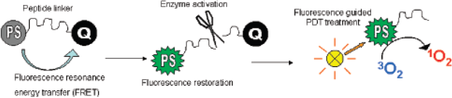
Abstract ID: 450 Poster board space: 9
Abstract ID: 451 Poster board space: 10
HER2 binding affibody is a 58 residue peptide that was selected for its binding to the extracellular domain of HER2/neu through phage display from a combinatorial protein library based on the IgG binding domains of staphylococcal protein A. We evaluated this probe for tumor labeling using HER2 expressing Skov3 ovarian tumor cells. In vitro labeling of Skov3 tumor cells was demonstrated through fluorescent microscopy using a dimerized HER2 affibody probe that was conjugated to Alexa Fluor 680 (HER2 affibody/AF680). Quantification of the fluorescent signal using an In Vivo Imaging System IVIS200 showed that the fluorescence signal was dose dependent when the probe concentrations were between 0-0.1 μg/ml. Preliminary experiments were performed to examine the in vivo tumor labeling property of the probe in a subcutaneous Skov3 tumor model. Accumulation of the HER2 affibody/AF680 probe at the tumor site was detectable at 1 hour after i.v. injection. In addition, the probe also accumulated in the kidney and other organs. Measurement of the tumor fluorescence signal at various time points showed a peak signal/background ratio at 3 hours in several groups of mice that were injected with the probe at doses between 10–50 μg/mouse. At a higher dose of 50 μg/mouse, the signal/background ratio was more than 10; at a lower dose of 10 μg/mouse, the signal/background ratio was less than 5. Ongoing work is characterizing the correlation between tumor mass and fluorescence labeling, and determining the sensitivity for detection tumors located in other body compartments.
Abstract ID: 452 Poster board space: 11
Kevin Groves, Sylvie Kossodo, Nara Narayanan, Jeffrey Peterson,
Nanomaterials can provide versatile platforms for constructing in vivo optical imaging probes. Properties required for extended in vivo use include biocompatible components, robust synthesis, abundant surface functional groups, stable formulation, lack of cytotoxicity, and significant absorbance and high near infrared fluorescence. To this end, we have developed a new family of superbright fluorescent nanoparticles termed NanoSPARKS, specifically designed for optical in vivo imaging applications. The nanoparticles are ≈30 nm in diameter and present a polymer surface with a high density of amino functionality suitable for modification. Approximately forty near infrared fluorochromes (VivoTag™680) can be attached without significant self quenching, yielding extremely high extinction coefficients (>4times106 M−1cm−1) and fluorescence brightness (Figure A). Importantly, more than 150 amino groups remain available for additional conjugation of molecules of choice, such as affinity ligands, peptides or PEGs. Biomolecules, including targeting peptides, were readily attached to these nanoparticles, directly using active ester coupling or via thiol reactive linkers. In order to provide long-circulating nanoparticles suitable for imaging vasculature in vivo, the surface amines were pegylated with 5 kDa mPEG-SS or mPEG-SPA. The pegylated versions were retained in the vasculature for extended periods of time, with a half-life of 12–16 hours as determined by intravital microscopy (Figure B: Image acquisition courtesy Dr. Rainer Kohler, MGH). These particles also provided a sensitive method for imaging subcutaneous tumor xenografts and deep tissue organs such as the heart using Fluorescence Molecular Tomography (FMT™), 24 hours after intravenous administration. Owing to the excellent fluorescent brightness and availability of 100-plus surface functional groups, we believe the NanoSPARK platform should be extremely useful for the development of multivalent targeting molecules for in vivo applications using fluorescence imaging.

Abstract ID: 453 Poster board space: 12
Split luciferase complementation allows for quantitative, real-time study of protein-protein interactions in vivo and in vitro. Current split complementation systems utilize firefly or Renilla luciferases and require juxtaposition of the split ends to create light. Herein we report novel split click beetle (CB) luciferases that emit green or red light. N-terminus and C-terminus fragments consist of amino acids 1–419 and 395-542, respectively, for both click beetle green (CBG) and red (CBR) luciferases. As previously reported with split firefly luciferase, FRAP/mTOR-rapamycin binding domain (FRB) and FK506 binding protein (FKBP), components of the mTOR pathway, were fused to the N and C termini, respectively, to test inducibility and measure spectral properties. We tested combinations of CB and firefly luciferase fragments and found that N-termini of CB luciferases reconstitute activity with C-termini of both CB and firefly luciferases. Firefly luciferase N-termini, however, will only reconstitute activity with firefly C-terminus. Overall color and color stability tracks with the N-terminus of all protein permutations. N-termini of CBR and CBG luciferases combine with either of the respective click beetle C-termini to give high photon output, though when combined with the firefly C-terminus, emit at much lower levels. Hetero-protein combinations show between 15% and 20% of total output of split firefly luciferase with comparable inducibility. However, compared to split firefly luciferase, split CBG emits greater than 6-fold higher and split CBR emits 2.5-fold higher photon outputs. Split CBG and CBR luciferases show 250% and 200% greater inducibility, respectively, than split firefly upon rapamycin induction. All functional split luciferase pairs yield rapamycin Kd values of 0.25 nM ± 0.05 and FK506 Ki values of 1.57 nM ± 0.86, consistent with previously cited literature values. These new split luciferases introduce new protein interaction imaging tools for deconvoluting cellular pathways in cells and animals.
Abstract ID: 454 Poster board space: 13
Angelika von Wallbrunn1, Carsten Höltke1, Michael Zühlsdorf2, Walter Heindel1, Michael Schaefers3,
Abstract ID: 455 Poster board space: 14
Novel fluorogenic probes for imaging enzyme activity were developed based on a newly synthesized water-soluble far-red (ex. 630 nm/em. 680 nm) fluorogenic dye, 9-di-3-sulfonyl-propylaminobenzo[a]phenoxazonium perchloride (2SPBO). The fluorescence of 2SBPO can be modulated by functionalization of the primary amino group on the dye. To report enzyme activities, enzyme substrates with custom-designed spacers were anchored onto the amino group, masking the fluorescence property. After hydrolysis of the enzyme substrates by their target enzymes, the fluorescence emission was recovered. Recently, two novel molecular probes for dipeptidyl peptidase IV (DPP-IV) and beta-galactosidase were developed based on this paradigm. DPP-IV is an exo-dipeptidyl protease. It recognizes the Gly-Pro dipeptide and cleaves the peptide after the Pro residue. After detailed studies of the proteolytic activation, a glycylprolylglycylprolyl tetrapeptide was chosen as the recognition sequence due to its stability under physiological conditions. Upon incubation with DPP-IV, a greater than 6-fold increase in fluorescence signal was observed in less than an hour. The molecular probe for beta-galactosidase was designed using a self-immolative mechanism. The specific substrate, beta-D-galactopyranoside, was conjugated to 2SBPO through a para-substituted benzyloxycarbonyl group as a bioresponsive linker. The fluorogenic probe is stable under physiological conditions, until reacting with beta-D-galactosidase. Following the enzymatic hydrolysis of the pyranose group, a spontaneous hydrolysis of the linker occurred, resulting a 10-fold increase in fluorescence signal.

Abstract ID: 456 Poster board space: 15
Dipicolylamine-Zinc(II) (DPA-Zn2+) coordination complexes were prepared and shown to associate with bacterial cell surfaces. The coordination complexes were conjugated to small organic fluorophores (fluorescein, NBD, and dansyl) and a biotinylated version was used in combination with streptavidin-coated quantum dots. Each fluorescent conjugate was evaluated for its ability to stain gram negative E. coli and gram positive S. aureus. All the conjugates stained the surface of E. coli cells, while binding to S. aureus membranes was limited to conjugates of organic fluorophores. A near infrared (NIR) probe, prepared by conjugating the DPA-Zn2+ affinity group to a NIR dye, was used for in vivo imaging of bacterial infection. Nude mice were given intramuscular injections of E. coli or S. aureus cells followed by a tail vein injection of NIR-DPA-Zn2+. Sites of S. aureus infection were readily detectable by small animal fluorescence imaging. E. coli sites of infection were also detected, but at lower signal/noise than the aforementioned gram-positive cells. The present study demonstrates that simple DPA-Zn2+ coordination complexes associate with bacteria in a potentially universal manner. Furthermore, these synthetically accessible compounds may be applied to a broad range of applications, from bacterial staining in a test tube to in vivo imaging of infection in living mice.
Abstract ID: 457 Poster board space: 16
XinJing Tang, Julia Richards,
Our lab has developed photoactive oligonucleotides for controlling gene expression within living cells and embryos with exquisite spatial and temporal resolution. Photoactivation produces a fluorescent signal that can be quantified in real-time. Several enzymes have been the targets of these studies, such as DNA polymerase, whose activity was modulated 25-fold using UV light. More recently, methods have been developed for controlling DNA and RNA hybridization. For example, hybridization of an antisense oligodeoxynucleotide (asODN) to a target mRNA can inhibit translation by sterically blocking the ribosome and/or recruiting endogenous ribonucleases. A series of light-activated DNA hairpins was synthesized by covalently attaching a 20-mer asODN to a complementary sense strand through a heterobifunctional photocleavable linker. The photoactive conjugates were stabilized by ≈ 1–4 kcal/mole compared to the corresponding asODN/sODN duplexes. These differences in stability made it possible to regulate asODN/RNA duplex formation. RNase H assays showed a 10-fold increase in RNA degradation upon photoactivation of the asODN/sODN hairpin. UV confocal microscopy experiments to photoregulate genes within leukemia cells, neurons, and zebrafish embryos will be described.
Abstract ID: 458 Poster board space: 17
The development of chemotherapeutic agents for the treatment of cancer has resulted in overcoming some tumors. Despite advances in the synthesis of new anti-tumor agents, these compounds possess inevitable, serious side effects like non-specific toxicity that limit the dose and uses of the drug. Attempts to decrease the toxicity of anti-tumor agents to normal tissues have involved localizing cytotoxicity at the tumor site and employing macromolecular pro-drugs to prolong duration of drug activity. Especially, the introduction of the concept “the enhanced permeability and retention (EPR) effect” to the cancer chemotherapy gave rise to extensive researches on polymeric drug carrier. Tumors have a unique feature which is greatly different from normal tissues. The tumoral angiogenesis renders tumor tissues to display several distinctive characters such as hyper-vasculature, defective vascular architecture, and a deficient lymphatic drainage system, which lead macromolecules to be accumulated preferentially and to be retained more in tumor tissues than in normal tissues. The superiority of macromolecular drug carriers to low molecular weight anti-cancer agents is based on this EPR effect. In this study, to obtain novel amphiphilic polymers that provide potential applications in biotechnology and medicine, hydrophobically modified glycol chitosans (HGCs) were prepared by covalent attachment of 5β-cholanic acid. The nanoaggregates composed of hydrophobically modified glycol-chitosan had a large diameter ranging from 150 to 800 nm. The tumor targeting efficiency, biodistribution their anti-tumor effect in a tumor-bearing animal model will be presented. Moreover, we also prepared a NIR polymer dot of self-aggregated HGC nanoparticles for tumor imaging. And thus this Cy5.5-labeled nanoparticle can be used to tumor imaging and quantify the biodistribution of tumor targeting nanoparticles, in vivo.
Abstract ID: 459 Poster board space: 18
Signal transduction systems allow living cells to respond and adapt to their environment. One of the most important and versatile transduction signals is protein phosphorylation. This is accomplished by protein kinases, which are enzymes that transfer phosphate groups from a donor molecule, usually ATP, to an amino acid residue of a protein. Protein kinases regulate a wide variety of cellular functions, including gene transcription, secretion, neuronal plasticity, cellular proliferation, differentiation, and death. This extraordinary activation of enzymes is closely related to various diseases such as melanoma, prostate cancer, and colon cancer. In vitro kinase assays are useful for general biological studies as well as for the assaying and screening of possible pharmaceutical or toxic chemicals that inhibit or facilitate cellular signaling pathways. Radiometric assay and enzyme-linked immunosorbent assay are commonly used to identify and measure protein kinase activity. Nonradioactive alternative methods based on microarray technology are currently emerging. In living cell imaging, various genetically encoded fluorescent protein reporters recently have been designed to probe phosphorylation by intramolecular fluorescence resonance energy transfer (FRET). We describe a poly-ion complexed nanoparticle probe for the measurement of protein kinase activity. Nanoparticles were prepared by formation of a polyelectrolyte ionic complex between a positive polyelectrolyte conjugated to fluorescein isothiocyanate (FITC) and a negative polyelectrolyte protein kinase substrate conjugated with tetramethyl rhodamine isothiocyanate (TRITC). In the nanoparticle, FITC and TRITC serve as the donor and acceptor fluorophores for intermolecular FRET, respectively. In the absence of phosphorylation, FRET occurs between FITC and TRITC, but when phosphorylated, the nanoparticles dissociate due to the addition of negatively charged phosphate groups, resulting in a loss of FRET and the appearance of FITC fluorescence. We used protein kinase A (PKA) as a model protein kinase because it has been studied extensively and it may be a useful biomarker.
Abstract ID: 460 Poster board space: 19
Fluorescence probes are excellent sensors for biomolecules, being sensitive, fast responding, and capable of affording high spatial resolution, via microscopic imaging. At present, majority of sensors signal the concentration of biomolecules by an increase or decrease of the emission intensity. However, emission intensity is also dependent on other factors, such as sensor concentration, bleaching, optical pathlength, and illumination intensity. So, it would be much better to have an indicator for ratiometric measurement that exhibits a spectral shift upon reaction or binding to the analyte of interest because the ratio between two fluorescence intensities avoids such factors.
Currently available ratiometric probes are very limited, using a few fluorophores, such as coumarin or benzofuran. These fluorophores are not satisfactory for microscopic imaging because of the use of UV excitation and low fluorescence quantum yield in aqueous environment. Therefore, the development of ratiometric probes with new fluorophore would be valuable.
For this purpose, we newly focused on the fluorophore “iminocoumarin”. Although coumarin derivatives have been one of the most widely studied fluorescent dyes, very few investigations have been carried out on iminocoumarins as a scaffold for fluorescence probes. We have designed and synthesized various iminocoumarin derivatives and found out that their excitation wavelength are long enough exceeding 500 nm, and that they have high quantum yields in aqueous environment. Furthermore, the modification of imino group allowed us to develop novel ratiometric probes. By this strategy, we succeeded in the development of a ratiometic probe for Zn2+, whose role in biological system has gained much attention lately (Figure). The biological applications of this probe for ratiometric imaging are currently underway.

Abstract ID: 461 Poster board space: 20
In recent years, there has been increased interest in the design of near infrared (NIR) fluorescence-based probes that incorporate innovative activation and targeting strategies. One area of active research is the development of probes that respond to their local environment. Such environment responsive probes, when coupled to nanoparticle scaffolds, can benefit from increased in vivo retention times, improved targeting, via multivalency effects, and enhanced signal resulting from the delivery of multiple fluorochromes to the desired target. In this report we describe the development of a nano-scale NIR fluorescence-based probe for ratiometric pH imaging. For effective imaging of acidic environments, careful design of a bright, water-soluble pH sensitive fluorochrome is critical. Dyes with a mildly acidic pKa are optimal for visualization of acidic microenvironments such as those found in acidic intracellular compartments and acidic environments often associated with tumors. The pH responsive dye, HCyC-646, with a pKa of 6.2, 668 nm fluorescence emission, and an extinction coefficient of 200,000 M−1cm−1 was designed to address these needs. To demonstrate the capability of this fluorochrome, a pH sensitive imaging agent, based on the recently validated bacteriophage M13 targeted imaging nano-scaffold, was assembled. The probe consists of several hundred copies of HCyC-646 and Cy7, a pH insensitive reference fluorochrome, conjugated to the bacteriophage particle. The functionality of the probe was shown by cell uptake experiments, where it was possible to visualize acidic intracellular compartments. Additional utility of the pH imaging particles was demonstrated by in vivo imaging of acidic environments in mouse model systems.

Abstract ID: 462 Poster board space: 21
Peripheral Benzodiazepine Receptor (PBR) has become an attractive target for cancer imaging and therapy. PBR is a mitochondrial protein with three subunits, including the 18 kDa isoquinoline-binding protein (IBP) and the 32 kDa voltage-dependant anion channel (VDAC) in the outer membrane, and the 30 kDa adenine nucleotide carrier (ANC) in the inner membrane. Over-expression of PBR has been observed in a variety of cancers, including brain, breast, colorectal and prostate cancers.
DAA1106 has recently been reported as a potent and selective PBR ligand [1]. DAA1106 displayed higher binding affinity than other PBR ligands, such as PK 11195, Ro5-4864 and FGIN-1-27. It has been shown that DAA1106 displaced PBR complexed PK 11195 and Ro5-4864 at very low concentration (10−15-10−12 M), while 0.1–1 μM amounts of PK 11195, Ro5-4864 or FGIN-1-27 were necessary to displace DAA1106. Therefore, DAA1106 can potentially provide enhanced sensitivity in cancer imaging and improved selectivity in cancer therapy. However, the application of DAA1106 has been limited because the current molecule is not readily functionalized.
In the current study, a conjugable analogue of DAA1106 has been synthesized and characterized. The analogue has a terminal amino group, facilitating coupling signaling moieties and therapy agents. It will be shown that the DAA1106 analogue conjugated to lanthanide chelates and fluorescence dyes can be used in MR, PET and optical imaging. The prospect of preparing the DAA1106 analogue conjugated to pharmaceuticals for selective therapy delivery and enhanced efficacy will also be considered.
Abstract ID: 463 Poster board space: 22
We have reported that genetically engineered bioluminescent E. coli and Salmonella typhimurium specifically target cancers. In contrast to the visible light range, the near-infrared (NIR) spectrum maximizes tissue penetration and minimizes autofluorescence in vivo. Rhodobacter sphaeroides has absorption and fluorescence peak in 850 nm and 900 nm, respectively. Because photon energy is absorbed by the electron acceptors in the reaction center (RC) of the photosynthetic membrane, the new idea is that membranes without the RC will produce more fluorescence than those containing the RC. We explored tumor targeting capacity of R. sphaeroides and the enhancement of fluorescence with RC defective mutant strain (LM1.1). Wild type (2.4.1) and RC defective (LM1.1) strain were cultured in sistron's minimal medium A (SIS) at 32°C. Tumor models has been established by subcutaneous injection of CT26, MCF-7, B16F10. R. sphaeroides (1×10⁸ cfu) were intravenously injected into tumor bearing mice. In vivo fluorescence imaging has been done by using indocyanine green (ICG) excitation (710-785 nm) and emission (810-885 nm) filters. LM1.1 mutant strain showed 5–10 fold stronger fluorescence than wild type (2.4.1) in vitro and in vivo. When R. sphaeroides were injected intravenously in tumor bearing mice, imaging signal was initially detected at the liver for first 3–6 days, then at the tumor area thereafter. The imaging signal continuously increased until 36 days after injection. The imaging findings corresponded to histology that showed localization of strong gram positive bacteria in the tumor tissue. To our knowledge, this is the first report to successfully target cancer cells with near infrared emitting fluorescent bacteria. RC defective mutant produced much stronger fluorescence than wild type possibly leading to development of a superior bio-probe for imaging cancer.
