The ability to visualize the immune response with radioligands targeted to immune cells will enhance our understanding of cellular responses in inflammatory diseases. Peripheral benzodiazepine receptors (PBR) are present in monocytes and neutrophils as well as in lung tissue. We used lipopolysaccharide (LPS) as a model of inflammation to assess whether the PBR could be used as a noninvasive marker of inflammation in the lungs. Planar imaging of mice administrated 10 or 30 mg/kg LPS showed increased [123I]-(R)-PK11195 radioactivity in the thorax 2 days after LPS treatment relative to control. Following imaging, lungs from control and LPS-treated mice were harvested for ex vivo gamma counting and showed significantly increased radioactivity above control levels. The specificity of the PBR response was determined using a blocking dose of nonradioactive PK11195 given 30 min prior to radiotracer injection. Static planar images of the thorax of nonradioactive PK11195 pretreated animals showed a significantly lower level of radiotracer accumulation in control and in LPS-treated animals (p < .05). These data show that LPS induces specific increases in PBR ligand binding in the lungs. We also used in vivo small-animal PET studies to demonstrate increased [11C]-(R)-PK11195 accumulation in the lungs of LPS-treated mice. This study suggests that measuring PBR expression using in vivo imaging techniques may be a useful biomarker to image lung inflammation.
Acute or chronic inflammation of the lungs afflicts millions of people each year. The increasing prevalence of asthma and other lung diseases associated with exposure to infectious agents or environmental air pollutants carries a significant social and economic burden. The ability to visualize the immune response in vivo with specific radioligands targeted to immune cells can greatly enhance our understanding, diagnosis, and monitoring of inflammatory lung disease and its treatment.
Investigations from a number of laboratories have documented the use of the peripheral benzodiazepine receptor (PBR) as an in vivo biomarker of brain inflammation and injury in experimental animals [1–6] and in human neurodegenerative diseases [7,8]. In the brain, the PBR is exclusively localized in microglia and astrocytes and PBR expression markedly increases following brain inflammation or injury [1,2,5,7]. Microglia are the immune-competent cells of the brain and PBR expression can be monitored noninvasively by imaging techniques. The specific PBR ligand [1-(2-chlorophenyl)-N-methyl-N-(1-methylpropyl)-3-isoquinoline carboxamide] also called PK11195 and its active enantiomer, (R)-PK11195, has been labeled with radioisotopes for in vivo imaging of brain inflammation and injury using positron emission tomography (PET) [7–10] or single photon emission computed tomography (SPECT) [11,12].
Several lines of evidence suggest that the PBR may also play an important role in the regulation of the immune response in peripheral organs. PBR is expressed in circulating blood cells with the highest concentrations in monocytes and polymorphonuclear neutrophils (PMNs) [13]. Specific PBR ligands have been shown to regulate human PMN chemotaxis [14] and PBR expression is increased in activated PMNs [15]. Further, PBR ligands inhibit macrophage production of several key immune response cytokines [16] and carrageenan-induced inflammation in a mouse model [17]. PBR is also expressed in cells intrinsic to the lungs such as epithelial cells of lung airways and in submucosal glands in intrapulmonary bronchi [18], although its function is not currently known.
In the present study, we assessed whether PBR could be a useful noninvasive biomarker of inflammation in the lungs using a murine model of lipopolysaccharide (LPS)-induced inflammation. The results show increased accumulation of [123I]-(R)-PK11195 activity in mice after LPS-induced inflammation of the lungs using planar in vivo imaging. Further, the in vivo findings were confirmed and extended using ex vivo methods. We also performed PET imaging using a dedicated small-animal scanner [19] to visualize the PBR response in the same LPS-induced mouse model of lung inflammation. The [11C]-(R)-PK11195 small-animal PET images also demonstrate a greater anatomic resolution of increased PBR expression than planar imaging in LPS-induced inflammation of the lungs. Thus, in vivo imaging of PBR levels in mice using small-animal PET could be a useful method to noninvasively study the progression of lung inflammation and the resulting effects of therapeutic interventions.
Materials and Methods
Synthesis of [123I]/[125I]-(R)-PK11195
(R)-1-(2-chlorophenyl)-N-methyl-N-(1-methyl-propyl)-3-isoquinoline (R−N-PK11195), the precursor for the radioisotope labeled (R)-PK11195, was purchased from ABX (Advanced Biochemical Compounds, Dresden, Germany). Synthesis of Iodine-123 or Iodine-125 labeled (R)-PK11195 was accomplished by a modification of the method of Gildersleeve et al. [20]. Briefly, [123I]/ [125I][NaI] in NaOH(aq) (0.1N, 20–40 µL) was added to a vial containing (NH4)2SO4 (6 mg), (R)-PK11195 (100 µg), EtOH/H2O (1:2) (300 µL), and 3 mm glass beads. The reactions contained 3 mCi of [125I] or 19 mCi of [123I]. The reaction mixture was evaporated at 230°C and then 10 mL of air was slowly injected into the vial. Heating at 230°C was continued for 20 min. After the reaction mixture had cooled to room temperature, it was extracted with EtOH/H2O (1:2, 3 × 100 µL). The [123I]/ [125I]-(R)-PK11195 contained in the combined crude extracts was purified by reversed-phase high-performance liquid chromatography (HPLC) (0.8 × 10 cm NovaPak C-18 radial Pak column, 50:50 CH3CN/H2O containing 0.1% trifluoroacetic acid). The radiochemical yield was 40–60%. The purified [123I]/[125I]-(R)-PK11195 contained in the eluate was extracted into ethanol by solid phase extraction techniques using C-18 Sep-Pak. Injectates were prepared in saline containing 5% ethanol. The purity for [123I]/[125I]-(R)-PK11195 is >95%. The specific activity of [125I]-(R)-PK11195 is over 2200 Ci/mmol. The specific activity of [123I]-(R)-PK11195 is over 25000 Ci/mmol.
Synthesis of [11C]-(R)-PK11195
(R)-1-(2-chlorophenyl)-N-methyl-N-(1-methyl-propyl)-3-isoquinoline (R−N-desmethyl PK11195), the precursor for the radioisotope labeled (R)-PK11195, was purchased from ABX. The synthesis of [11C]-(R)-PK11195 was accomplished by a modification of the method of Camsonne et al. [21]. Briefly, [11C]methyl iodide ([11C]CH3I) was transferred by a stream of nitrogen at a flow rate of 10 mL/min into the reaction medium containing 1.2 µmol of R−N-desmethyl PK11195 previously dissolved in 120 µL dimethylsulphoxide (DMSO) and 20 mg potassium hydroxide. The reaction began at room temperature. The bubbling of [11C]CH3I was continued over 6–10 min. Purification was carried out by HPLC using a reversed-phase C-18 column and an ethanol/water (70:30) mixture as the mobile phase. Radiochemical yields were consistently over 50%. [11C]-(R)-PK11195 was obtained with a specific radioactivity of over 814 GBq/µmol (22,000 Ci/mmol).
In Vivo Planar Imaging of [123I]-(R)-PK11195 Distribution in Mice
Adult CD-1 mice (Charles River, Wilmington, MA) received a single intraperitoneal injection of phosphate-buffered saline (PBS vehicle), 10 mg/kg, or 30 mg/kg LPS (Sigma, St. Louis, MO). Forty-eight hours after vehicle or LPS treatments, mice were given tail-vein injections of approximately 3.7 MBq (100 µCi) [123I]-(R)-PK11195 (0.2 mL injection volume) immediately followed by an intraperitoneal injection of 25% urethane. Anesthetized mice were then placed on a disposable underpad (supine position) on one head of the IRIX3 VT triple-head SPECT (Philips Medical Systems, The Netherlands). Three hours after [123I]-(R)-PK11195 injection, static images were acquired for 10 min. Images were saved and analyzed using the Odyssey FX810 computer using the Odyssey FX 8.9C software (Philips). Rectangular regions of interest (ROIs) were drawn over the thorax area of the mouse images and counts per pixel measured by the software PIXIE Interactive Functions (Philips). In animals used for blocking of [123I]-(R)-PK11195 binding in the lung, an intraperitoneal injection of nonradioactive racemic PK11195 (1 mg total dose) was given 30 min prior to injection of [123I]-(R)-PK11195. All animal studies were approved by the Animal Care and Use Committee of the Johns Hopkins Medical Institutions.
Ex Vivo Radiotracer Activity in Peripheral Organs
Immediately after the acquisition of the planar image, animals were euthanized and the lungs were harvested and were placed on dry ice, weighed, frozen, and stored at −80°C to allow for decay of radioactivity (2 days). The lungs were then counted on the 1480 WIZARD 3-in. Gamma Counter (Wallac Turku, Finland) with three diluted [123I]-(R)-PK11195 tracer samples (1:50) of injected dose serving as standards.
[125I]-(R)-PK11195 Autoradiography of Lung PBR Levels
Mice were injected with either PBS or 30 mg/kg LPS. Forty-eight hours after vehicle or LPS treatment, mice were anesthetized with 25% urethane and lungs inflated with 20% sucrose under pressure. The lungs were frozen in isobutane in liquid nitrogen and stored at −80°C. Frozen lungs were sectioned longitudinally using a cryostat, mounted onto Poly-Prep slides (Sigma) and sections were warmed at 65°C for 15 min and stored at −20°C. On the day of the assay, lung sections in slides were brought to room temperature (RT) for 30 min and washed in buffer (0.05M Tris-HCl, pH 7.2) for 5 min at RT. Slides were then incubated for 30 min at RT in 0.5 nM [125I]-(R)-PK11195 in buffer either with or without 10 µM unlabeled R-PK11195 to measure total and nonspecific binding, respectively. Following incubation, slides were washed twice in ice-cold buffer for 3 min and then twice in ice-cold distilled H2O for 5 sec. Slides were then dried overnight at RT and apposed onto Kodak Bio-Max MR film (Eastman Kodak, Rochester, NY) for 3 hr at RT with [125I]-standards (Amersham, Piscataway, NJ) to assess film linearity and for quantitative analysis of images. All images were captured with Inquiry Quantitative Autoradiography, version 3.08 (Loats Associates, Westminster, MD) and analyzed with NIH Image, version 1.62 (NIH, Bethesda, MD).
In Vivo [11C]-(R)-PK11195 PET Imaging in Mice
Adult CD-1 mice received a single intraperitonial injection of PBS (PBS vehicle) or 30 mg/kg LPS. Two days after vehicle or LPS treatments, mice were anesthetized by an intraperitoneal injection of a mixture of Acepromazine Maleate (10 mg/mL; Phoenix Pharmaceutical, St Joseph, MO), Ketaject (ketamine HCl, 100 mg/mL; Phoenix Pharmaceutical), and normal saline (0.9%) (ratio 1:1:2, dosage 2.3 µL/g). Approximately 37 MBq (1 mCi) [11C]-(R)-PK11195 (0.2 mL injection volume) was administered by tail-vein injection. Anesthetized mice were then fixed on a platform (prone position) in the gantry of the ATLAS small-animal PET scanner (NIH), which has an 11.8-cm ring diameter, an 8-cm aperture, a 6-cm effective transverse field of view, and a 2-cm axial field of view. The imaging system, comprised of 18 depth-of-interaction detector modules, surrounds the mouse. Radial and tangential resolutions of the reconstructed image (pixel size = 0.56 mm) using a three-dimensional ordered subset expectation maximum (3-D OSEM) algorithm (10 iterations) were 1.36 mm at the center and 1.96 (radial) and 2.13 (tangential) at 2 cm radial offset. Sensitivity was >2.0% after correcting for position escape. Maximal noise equivalent count rate was 10.3 kcps at 52.2 MBq (1.41 mCi) total activity for the rat phantom. Ten minutes after [11C]-(R)-PK11195 injection, static images were acquired for 45 min in three bed positions covering from neck to pelvis (15 min for each position). Images were reconstructed by 3-D OSEM using the ATLAS software interface. ROIs were drawn (n = 7-8) manually over the lungs and kidneys in a series of axial slices, and averaged signal intensity per pixel was measured using the analysis mode of the ATLAS software interface. The lung/kidney ratio was measured as an index for [11C]-(R)-PK11195 accumulation. The accumulation of radioactivity may be affected either by the dose of injection or how well the tracer goes to the target organ that is affected by blood flow. Because the LPS-treated mice lost some weight, we used the lung/kidney ratio to normalize potential variations for a better comparison among mice. Kidney levels of PK11195 binding do not change and are a good internal standard.
Statistics
Multiple mean comparisons used a one-way analysis of variance (ANOVA) with the Student-Newman-Keuls post hoc test. In cases where comparison between two means was performed, Student's t tests were used.
Results
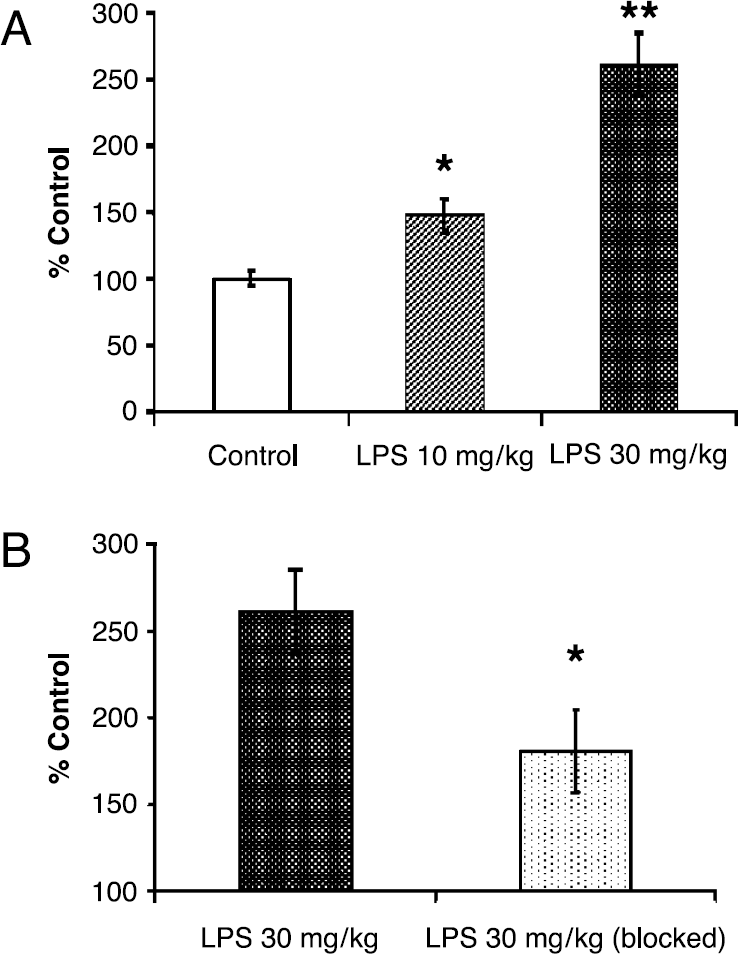
Administration of LPS in mice is known to induce an inflammatory response in the lungs involving a number of inflammatory cells [22,23]. Mice injected with vehicle, 10 or 30 mg/kg LPS were given tail-vein injections of 3.7 MBq (100 µCi) [123I]-(R)-PK11195 at 48 hr after treatment. Static planar images taken 180 min after [123I]-(R)-PK11195 injection revealed significant increases in [123I]-(R)-PK11195 accumulation in the thorax in both the 10- and 30-mg/kg LPS animals relative to vehicle-treated controls (Figure 1A). The in vivo accumulation of [123I]-(R)-PK11195 in the thorax of the control, 10 and 30 mg/kg LPS mice were dramatically decreased by the preadministration of a blocking dose of nonradioactive racemic PK11195 (Figure 1B). Significant increases in [123I]-(R)-PK11195 accumulation in the thorax were measured in both the 10- and 30-mg/kg LPS animals relative to vehicle-treated controls (48% ± 13% and 161% ± 24%, respectively; Figure 2A). The in vivo accumulation of [123I]-(R)-PK11195 in the thorax of 30 mg/kg LPS-treated mice was significantly decreased (80.3% control) by the preadministration of a blocking dose of nonradioactive racemic PK11195 (Figures 2B). This suggests that LPS-induced [123I]-(R)-PK11195 radioactivity accumulation in the thorax is specific and associated with increased PBR expression.
(A) Posterior view of [123I]-(R)-PK11195 planar images of LPS-treated mice at 180 min postinjection of radiotracer. Total acquisition time was 10 min. Because [123I]-(R)-PK11195 is mainly excreted and metabolized through the gastrointestinal tract, the abdominal regions show the most intense increase of radioactivity in all treatment groups. In the thorax, there is a significant difference of radioactivity accumulation among the three groups. (B) Posterior view of [123I]-(R)-PK11195 planar images in the presence of 1 mg PK11195 to determine specificity of the in vivo binding. The images show that the binding of [123I]-(R)-PK11195 in the thorax is blocked by pretreatment (30 min prior) with unlabeled PK11195 in all treatment groups. The color scale on the left-hand side represents the magnitude of the radioactivity in these images (0-200 counts/pixel). Images are scaled to the highest radioactivity.
(A) Analysis of [123I]-(R)-PK11195 radioactivity on the planar images shows that PBR levels in the thorax are significantly increased in the 10-mg/kg LPS (48% ± 13%, *p<.05; n = 5) and the 30-mg/kg LPS groups (161% ± 24%, **p .01; n = 9) relative to controls (n = 11). Each value is the mean ± SEM. (B) Analysis of [123I]-(R)-PK11195 planar images in the 30-mg/kg LPS group in the presence of unlabeled PK11195 shows a significant decrease in binding (*p .05; n = 3) in the thorax (180.7% control) compared to the 30-mg/kg LPS group (261.0% control) (n = 9). Each value is the mean ± SEM.
In order to confirm the in vivo findings, animals were sacrificed and the lungs were harvested immediately after imaging. Ex vivo counting of [123I]-(R)-PK11195 in the lungs indicated significantly increased accumulation of radioactivity (p < .05) in the 30-mg/kg LPS group (6.08 ± 0.72% ID/g, n = 11) relative to controls (2.23 ± 0.27% ID/g, n = 12) (Figure 3). There is an increase in the 10-mg/kg LPS group (4.56 ± 0.84% ID/g, n = 5) relative to control, but the difference did not reach statistical significance. There is also no statistically significant difference between the 10-mg/kg and 30-mg/kg LPS groups (Figure 3). Ex vivo results also show that the effect of LPS treatment on the accumulation of [123I]-(R)-PK11195 was significantly decreased (p < .05) in the thorax by the preadministration of a blocking dose of nonradioactive racemic PK11195 in the 30-mg/kg LPS group (2.81 ± 0.88% ID/g, n = 4) compared to the 30-mg/kg LPS group (6.08 ± 0.72% ID/g, n = 11) (Figure 3).
Ex vivo [123I]-(R)-PK11195 radioactivity in the lungs shows significant increases (172%, p<.05) in the 30-mg/kg LPS group (6.08 ± 0.72% ID/g, n= 11) relative to controls (2.23 ± 0.27% ID/g, n = 12). There is an increase but this was not statistically significant in the 10-mg/kg LPS group (4.56 ± 0.84% ID/g, n = 5) relative to control. There is also no statistically significant difference between the 10- and 30-mg/kg LPS groups. The pretreatment with racemic PK11195 (1 mg) 30 min before [123I]-(R)-PK11195 injection significantly blocked [123I]-(R)-PK11195 radioactivity in the lungs (2.81 ± 0.88% ID/g, n = 4, p<.05). Each value is mean ± SEM; different letters are significantly different at level of p<.05.
To examine further the PBR response in the lungs, we performed [125I]-(R)-PK11195 autoradiography in animals treated with PBS vehicle or 30 mg/kg LPS (Figure 4A). The [125I]-(R)-PK11195 lung autoradiograms revealed a 36% increase in specific binding in LPS-treated mice relative to controls (p < .01) (Figure 4B).
(A) [125I]-(R)-PK11195 autoradiograms of longitudinal lung sections from the 30-mg/kg LPS and control groups. The color scale on the left represents the magnitude of the radioactivity in those images (0-220 fmol/mg tissue). There is a significant increase in PBR levels in the lungs of LPS-treated animals relative to controls. (B) Analysis of [125I]-(R)-PK11195 specific binding in lung sections shows a 36% increase in the 30-mg/kg LPS group (n = 4) relative to controls (n = 3) (p<.01). Each value is the mean ± SEM.
During the course of these studies, the ATLAS small-animal PET scanner became available at the Johns Hopkins Medical Institutions to perform tomographic rather than planar imaging. Using that system, our studies also showed a significant accumulation of [11C]-(R)-PK11195 radioactivity in the lungs in LPS-treated mice compared to controls (Figure 5A and B).
(A) [11C]-(R)-PK11195 small-animal projection imaging from the 3-D whole-body reconstructed PET in the 30-mg/kg LPS and control group. There is a significant increase in PBR levels in the lungs of LPS-treated animals relative to controls. (B) Analysis of [11C]-(R)-PK11195 accumulation in both lungs (lung/kidney ratio). There is a significant increase (146%) of [11C]-(R)-PK11195 accumulation in the right lung in the 30-mg/kg LPS group (0.96 ± 0.19) (n = 3) relative to controls (0.39 ± 0.07) (n = 3) (p< .05). There is also a significant increase (126%) of [11C]-(R)-PK11195 accumulation in the left lung in the 30-mg/kg LPS group (0.97 ± 0.11) (n = 3) relative to controls (0.43 ± 0.04) (n = 3) (p<.01). Each value is the mean ± SEM.
Discussion
The present study shows that LPS administration in mice induces increased levels of radiolabeled (R)-PK11195 binding to PBR in lungs measured in vivo and ex vivo. Further, these preliminary findings indicate that the PBR is a promising noninvasive marker for imaging lung inflammation. This study did not examine the cellular sources of increased [123I]-(R)-PK11195 or [11C]-(R)-PK11195 accumulation in lungs. Increased levels of radioactivity in the lungs may be due to increased infiltration of circulating inflammatory cells that express PBR [13]. However, we cannot rule out the possibility that other PBR expressing cells intrinsic to the lungs may also contribute to the increased accumulation of radioactivity in LPS-treated mice. Future efforts will need to define the cellular sources of increased PBR levels in lungs following inflammation.
There is experimental evidence to suggest that LPS-induced increases in lung PBR may be due to an influx of circulating monocytes and neutrophils. LPS is known to induce neutrophil accumulation in lung capillaries [23] and PBR is highly expressed in neutrophils relative to most other leukocytes [13]. Macrophages are another possible source of increased PBR levels as administration of LPS is also known to induce activation of macrophages in mice [22]. In this context, it should be noted that during the time that our studies were being performed, a study was published describing the use of [11C]-(R)-PK11195 PET to monitor lung macrophages in vivo following particulate challenge in rabbits [24]. More recently, another study from the same group used [11C]-(R)-PK11195 PET to monitor inflammatory cells in chronic obstructive pulmonary disease (COPD) and asthma [25]. In the first study, the investigators used direct administration of silica into the upper pulmonary lobes to induce an inflammatory response and measured increased [11C]-(R)-PK11195 labeling in the site of injection. In the same study, [3H]-(R)-PK11195 microautoradiography was performed on a mixed population of inflammatory cells obtained by lavage from the interarticular space of the rabbit knee joint in which inflammation was induced by bacterial injection. Their findings indicate that the majority of the radioactivity was associated with macrophages [24]. Although this study supports the notion that increased PBR levels occurs in macrophages due to inflammation, caution must be taken to extrapolate from this type of inflammatory response to the one that may occur in the lungs. Direct analysis of inflammatory cells and PBR levels in lung tissue needs to be performed. In the second study, the authors concluded that PET might be useful to assess PMN using [18F]FDG and macrophages using [11C]-(R)-PK11195 in COPD and asthma patients even though there is lack of correlation with sputum macrophages. Thus, continued validation of the PBR as a biomarker of lung inflammation necessitates a better understanding of the cellular sources of increased PBR levels.
In summary, the data presented herein show that LPS administration induces specific increases in PBR levels in mouse lungs. These findings suggest that specific [123I] or [11C]-(R)-PK11195 binding to PBR is a viable and promising noninvasive marker for imaging inflammatory responses in the lungs. Our ability to obtain tomographic images of the lungs in small-animal models of lung inflammation will facilitate future studies to visualize regional inflammation and the effects of different therapies [26]. Further advances in small-animal imaging instrumentation will provide higher spatial resolution in order to visualize regional lung inflammation, and will enable anatomic coregistration to facilitate a more quantitative approach.
Footnotes
Acknowledgments
This work was supported by NIH grants ES07062 (to T.R.G.), CA32845 (to KB), SAIRP grant CA92871 (to MGP) and training grant CA09199 (to MJH). Resources for NIEHS center in Urban Environmental Health (P30ES03819) were also used for animal imaging. We thank Mr. James Fox, Andrew Luu, and Kenneth Brenneman for the assistance of mouse injection and image acquisition. We also thank Mr. Shawn Soutiere for assistance in inflating the lungs for PBR autoradiography.
Abbreviations:
References
1.
BanatiRBMyersRKreutzbergGW (1997). PK (“peripheral benzodiazepine”)-binding sites in the cns indicate early and discrete brain lesions: Microautoradiographic detection of [3H]PK11195 binding to activated microglia. J Neurocytol.26:77–82.
2.
ChenMKBaidooKVerinaTGuilarteTR (2004). Peripheral benzodiazepine receptor imaging in CNS demyelination: Functional implications of anatomical and cellular localization. Brain.127:1379–1392.
3.
KuhlmannACGuilarteTR (1997). The peripheral benzodiazepine receptor is a sensitive indicator of domoic acid neurotoxicity. Brain Res.751:281–288.
4.
KuhlmannACGuilarteTR (1999). Regional and temporal expression of the peripheral benzodiazepine receptor in mptp neurotoxicity. Toxicol Sci.48:107–116.
5.
KuhlmannACGuilarteTR (2000). Cellular and subcellular localization of peripheral benzodiazepine receptors after trimethyltin neurotoxicity. J Neurochem.74:1694–1704.
BanatiRBNewcombeJGunnRNCagninATurkheimerFHeppnerFPriceGWegnerFGiovannoniGMillerDHPerkinGDSmithTHewsonAKBydderGKreutzbergGWJonesTCuznerMLMyersR (2000). The peripheral benzodiazepine binding site in the brain in multiple sclerosis: Quantitative in vivo imaging of microglia as a measure of disease activity. Brain.123:2321–2337.
8.
CagninABrooksDJKennedyAMGunnRNMyersRTurkheimerFEJonesTBanatiRB (2001). In-vivo measurement of activated microglia in dementia. Lancet.358:461–467.
9.
CagninAMyersRGunnRNLawrenceADStevensTKreutzbergGWJonesTBanatiRB (2001). In vivo visualization of activated glia by [11C] (R)-PK11195-PET following herpes encephalitis reveals projected neuronal damage beyond the primary focal lesion. Brain.124:2014–2027.
10.
PappataSLevasseurMGunnRNMyersRCrouzelCSyrotaAJonesTKreutzbergGWBanatiRB (2000). Thalamic microglial activation in ischemic stroke detected in vivo by pet and [11C]PK11195. Neurology.55:1052–1054.
11.
DumontFDe VosFVersijptJJansenHMKorfJDierckxRASlegersG (1999). In vivo evaluation in mice and metabolism in blood of human volunteers of [I23I]iodo-PK11195: A possible single-photon emission tomography tracer for visualization of inflammation. Eur J Nucl Med.26:194–200.
12.
GildersleeveDLVan DortMEJohnsonJWShermanPSWielandDM (1996). Synthesis and evaluation of [I23I]-iodo-PK11195 for mapping peripheral-type benzodiazepine receptors (omega 3) in heart. Nucl Med Biol.23:23–28.
13.
CanatXCarayonPBouaboulaMCahardDShireDRoqueCLe FurGCasellasP (1993). Distribution profile and properties of peripheral-type benzodiazepine receptors on human hemopoietic cells. Life Sci.52:107–118.
14.
RuffMRPertCBWeberRJWahlLMWahlSMPaulSM (1985). Benzodiazepine receptor-mediated chemotaxis of human monocytes. Science.229:1281–1283.
15.
ZavalaFVeberFDescamps-LatschaB (1990). Altered expression of neutrophil peripheral benzodiazepine receptor in x-linked chronic granulomatous disease. Blood.76:184–188.
16.
ZavalaFTaupinVDescamps-LatschaB (1990). In vivo treatment with benzodiazepines inhibits murine phagocyte oxidative metabolism and production of interleukin 1, tumor necrosis factor and interleukin-6. J Pharmacol Exp Ther.255: 442–450.
17.
TorresSRFrodeTSNardiGMVitaNReebRFerraraPRibeiro-do-ValleRMFargesRC (2000). Anti-inflammatory effects of peripheral benzodiazepine receptor ligands in two mouse models of inflammation. Eur J Pharmacol.408:199–211.
18.
MakJCBarnesPJ (1990). Peripheral type benzodiazepine receptors in human and guinea pig lung: Characterization and autoradiographic mapping. J Pharmacol Exp Ther.252:880–885.
19.
SeidelJVaqueroJGreenM (2001). Resolution uniformity and sensitivity of the NIH ATsmall animal pet scanner: Comparison to simulated LSO scanners without depth-of-interaction capability. IEEE Nucl Sci Symp Conf Rec.1555–1558.
20.
GildersleeveDLLinTYWielandDMCiliaxBJOlsonJMYoungAB (1989). Synthesis of a high specific activity 125I-labeled analog of PK 11195, potential agent for SPECT imaging of the peripheral benzodiazepine binding site. Int J Rad Appl Instrum B.16:423–429.
21.
CamsonneRCrouzelCComarDMaziereMPrenantCSastreJMoulinMSyrotaA (1984). Synthesis of N-(11C)methyl, N-(1-methylpropyl), 1-(2-chlorophenyl)isoquinoleine carboxamide-3 (PK 11195): A new ligand for peripheral benzodiazepine receptors. J Labelled Compd Radiopharm.21:985–991.
22.
HarmsenAG (1988). Role of alveolar macrophages in lipopolysaccharide-induced neutrophil accumulation. Infect Immun.56:1858–1863.
23.
LefortJSingerMLeducDRenestoPNahoriMAHuerreMCreminonCChignardMVargaftigBB (1998). Systemic administration of endotoxin induces bronchopulmonary hyperreactivity dissociated from TNF-alpha formation and neutrophil sequestration into the murine lungs. J Immunol.161: 474–480.
24.
JonesHAValindSOClarkICBoldenGEKrauszTSchofieldJBBoobisARHaslettC (2002). Kinetics of lung macrophages monitored in vivo following particulate challenge in rabbits. Toxicol Appl Pharmacol.183:46–54.
25.
JonesHAMarinoPSShakurBHMorrellNW (2003). In vivo assessment of lung inflammatory cell activity in patients with COPD and asthma. Eur Respir J.21:567–573.
26.
PomperMG (2002). Can small animal imaging accelerate drug development?J Cell Biochem Suppl.39:211–220.