
Abstract
Pompe disease (glycogenosis type II) is a rare lysosomal disorder caused by a mutational deficiency of acid α-glucosidase (GAA). This deficiency leads to glycogen accumulation in multiple tissues: heart, skeletal muscles, and the central nervous system. A knockout mouse model mimicking the human condition has been used for histological evaluation. Currently, the best method for preserving glycogen in Pompe samples uses epon–araldite resin. Although the preservation by this method is excellent, the size of the tissue is limited to 1 mm3. To accurately evaluate brain pathology in the Pompe mouse model, a modified glycol methacrylate (JB-4 Plus) method was developed. This approach allowed the production of larger tissue sections encompassing an entire mouse hemisphere (8 × 15 mm) while also providing a high level of morphological detail and preservation of glycogen. Application of the JB-4 Plus method is appropriate when a high level of cellular detail is desired. A modified paraffin method was also developed for use when rapid processing of multiple samples is a priority. Traditional paraffin processing results in glycogen loss. The modified paraffin method with periodic acid postfixation resulted in improved tissue morphology and glycogen preservation. Both techniques provide accurate anatomic evaluation of the glycogen distribution in Pompe mouse brain.
P
Materials and Methods
Tissue Harvesting and Fixation
Animal experiments were conducted in the Department of Comparative Medicine (Genzyme Corporation; Framingham, MA) according to Institutional Animal Care and Use Committee standards. GAA-KO 6neo/6neo mice (Raben et al. 1998) at various ages (3, 4, 10, 13, 15, and 22 months old) and age-matched C57Bl/6 wild-type (WT) mice (the background of the GAA-KO mouse obtained from Charles River Laboratories; Wilmington, MA) were sacrificed for use in the development and validation of these processing methods. The mice were euthanized by CO2 asphyxiation and brains were quickly removed and immediately immersion fixed in 10% NBF for 48 hr (∼1.5 min elapsed between the death of the mice and the fixation of brains). After fixation, WT and KO brains were bisected at midline, and right hemispheres were divided into sagittal slabs 1 mm apart from each other using a mouse brain blocker (David Kopf Instruments; Tujunga, CA) for processing into JB-4 Plus media (Electron Microscopy Sciences; Hatfield, PA) followed by staining with PAS and Richardson's counterstain. Whole left hemispheres were used for paraffin processing, followed by PAS staining and hematoxylin counterstain.
Epon–Araldite Processing
Epon–araldite processing and staining methods were performed according to previously described methods (Lynch et al. 2005).
Traditional JB-4 Plus Processing
After primary immersion fixation in 10% NBF, slabs of right hemispheres were washed twice in PBS (pH 7.4) for 30 min each, dehydrated in ascending grades of ethyl alcohol (30%, 50%, 70%, 80%, and 90%) for 30 min each, and infiltrated with infiltration solution [100 ml of JB-4 Plus (JB-4 Plus embedding kit) solution A (monomer) mixed with 1.25 g benzoyl peroxide, plasticized (catalyst)] overnight at 4C. Once the slabs sank to the bottom of the 12-well plate indicating complete infiltration, they were oriented in the molding cup tray capped by an EBH-2 Block Holder (Electron Microscopy Sciences), embedded in a fresh mixture of embedding solution of JB-4 Plus [25 ml of infiltration solution mixed with 1 ml of solution B (accelerator)], and polymerized at 4C overnight according to the manufacturer's directions. Polymerized blocks were cut at 1–2 μm using a Leica RM2255 microtome (Leica Microsystems; Vienna, Austria) with glass knives made from 400 × 25 × 8.0 mm glass strips (Leica Microsystems) on a Leica EM KMR2 Knifemaker (Leica Microsystems). Sections were collected onto charged glass slides and dried overnight at room temperature.
Modified JB-4 Plus Processing
After primary immersion fixation in 10% NBF, slabs were washed twice for 10 min each in 0.2 mol/L sodium cacodylate buffer (pH 7.3) and postfixed in a 1:1 mixture of 2% osmium tetroxide in 0.2 M cacodylate buffer and 5% potassium dichromate for 1 hr followed by multiple (four to five) rinses, 5 min each, with distilled water until clear. The wash steps prevent formation of an oxide in alcohol, which cannot be removed later (Luna 1968). After the postfixation step, slabs were dehydrated, infiltrated, embedded, and sectioned as described above.
Traditional Paraffin Processing
Left brain hemispheres were immersion fixed in 10% NBF for 48 hr, dehydrated with ascending grades of reagent alcohol, cleared with xylene, and infiltrated with paraffin using a Leica TP-1050 tissue processor (Leica Microsystems). Hemispheres were embedded in paraffin sagittally, cut at 5 μm, and mounted on charged slides.
Modified Paraffin Processing
After primary immersion fixation for 48 hr with 10% NBF, left brain hemispheres were postfixed with 1% PA/10% NBF for 48 hr at 4C, rinsed with PBS (three washes, 10 min each), and placed on a Leica tissue processor (Leica Microsystems) for dehydration, clearing, and infiltration as described above. Four-μm sections were taken and mounted on charged slides.
PAS Reaction on JB-4 Plus-embedded Tissue
Slides were hydrated in distilled water for 5 min, and identification of glycogen was performed using the PAS reaction. Slides were oxidized in 0.5% PA solution for 10 min, rinsed for 1 min in distilled water, and immersed in Schiff's reagent for 20 min at room temperature. Slides were then washed in running water for 10 min in order for the pink color to completely develop. Slides were dried on a warming plate at 45C for 10 min and counterstained with 1:10 dilution of Richardson's (Richardson et al. 1960) stock solution (1% methylene blue made up in 1% borax, mixed in equal parts with 1% azure II, and filtered before use) for 1 min. Once stained, the slides were rinsed with tap water until clear, dehydrated in 95% alcohol, absolute alcohol, and cleared in xylenes, two changes each for 2 min. Slides were coverslipped with Acrytol mounting medium (SurgiPath; Richmond, IL). When performed successfully, glycogen stains dark pink to magenta, nuclei stain dark blue, and cytoplasm and other elements stain with a light blue background.
PAS Reaction on Paraffin-embedded Tissue
For tissues subjected to traditional paraffin processing, slides were deparaffinized, hydrated to distilled water for 5 min, and identification of glycogen was performed using the PAS reaction. Slides were oxidized in freshly made 0.5% PA for 5 min and rinsed in deionized water for 1 min. After 15 min in Schiff's reagent (SurgiPath) at room temperature, slides were washed in running tap water for 10 min for pink color to develop. Slides were counterstained with Hematoxylin 1 (Richard Allan Scientific; Kalamazoo, MI) for 1 min, rinsed with tap water, dipped in bluing reagent (Richard Allan Scientific) for 30 sec, dehydrated to xylene, and coverslipped with Acrytol mounting medium (SurgiPath).
For tissues subjected to the modified paraffin processing, the oxidizing step with PA was eliminated, and slides were treated with the Schiff's reagent only, for 15 min at room temperature. Slides were counterstained, dehydrated, and coverslipped as described above. All post-fixation, processing, and staining techniques employed to optimize preservation of glycogen in Pompe mouse brain tissue are summarized in Table 1.
Summary of the various postfixation, processing, and staining techniques used to optimize preservation of glycogen in Pompe mouse brain tissue
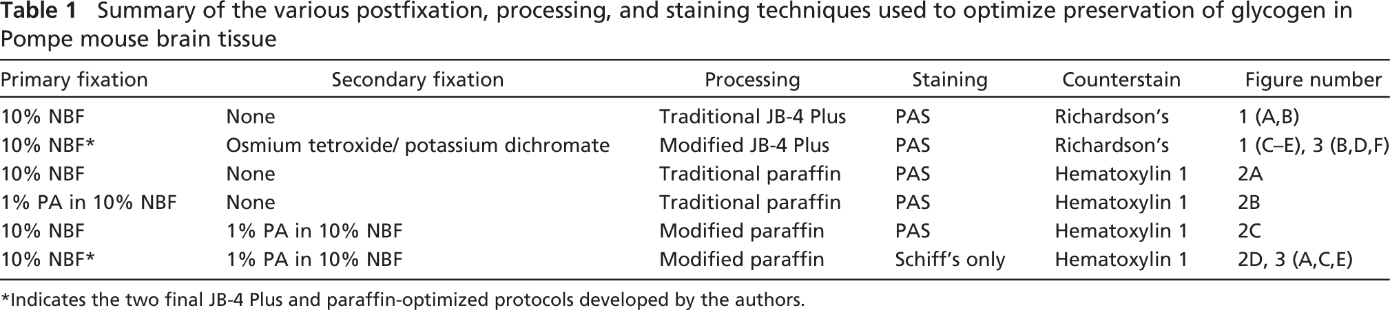
Indicates the two final JB-4 Plus and paraffin-optimized protocols developed by the authors.
Results
Plastic Processing
The epon–araldite method resulted in superior glycogen preservation of mouse brain sections; however, the size of the sample was limited to 1 × 1 × 1 mm (Figure 1C, inset). Traditional JB-4 Plus processing (as recommended by the manufacturer) resulted in chatter and histological artifacts on the large brain tissue sections (Figures 1A and 1B). A modified JB-4 Plus processing method using a postfixation step with potassium dichromate/osmium tetroxide improved the quality of sectioning and the ability to process an entire brain hemisphere measuring 5 × 12 mm with optimal glycogen preservation within the tissue (Figure 1C). When PAS was combined with Richardson's counterstain, glycogen deposits stained magenta were seen most prominently in the swollen neuronal bodies of the medulla (Figure 1D) and throughout areas of the cerebellum (Figure 1E) of 10-month-old Pompe mouse brain.
Paraffin Processing
Traditional paraffin processing using immersion fixation with 10% NBF followed by paraffin embedding resulted in severe glycogen loss, leaving empty spaces (vacuoles) and uneven PAS staining, particularly in swollen neurons (Figure 2A, arrows). When PA was combined with 10% NBF for the primary immersion fixation as described by Kinsley et al. (2000) for improving glycogen preservation in paraffin-embedded mouse liver, the result was disappointing, exhibiting an uneven staining pattern on mouse brain tissue (Figure 2B). Glycogen was well preserved on the edges, but staining was pale in the middle of the section. Primary fixation in 10% NBF alone, followed by the addition of a postfixation step with 1% PA/10% NBF, improved penetration throughout the tissue, resulting in well-preserved glycogen in paraffin-processed mouse brain tissue (Figure 2C). In addition, the oxidation step (with 1% PA) of the PAS reaction could be eliminated due to the fact that tissue was already oxidized during postfixation. After performing the PAS reaction, there was no significant difference between the serial sections stained with PA (Figure 2C) and without PA (Figure 2D); glycogen was well visualized, and the procedure was shortened.
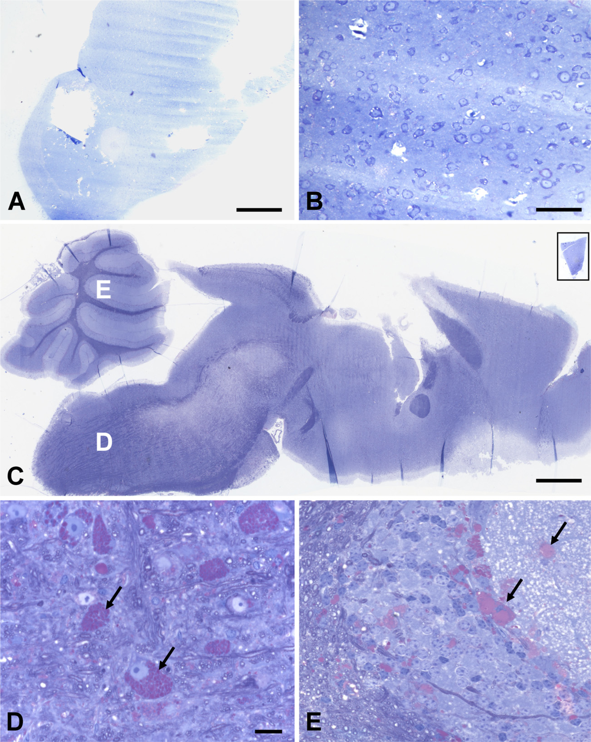
Comparison of different plastic processing of Pompe brain tissue samples: traditional JB-4 Plus processing (
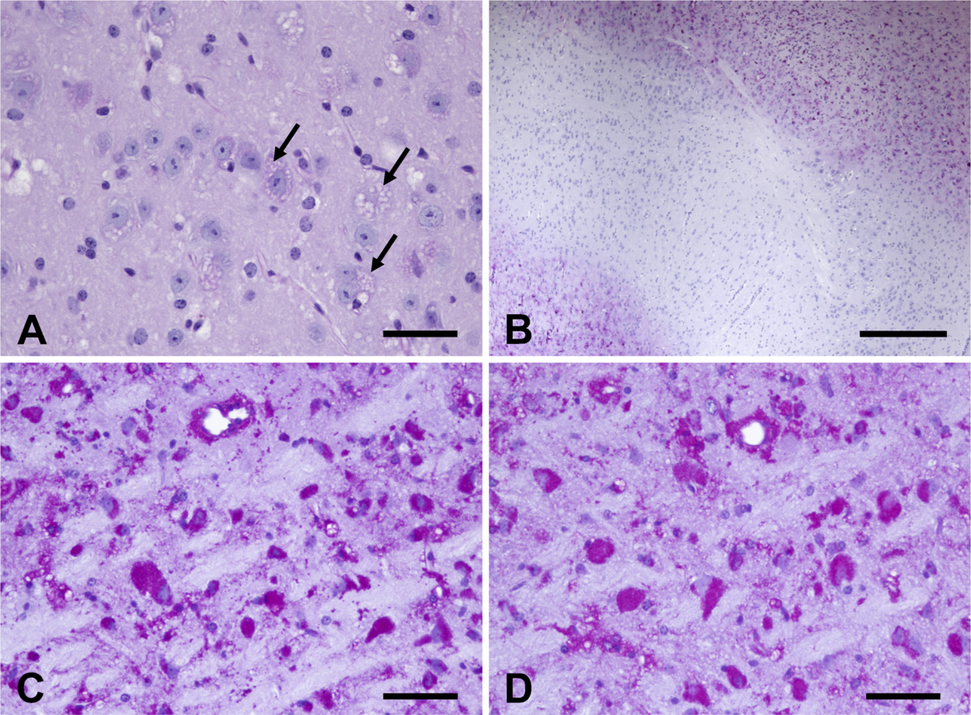
Comparison of traditional vs modified paraffin processing and staining. (
Progression of Cerebellar Neuropathology Using Modified Paraffin and Modified JB-4 Plus Methods
Using the same universal fixative (10% NBF), brain hemispheres were fixed and processed for modified paraffin and modified JB-4 Plus methods. Cerebella from WT mice showed relatively good morphology on a 4-μm paraffin-processed section (Figure 3A) and even better morphological detail on a 2-μm JB-4 Plus section (Figure 3B). WT glycogen levels were relatively undetectable by histological staining, in contrast to age-matched Pompe cerebella where glycogen accumulation was evident at 4 months of age. At this age, glycogen accumulation can be seen in the granular layers and in Purkinje cells of Pompe mice (Figures 3C and 3D). By 15 months of age, the pathology of Pompe cerebella progressed dramatically with accumulation of glycogen in every layer including the molecular layer (Figures 3E and 3F). Staining yielded high contrast between the glycogen and surrounding tissue.
Discussion
Preservation and demonstration of glycogen in tissue for histochemical examination has long been a two-sided problem. First, the glycogen must be adequately fixed in the tissue so that subsequent processing removes as little glycogen as possible. Second, the staining method must show the glycogen clearly and specifically (Trott 1961). The well-established method for optimal processing of Pompe tissue is epon–araldite (Lynch et al. 2005), but size limitations for this processing restrict its application to brain tissue due to the heterogeneity of CNS tissue. JB-4 Plus medium is recognized as a superior method for obtaining greater morphological detail in large (up to 2 cm) tissue sections. However, the processing schedule suggested by the manufacturer (Electron Microscopy Sciences) resulted in chatter on 10% NBF-fixed brain tissue, probably from over-dehydration of the tissue during processing. To improve the quality of sectioning and at the same time to preserve lipid components of the brain, mainly myelin, a post-fixation step with osmium tetroxide and potassium dichromate was added to the JB-4 Plus processing. Osmium tetroxide is soluble in fats and forms a black reduction compound at the double carbon-to-carbon bond by the addition of one molecule of osmium tetroxide to one double bond. The resulting ester may, in turn, attach to other double bonds forming double coordinate linkages. Complexes formed are no longer soluble in the usual fat solvents (Sheehan and Hrapchak 1980). At the same time, potassium dichromate renders lipids insoluble in lipid solvents and acts as an effective mordanting reagent during staining. Immersion fixation in 10% NBF followed by the postfixation step with osmium tetroxide and potassium dichromate allowed the processing of large brain hemispheres (up to 15 mm; Figure 1C) and dramatically improved cellular preservation and retention of glycogen confirmed by staining with the PAS reaction. Using Richardson's solution for counterstaining showed the visible contrast between glycogen and surrounding tissue (Figures 3D and 3F). Although the JB-4 Plus procedure proves to be an excellent method for characterizing Pompe disease in the CNS, it is time-consuming, especially when a large number of samples need to be processed. In clinical settings, paraffin processing is more practical for light microscopic investigation of Pompe tissue, but glycogen loss during traditional paraffin processing makes it difficult to characterize the neuropathology of GAA-KO 6neo/6neo mice (Figure 2A). Superior glycogen preservation with optimal tissue morphology has been demonstrated by others on 3-to 7-mm liver samples fixed in 1% PA in 10% NBF and embedded in paraffin (Kinsley et al. 2000). In our hands, when this primary fixative was applied to large pieces (15 mm) of mouse brain tissue, results were disappointing. Inadequately stained glycogen in the center of the tissue section indicated incomplete penetration and preservation (Figure 2B). Primary immersion fixation with 10% NBF followed by secondary fixation with 1% PA/10% NBF dramatically improved the result on 15-mm paraffin-embedded brain tissue. Generally, formalin does not fix glycogen but fixes proteins where glycogen is trapped. If the protein is not fixed, the glycogen granules move and escape (Kinsley et al. 2000). Thus, fixation of proteins is an important step prior to implementing any processing methods. Postfixation with PA/NBF preserved glycogen well, due to the formation of dialdehydes by PA in the fixative (Trott 1961), resulting in little glycogen lost. Not only did this postfixation step prove to be an excellent preservative of glycogen, but the oxidation step (using 1% PA) of the PAS reaction (McManus and Mowry 1960) could be eliminated because the tissue was already oxidized during the postfixation step. Sections from tissue that had been fixed initially in 10% NBF and postfixed in 1% PA/10% NBF were passed directly into Schiff's reagent, omitting the PA step. Glycogen was well visualized, and the procedure was shortened (Figure 2D). Comparison of serial sections stained for PAS with and without PA showed no difference in staining, thus demonstrating that glycogen in Pompe mouse tissue had been oxidized during the postfixation step. This modified procedure showed superior glycogen preservation and evenly distributed staining through the entire brain hemisphere. The progression of Pompe cerebellar pathology in progressively aged mice was visualized by using both modified paraffin and modified JB-4 Plus methods. WT mouse brains used as controls showed negative glycogen storage using either method (Figures 3A and 3B), whereas GAA-KO 6neo/6neo brains stained positive for glycogen (Figures 3C and 3D). This comparison illustrated that both methods were suitable for identification of glycogen in Pompe mouse brain tissue with superior tissue morphology and optimal glycogen preservation. Even at an early stage of disease (4 months old), significant differences can be visualized between GAA-KO (Figures 3C and 3D) and WT brains (Figures 3A and 3B). The disease progressed dramatically by 15 months of age (Figures 3E and 3F). With the use of either modified method it is possible to accurately and more easily evaluate entire brain hemispheres for disease progression, as well as to assess the brain's response to experimental therapeutic treatments.
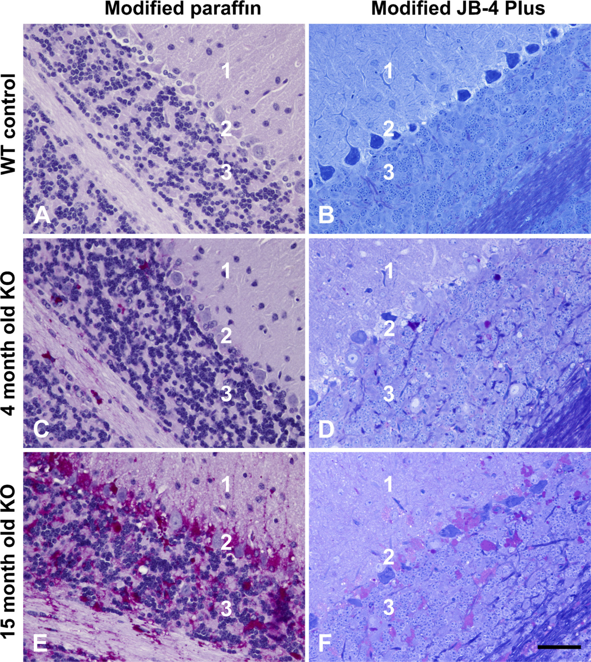
Comparison of modified paraffin and modified JB-4 Plus morphology: the progression of cerebellar pathology in Pompe disease. Four-month-old wild-type (WT) control cerebella (
Footnotes
Acknowledgements
We would like to express our appreciation to Robin Ziegler and Scott Bercury from the Department of Gene Transfer for providing us with animals for this experiment, to Trent Richardson from Biomedical Media Services of Genzyme Corporation for technical support, and Douglas Matthews from the Pathology Department for assistance with image processing. All authors are employees of Genzyme Corporation, which supported this research.