
Abstract
Because of its sensitivity, immunohistochemistry (IHC) of abnormal prion protein (PrPsc) is used more often in the diagnosis of transmissible spongiform encephalopathies (TSEs), such as scrapie and bovine spongiform encephalopathy (BSE). PrPsc IHC requires a combination of pretreatments (chemical, heating, and enzymatic). The method of application may depend on the anti-prion antibody considered. If these pretreatments are efficient for diagnostic purpose, it may, however, be interesting to use an alternative method to efficiently detect PrPsc IHC immunohistochemically using chemical pretreatments solely. Here we describe such pretreatments reporting the difficulty (section adhesion) but also the potential advantages of such methods (easy, quick, inexpensive, and amplifying effect).
Remarkably, because antibodies recognizing only the pathological form (PrPsc) are not available, several pretreatments (a combination of chemical, heating, and enzymatic treatments) are essential for PrPsc IHC because it enhances antigen retrieval, while at the same time suppresses the recognition of normal cellular PrP (PrPc). Although very efficient, it may be interesting to use only chemical pretreatments. In particular, PrPsc IHC may not be only easier, less expensive, and quicker but also potentially as efficient as using the current pretreatment. For this reason, we compared the PrPsc detections obtained using classical and only chemical pretreatments on brain samples originating from BSE- or scrapie-affected animals as well as from unaffected animals. We showed that, in all tested species, PrPsc is detectable in both cases as well, without generating any unspecific background staining. Here we demonstrate the possibility to use this new pretreatment at least with the scrapie-associated fibrils (SAF)-84 antibody on brain samples for diagnostic purposes in animal TSEs with very good efficacy, despite evident increase of adhesion of brain slices.
Materials and Methods
Histological Samples
Brain slices of the obex area from eight cows (two BSE negative and six BSE positive) and eight sheep (one scrapie negative and seven scrapie positive) were selected for this study on the basis of having been previously diagnosed as TSE affected or not, using routine IHC and Western blot analysis. For one cow, other brain levels (from the midbrain to the medulla caudal to the obex) were also added. Briefly, these slides were prepared from formalin-fixed samples (10% in PBS 0.1 M) for 48 hr that were placed in a bath of formic acid (98-100%; Merck Eurolab, Darmstadt, Germany) for 1 hr at room temperature (RT) to reduce infectivity (Taylor et al. 1997) and then automatically processed through graded alcohols to chloroform, and embedded in paraffin (Bayer; Cergy-Pontoise, France). Five-μm-thick brain sections were collected on treated glass slides (Starfrost, Medite Histotechnic; Burgdorf, Germany) and kept overnight in an oven at 55C.
PrP IHC Pretreatments
Brain slices were first dewaxed and rehydrated in graded alcohols. We used the combination of pretreatments already published (Debeer et al. 2001, 2002) that consisted of 98-100% formic acid bath for 10 min at RT, followed by hydrated autoclaving for 20 min at 121C in water (Prestige Medical; Blackburn Lane, UK), and finally a digestion using proteinase K at 20 μg/ml (Roche Diagnostics; Meylan, France) for 15 min at 37C.
Additionally, we analyzed pretreatments performed at RT and based on a chemical sequence defined as follows: 10-min incubation in a solution of KMnO4 0.5% (in 0.1 M PBS, pH 7.0), three washes in distilled water, then 2-min incubation in a bath of Na2S2O5 1%, a wash in distilled water, and finally a 10-min incubation in a solution of sarkosyl (N-lauroylsarcosine) 0.1%, NaOH 75 mM, NaCl 2%. After a 5-min wash in tap water, the slides were ready for the PrP IHC procedure.
PrP Immunohistochemistry
PrPsc IHC was then performed as described previously, using SAF-84 monoclonal primary antibody (0.5 μg/ml in PBS 0.1 M, pH 7.4/0.1% Triton X-100) (Demart et al. 1999; Debeer et al. 2001, 2002). Without any modification for the usual pretreatment, the chemical-only pretreatment required a little adjustment that was indeed interesting to note. Because KMnO4 reagent strongly inhibits endogenous peroxidase, there was no need for that separate inhibition step in the case of the chemical pretreatment. Final revelation was achieved using diaminobenzidine intensified with NiCl2 (Biosys GmbH; Karben, Germany). Finally, the sections were dehydrated, mounted using Eukitt (Microm; Franchville, France), and observed under a microscope (Olympus; Rungis, France) coupled to an image analysis workstation (Biocom; Les Ulis, France). The omission of the primary antibody that was substituted by control serum was used to check the nonspecific background staining in scrapie and BSE cases. Specificity of positive PrPsc immunolabeling was also assessed using the scrapie- and BSE-negative brains. These pretreatment procedures were repeated five times on these brain sections.
Results
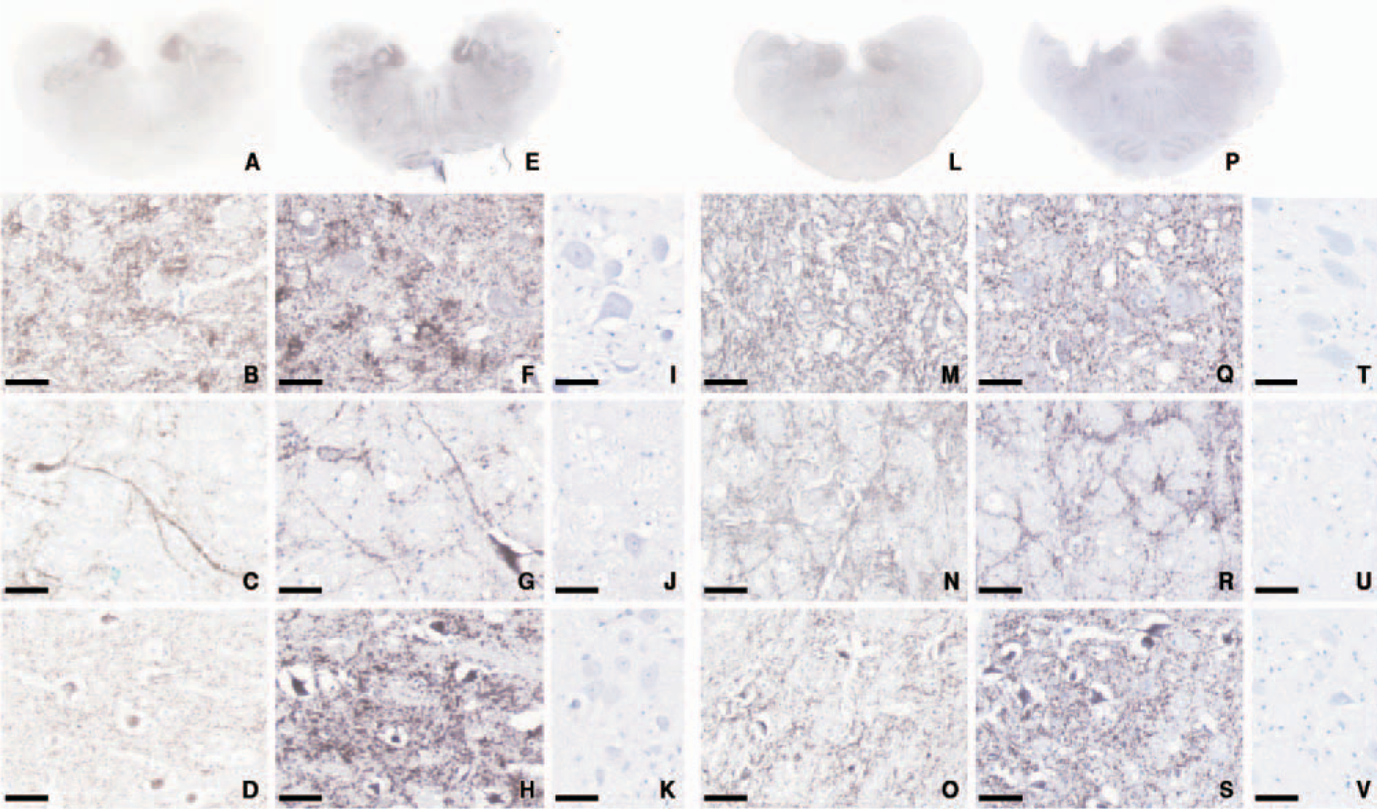
Typical topographical repartition of PrPsc deposits seen in the obex of BSE-affected cows, more precisely in gray matter nuclei such as the dorsal nucleus of the vagus nerve, the nucleus of the solitary tract, the olivary nucleus, the spinal tract of the trigeminal nerve, and in the reticular formation illustrated in Figure 1A using usual PrP IHC pretreatments, was seen as well after the chemical-only pretreatment (Figure 1E). At higher magnification it was possible to inspect the different typical PrPsc depositions that were detected equally in both cases (Figures 1B–1D, 1F–1H). In unaffected animals, the chemical-only pretreatment did not generate any nonspecific background staining as shown in Figures 1I–1K. In addition, the result of the omission of primary antibodies confirmed the specificity of the PrPsc staining.
The results in sheep brain samples were similar to the results seen in bovine species. PrPsc distribution and type of deposits were alike, regardless of the applied pretreatments (Figures 1L–1S). Again, in unaffected sheep, unwanted labeling was not seen after chemical pretreatments (Figures 1T–1V).
Interestingly, this new pretreatment compared with the usual pretreatment repeatedly gave a more intense and contrasted immunolabeling using SAF-84 monoclonal antibody in both species in each of the different brain areas analyzed. For two sheep, we repeatedly observed equal immunolabeling intensity with both pretreatments, without obvious intensification as seen in the five other cases.
Discussion
The current diagnosis of animal TSEs is becoming increasingly based only on PrPsc detection, which appears to be the most reliable marker of TSEs (Bolton et al. 1982). Although PrPsc IHC is considered as a highly specific and sensitive tool for PrPsc detection (Miller et al. 1994; Debeer et al. 2001), it is usually performed after indispensable pretreatments (Van Everbroeck et al. 1999) that make the IHC procedure more complex because of longer time involved and higher cost, as well as requiring the availability of an unfailing heating system such as an autoclave. Various kinds of pretreatments in different possible combinations were described for PrP IHC and, depending on the PrP antibody used, none of the described pretreatments was as simple, inexpensive, and easy as the one we propose here.
To explore their possibilities we applied these pretreatments on brain slices prepared routinely for TSE diagnostic purpose. In the present analysis, we showed that PrP IHC analysis using only the chemical pretreatments retained all its abilities to satisfactorily detect PrPsc at cellular and histological levels (midbrain and brainstem regions) in both bovine and ovine species affected with TSEs. Furthermore, it regularly showed stronger staining in the bovine samples. In sheep staining it was at least of equal intensity, and an enhancement of PrP detection was obvious in five of seven cases. This may reflect the known variety of scrapie strains as well as the various PrP genotypes found in this species.
Immunohistochemical detection of PrP in bovine
Based on strong oxidation of the brain sections followed by a neutralization stage and the increase of the accessibility of PrP epitopes with a last detergent step, this pretreatment is cheaper and accessible for every routine histological laboratory. Because IHC labeling intensity appeared to be increased, it would also be possible to increase SAF-84 dilution, which represents an extra cost. However, there is a noticeable constraint found in the performance of this new pretreatment, which is a great incidence on the tissue slices adhesion. It is possible to play on some subtle changes in the composition of the chemical solutions (Onnasch et al. 2005) to avoid a too-strong tissue-loss effect, as well as using glass slides that allow a strong adhesion of the brain sample or, if necessary, 3-aminopropyltriethoxysilane-coated slides (Rentrop et al. 1986). Actually, this chemical sequence is quite drastic and leads to a certain fragility of the sections that may represent an obstacle, but even if this method may not work with every PrP antibody, or in every species, in our study it worked as well in mice experimentally affected with TSEs (data not shown).
Thus, even if additional studies may be warranted to test it with other PrP antibodies, and even if it requires some technical cautions, we clearly showed the potentiality of this new method of PrPsc IHC, using SAF-84 antibody. This study opens new possibilities in the diagnosis of ruminant TSEs as an amplifying effect is also suggested.
Footnotes
Acknowledgements
This work was financially supported by grants from European FAIR 98–7021 and the French Ministry of Agriculture and Fishery. S.D. was financially supported by the European FAIR 98–7021.
The authors would like to thank Dr. Eoin Monks, without whom this study would not have been possible. The chemical pretreatment was developed by E. Monks, A. Roche, and A. Church (unpublished data), Department of Agriculture, Food and Rural Development, Central Veterinary Research Laboratory, Abbotstown, Castleknock, Dublin, Ireland.