
Abstract
This work was directed to evaluate immunoexpression of markers for apoptosis, resistance to apoptosis, and cell proliferation, as well as estimates of nuclear size in ventral prostate of rats treated with cadmium chloride and cadmium + zinc chloride because a possible protective effect of zinc has been postulated. The following variables were studied: volume fraction (VF) of Bcl-2 immunostaining, percentage of cells immunoreactive to proliferating cell nuclear antigen (LIPCNA) and p53 (LIp53), numerical density of caspase-3 immunoreactive cells (NV caspase-3), and estimates of volume-weighted mean nuclear volume (υV). The LIPCNA and VF of Bcl-2 were significantly increased in the treated animals. The dysplasias (independent of their origin) showed a significant increase of the LIp53, NV caspase-3, and υV in comparison with normal acini from treated and control animals. It can be concluded that cell proliferation is enhanced in long-term cadmium-exposed rats, and exposure to zinc combined with cadmium had no effect on any of the variables studied when comparing with normal acini. The increase of nuclear υV could indicate a more aggressive behavior for pretumoral lesions.
P
Molecular and immunohistochemical markers have been found to be useful in characterizing the progression of human PIN. These markers include changes in several genes that encode the expression of cell proliferation regulators (e.g., bcl-2, p53, p16, p22, and Cerb-B2) (Bostwick 1996; Bostwick et al. 1996; Martin et al. 2001; Nicholson and Theodorescu 2003).
Bcl-2 is a small intracellular non-glycosylated protein that is able to inhibit the apoptotic pathway when overexpressed in cells (Knillova and Kolar 2003). p53 protein regulates cell-cycle inhibition and apoptosis in response to DNA damage, and mutated forms of p53 gene are commonly found in human cancers (Knillova and Kolar 2003; Sirvent et al. 2004). Caspases are aspartate-specific cysteine proteases existing as latent intracellular zymogens (O'Neill et al. 2001). They are fundamental components of the mammalian apoptotic machinery (Cohen 1997), but the precise contribution of individual caspases is controversial. Caspase-3 is one of the key executioners that becomes activated during apoptosis (Cohen 1997).
Some epidemiological and animal studies provide substantial evidence implicating cadmium, a known human carcinogen, as a prostate carcinogen (Waalkes 2000; Nakamura et al. 2002). It has been stated that cadmium chloride produces premalignant and/or invasive epithelial lesions in ventral rat prostate when administered in drinking water (Waalkes 2000; Martin et al. 2001). Zinc may have a relevant impact on cadmium carcinogenesis. Because of the similarity in the transport characteristics between zinc and cadmium, it has been suggested that these metals share a common transport pathway (King et al. 1999; Waalkes 2000), and zinc treatments should reduce or abolish the adverse effects of cadmium, depending on the tissue and circumstances (Waalkes 2000).
The main experimental model for human prostate cancer is the rat (Pollard and Wolter 2000; Shirai et al. 2000; Pollard et al. 2001; Morrissey et al. 2002), and morphological similarities between human PIN and dysplastic changes experimentally promoted in rodent prostate have been reported (Bosland 1999; Martin et al. 2001).
The aim of this study was to investigate (a) the presence and morphology of preneoplastic lesions caused by exposure to cadmium and the influence of zinc on these lesions in rat prostate and (b) the immunohistochemical protein expression of apoptosis promoter and suppressor genes and cell proliferation.
Materials and Methods
Animals
Sixty male Sprague Dawley rats, 30 days old at the beginning of the study, were used for immunohistochemical and stereological studies. The animals were fed with Panlab Lab Chow (Barcelona, Spain) and water ad libitum. Animal protocols are in compliance with the guidelines for the care and use of research animals adopted for the Society of Reproduction. The animals were classified into three groups according to treatment (20 rats/group). Cadmium (Panreac; Madrid, Spain) was added to the drinking water of the first group at a concentration of 60 ppm during the time course of the experiment (24 months). The second group received zinc chloride (Panreac) at a concentration of 50 ppm plus cadmium (60 ppm) in the drinking water. The third group was used as control and received drinking water free of these metals.
Five rats per treatment group were killed each 6 months until the end of the experiment (24 months). All animals were euthanized by exsanguination after CO2 narcosis. Although body weight at the end of the experiment was slightly higher in controls than in treated rats, no significant differences in the size of the animals were detected.
The prostate complex was dissected from the abdominal cavity of each animal. Dorsolateral prostate lobes were routinely examined in all groups studied, and no dysplastic changes were detected. Only the ventral lobe was employed in the study. Afterwards, the ventral prostate was exhaustively sectioned into 2-mm-width slices. The section plane was perpendicular to the sagittal axis of the gland. All specimens were fixed by immersion in 4% paraformaldehyde in PBS, pH 7.4, for 24 hr and afterwards embedded in paraffin.
Sampling Procedure
For each prostate, the slices obtained were embedded in a paraffin block. The blocks were serially sectioned, and 5-μm-thick sections (for immunohistochemistry and routine hematoxylin-eosin staining) alternating with 10-μm-thick sections (for stereological cell counting) were obtained from each block. Ventral prostate was included in every section.
Evaluation of immunostainings was performed on five sections selected by random systematic sampling (Gundersen et al. 1988; Nevalainem et al. 1996) from each block obtained from each animal in each group for each immunostaining.
Histologic Definition of the Lesions
Morphological criteria used for identifying the lesions as dysplastic were (a) enlargement of the epithelial lining of the acinus usually associated with nuclear pseudostratification, (b) increase in the basophilic appearance of the epithelium with nuclear crowding, (c) nuclear pleomorphism, and (d) cribriform pattern.
Immunohistochemical Methods
In all groups, at least five selected slides per animal (per prostate) and per antigen were immunostained. Deparaffinized and rehydrated tissue sections were treated for 30 min with 0.3% hydrogen peroxide in phosphate-buffered saline (PBS), pH 7.4, to block endogenous peroxidase. To detect Bcl-2, p53, caspase-3, and proliferating cell nuclear antigen (PCNA) immunoreactivities, sections were incubated with the following monoclonal antibodies: anti-Bcl-2 antibody (1:10; Dako, Copenhagen, Denmark), anti-p53 antibody (1:50; Cell Signaling, Beverly, MA), anti-caspase-3 antibody (1:50; Cell Signaling), and anti-PCNA antibody (1:100; Biomeda, Foster City, CA). Pretreatment of sections by heating in citrate buffer, pH 6.0 (using a pressure cooker) (Martin et al. 2001), was performed to enhance Bcl-2, p53, and caspase-3 immuno-staining. All primary antisera were diluted in PBS, pH 7.4, containing 1% BSA (Merck; Darmstadt, Germany) plus 0.1% sodium azide. All incubations with primary antisera were done overnight at 4C. Secondary antibodies employed were biotin-caproyl anti-mouse immunoglobulins (Biomeda) diluted 1:400 in PBS containing 1% BSA without sodium azide. All incubations with secondary antibodies were done for 30 min at room temperature. Thereafter, sections were treated with a streptavidin-biotin-peroxidase complex (Biomeda). The immunostaining reaction product was developed using 0.1 g diaminobenzidine (DAB) (3,3′,4,4′-tetraminobiphenyl; Sigma, St Louis, MO) in 200 ml of PBS plus 40 μl 30% hydrogen peroxide.
After immunoreactions, slides were counterstained with either acetic carmine, Harris hematoxylin, or methyl green, dehydrated in ethanol, and mounted in a synthetic resin (Depex; Serva, Heidelberg, Germany). Specificity of the immunohistochemical procedures was checked by incubation of sections with non-immune serum instead of the primary antibody.
Quantitative Evaluation of Cell Proliferation and p53
Percentages of PCNA-immunostained nuclei (PCNA labeling index, LIPCNA) (Martin et al. 2001; Arriazu et al. 2005a) and p53-immunostained nuclei (p53 labeling index, LIp53) were calculated in each selected section for control rats, non-dysplastic acini of treated rats, and dysplastic acini of treated animals, using the following formula: number of labeled nuclei × 100/total number (labeled + unlabeled) of nuclei. Measurements were carried out using an Olympus (Hamburg, Germany) microscope equipped with a X100 oil-immersion lens (numerical aperture 1.4) at a final magnification of X1200 and using the stereologic software GRID (Interactivision; Silkeborg, Denmark). This program allows the selection of fields to be studied by random systematic sampling after the input of an appropriate sampling fraction. An average of 100 fields per section was scanned, and a total of 500 epithelial nuclei were evaluated per section in each group (controls, non-dysplastic, or dysplastic) per animal. The systematic field selection with a random starting point (random systematic sampling method) assures that p53 and PCNA estimates were representative of all the prostate tissue (Martin et al. 2001; Arriazu et al. 2005a). p53- and PCNA-immunostained nuclei were considered positive regardless of staining intensity.
Quantitative Evaluation of Bcl-2 Immunostaining
To quantify the immunoreactivity of Bcl-2 protein, its volume fraction (VF) was measured and expressed as percentage of immunostained epithelium. Estimation of the VF was performed using the Scion Image Beta 4.02 program, available at http://www.scioncorp.com/frames/fr_scion_products.htm. The systematic randomly selected fields were photographed at a final magnification of X500 and were then captured by image program, thresholded, and binarized, and the percentage of epithelial area was automatically measured by the program to obtain the VF (Arriazu et al. 2005b). The VF of Bcl-2 immunoreactivity was estimated in ventral prostates from control and experimental animals to ascertain if the treatment changes the total amount of Bcl-2 immunostaining in the epithelium. Bcl-2 was measured in each selected section from control rats, non-dysplastic acini of treated rats, and dysplastic acini of treated rats.
Evaluation of Numerical Cell Density
Estimation of the number of epithelial caspase-3-positive (caspase-3+) cells was performed using the optical dissector technique, an unbiased stereological method (Wreford 1995; Howard and Reed 1998). Three 10-μm-thick caspase-3 immunostained sections of control, treated, and dysplastic acini were chosen by systematic random sampling from each tissue block selected in each specimen. An average of 100 fields/section was systematically randomly sampled and used to count the number of cells by applying the GRID stereologic software. The amount of cells was evaluated as numerical density (NV), i.e., the number of epithelial caspase-3+ cells/mm3 of epithelial acinar volume.
Volume-Weighted Mean Nuclear Volume (υV)
Estimation of the nuclear υV was carried out on three systematically random sampled hematoxylin-eosin sections per slice in each study group using the stereologic software GRID. An average of 120 nuclei were point sampled per case because the number of nuclei to be estimated to obtain reliable results is considered within the range of 70-150 (Martin et al. 1999). All measurements were performed using an Olympus microscope equipped with a X100 oil-immersion lens at a final magnification of X1200. The program used to evaluate the nuclear υV enables the generation of random test-line directions that were superimposed onto the microscopic images. Nuclear intercepts can be measured along these test lines (Gundersen and Jensen 1985; Sorensen and Ottosen 1991). The length of nuclear intercepts (IO) was processed to obtain πO 3/3, an unbiased estimate of nuclear υV independent of nuclear shape, which because of point sampling emphasizes larger nuclei rather than smaller ones. In addition, estimates of nuclear υV combine information about the three-dimensional nuclear size with knowledge of variability of nuclear size.
Statistical Analysis
For each parameter studied, mean ± SD was calculated. Three analyses by ANOVA were performed to: (a) investigate the influence of addition of zinc over the quantitative parameter considered (controls, cadmium-exposed rats, and cadmium + zinc-treated rats); (b) evaluate differences among controls, dysplastic acini from cadmium-treated animals, and dysplastic acini from cadmium + zinc-exposed rats; and (c) investigate changes in acini from treated and non-treated groups and normal/dysplastic changes within groups. Comparison between each pair of means was performed using the Student-Newmann-Keuls test; p<0.05 was considered significant.
To evaluate differences between age groups independent of the treatment (6 months, >6 months), Student's t-test was used; p<0.05 was considered significant.
Results
Histological Findings
Fifteen dysplastic focal lesions were detected at the end of the experiment in the acinar epithelium of ventral prostate from cases treated with cadmium alone (six rats) and eight dysplastic focal lesions were detected in animals treated with cadmium + zinc (five rats). The acini from the controls showed a columnar mono-stratified epithelium (Figures 1A and 1B), whereas the dysplastic acini showed an irregularly enlarged epithelial lining (Figures 1C and 1D). The first dysplastic changes appeared after 12 months of treatment and are early, isolated, and small lesions (Figure 1C), whereas those appearing at 24 months are larger. Distribution of the dysplastic acini of long evolution was multicentric for the group of cadmium-exposed rats and single for the cadmium + zinc-treated animals. The non-dysplastic acini from treated rats had a similar morphology to that of the acini of control rats. In comparison with control acini, dysplastic glands showed a remarkable enlargement with frequent cribriform pattern (Figures 1C and 1D). Occasional polypoid formations were seen. The nuclei presented important size heterogeneity, and nucleoli were usually prominent (Figure 1D). Occasional mitosis was observed (Figure 1D). No tumoral infiltration in the surrounding stroma was demonstrated.
PCNA and p53 Immunostaining
PCNA immunostaining has been detected in the nuclei from all specimens studied (Figures 2A and 2B). Dysplastic acini showed remarkable immunoreactivity to PCNA in comparison with controls (Figure 2C).
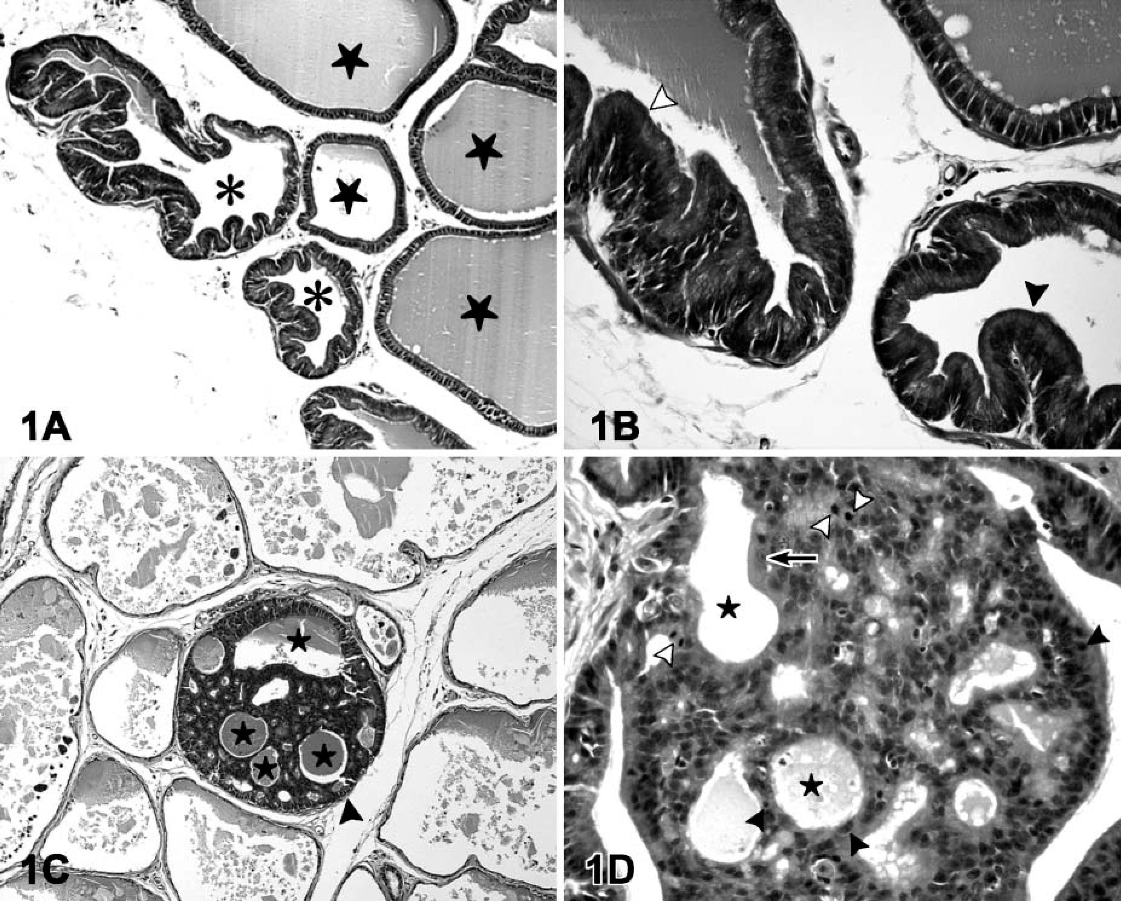
Hematoxylin-eosin staining of ventral prostate from control and rats exposed to metals. (
Immunoreactivity to p53 was detected in both cytoplasm and nucleus of epithelial cells from prostate acini throughout all groups (Figures 2D and 2E). Nuclear p53 immunostaining was also remarkable in the acini with dysplastic changes (Figure 2F).
Bcl-2 and Caspase-3 Immunostaining
Immunoreactivity to Bcl-2 protein was observed in all groups of rats. It was granular and expressed in the apical border of the epithelium in controls and non-dysplastic acini (Figures 2G and 2H). Bcl-2 immunostaining was more widely distributed throughout the cytoplasm in dysplastic glands but was more prominent near the lumen of the cribriform structures (Figure 2I).
Immunostaining to caspase-3 was not detected in the prostate epithelium of controls (Figure 2J). Caspase-3 immunoreactivity was observed in the cytoplasm from isolated epithelial cells and was frequent in those cells detached to the lumen of the acinus. This immunoreactivity was occasionally shown in the non-dysplastic epithelium from treated rats (Figure 2K) and remarkably increased in the dyplastic lesions (Figure 2L).
Quantitative Results
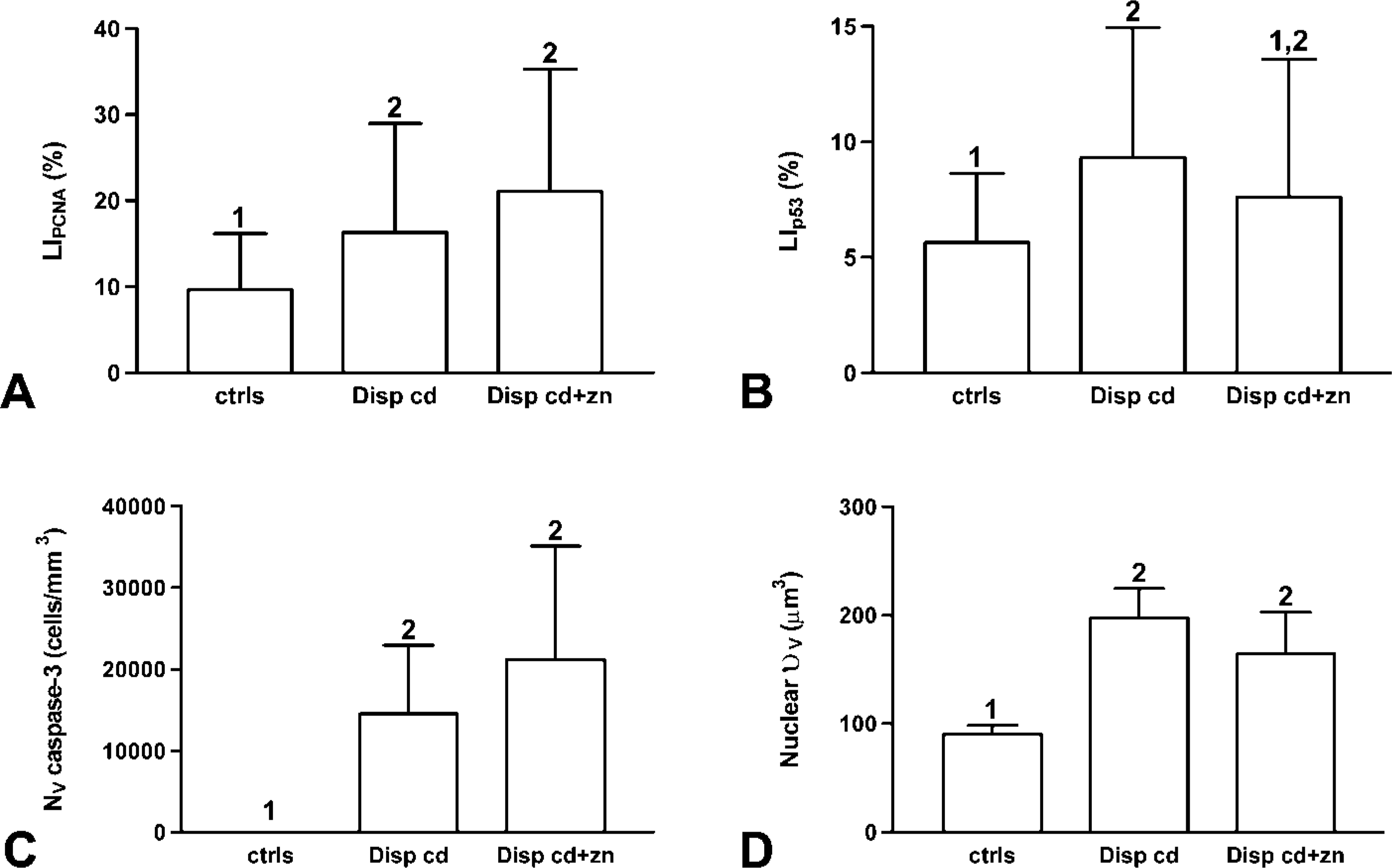
Significance of ANOVA carried out to evidence influence of the type of treatment on the quantitative parameters is shown in Table 1. A significant increase for the LIPCNA and VF Bcl-2 occurs in both cadmium-and cadmium + zinc-treated animals when compared with the controls. No significant differences were observed between treatment types (Figures 3A and 3B). Nuclear υV did not show significant differences between treatment types, but the cadmium-exposed rats showed a significant increase in this parameter in comparison to controls (Figure 3C).
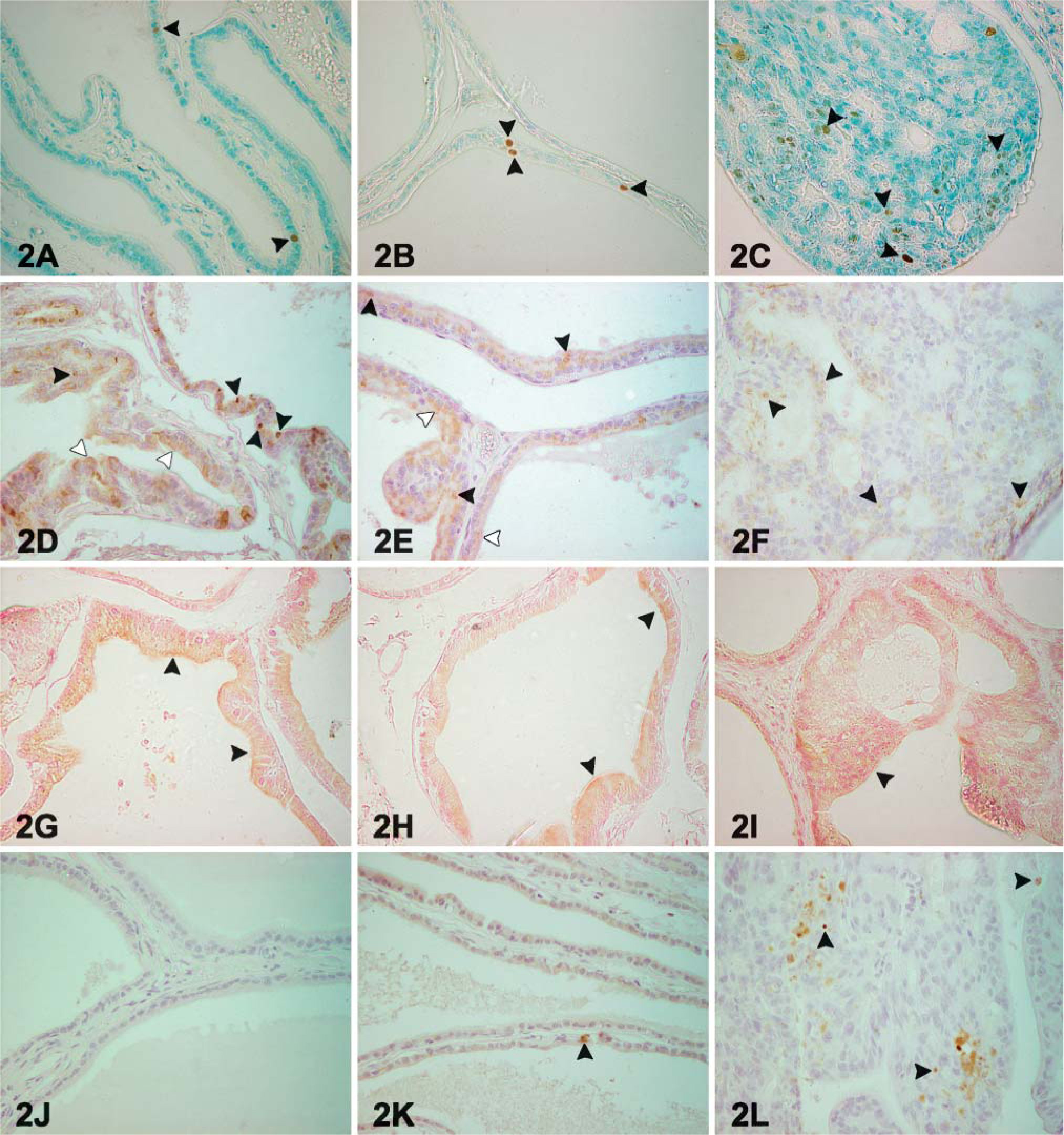
(A) A representative acini from an 18-month-old control animal immunostained to proliferating cell nuclear antigen (PCNA). Immunoreactive nuclei are observed in the epithelium (arrowheads). (
Significance of ANOVAs carried out to investigate the influence of treatments (p treatment) and the influence of the type of dysplastic acini according to their origin (cadmium or cadmium + zinc) (p dysplastic acini)
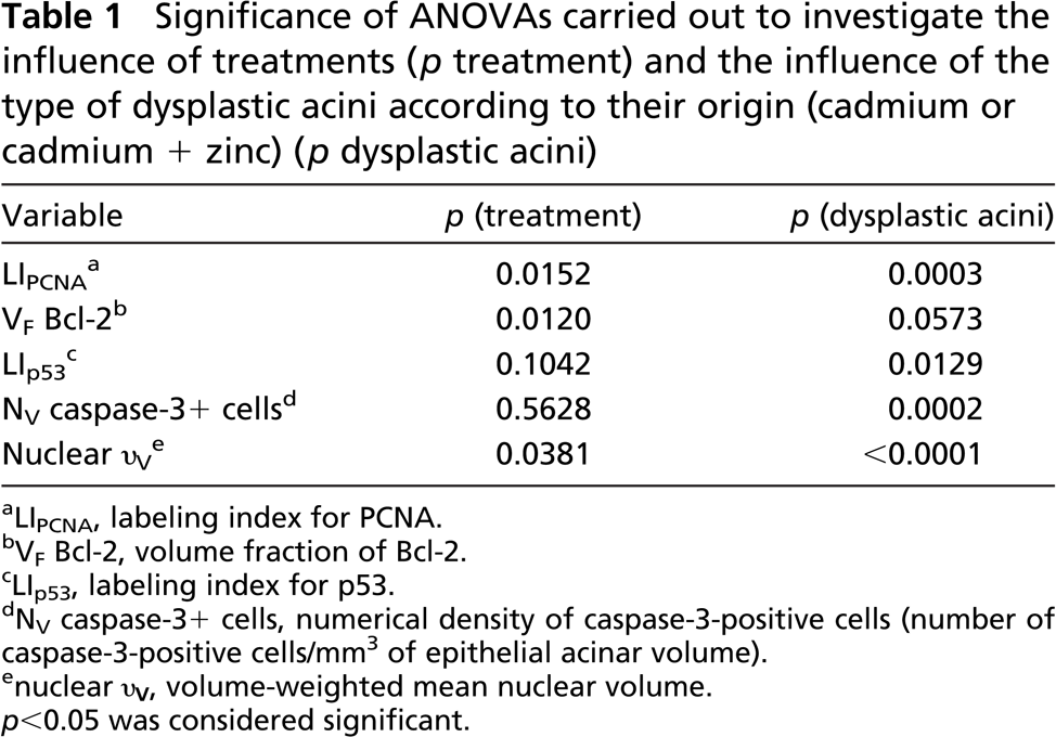
LIPCNA, labeling index for PCNA.
VF Bcl-2, volume fraction of Bcl-2.
LIp53, labeling index for p53.
NV caspase-3+ cells, numerical density of caspase-3-positive cells (number of caspase-3-positive cells/mm3 of epithelial acinar volume).
nuclear υV, volume-weighted mean nuclear volume.
p<0.05 was considered significant.
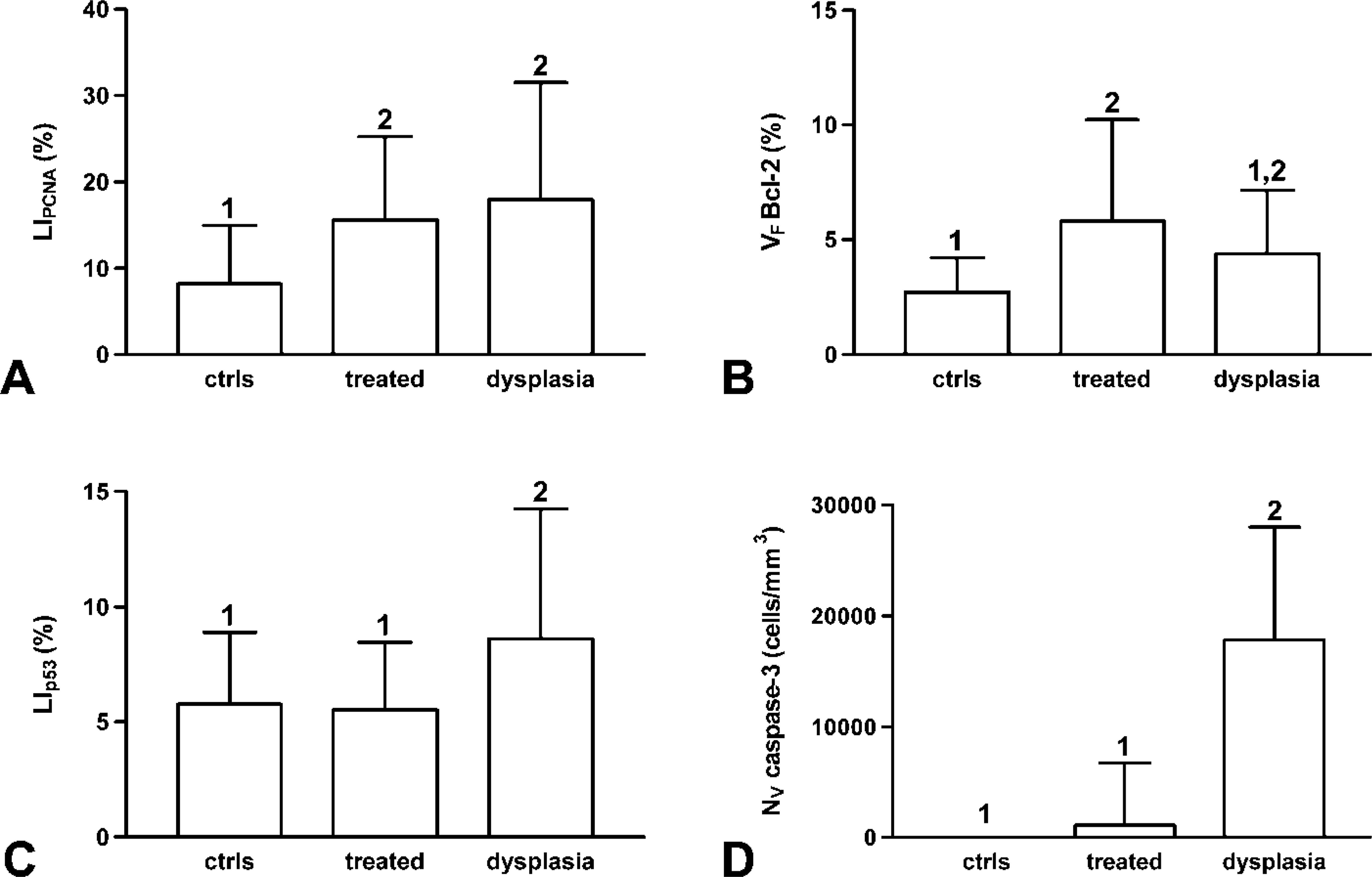
ANOVA results regarding the influence of the type of dysplasias according to their origin (cadmium- or cadmium + zinc-exposed rats) are also shown in Table 1. LIPCNA showed a significant increase in the dysplasias in comparison with control acini. The two treatment groups showed no significant differences (Figure 4A). For LIp53, no significant differences between types of dysplasias could be detected, but the dysplasias caused by cadmium exposure showed a significant increase of LIp53 in comparison with the controls (Figure 4B). However, when the numerical density for caspase-3+ cells was considered, this parameter showed a significant increase in the dysplasias. Dysplastic regions from both treatment groups displayed no significant differences. No significant differences were also observed between the sources of dysplasias (Figure 4C). Nuclear υV showed a significant increase in both cadmium and cadmium + zinc dysplasias when compared with controls. No significant differences were ascertained between the sources of dysplasias (Figure 4D).
When the two types of dysplastic lesions were considered as a whole, LIPCNA was significantly increased in both treated non-dysplastic and dysplastic acini when compared with controls. Nevertheless, no significant differences were observed between treated non-dysplastic and dysplastic glands (Figure 5A).
There was a significant increase in the VF Bcl-2 in dysplastic acini when compared with controls, but no differences were observed between treated non-dysplastic and dysplastic glands (Figure 5B).
LIp53 and the numerical density for caspase-3+ cells were significantly increased in dysplastic acini in comparison with controls and treated non-dysplastic acini (Figures 5C and 5D).
There was a significant increase in nuclear υV from dysplastic acini, in comparison with both controls and treated non-dysplastic acini (Figure 6).
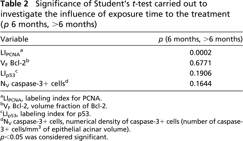
The significance of Student's t-test carried out to ascertain influence of the time of exposure to the treatment is shown in Table 2. Significant differences were detected only for LIPCNA. A significant increase was also observed for the LIPCNA of animals treated for >6 months (Figure 7).
No significant histologic lesions were observed during the first 6 months of treatment with cadmium chloride. These results are in accordance with the findings of Visser and Dekler (1979), who reported no significant changes in the rat prostate epithelium after 6 and 10 months of cadmium treatment. In the specimens treated for 12 and 18 months with cadmium or cadmium + zinc, structural alterations observed were considered as preneoplastic lesions by several authors (Parkinson 1995; Bostwick et al. 1996). Evident morphological changes (dysplasia) were observed in the present study in prostate acini after 24 months of cadmium and cadmium + zinc exposure, and these results confirm the carcinogenic effects of cadmium (Shirai et al. 1993; Waalkes 2000). Our results suggest that the oral administration of zinc might reduce the occurrence of dysplastic changes mediated by cadmium. The isolate character of the appearance of the lesions in rats treated with zinc, in comparison to the multifocal dysplasias observed in animals exposed to cadmium alone, could indicate a lesser aggressiveness when zinc was added. It has been demonstrated that prostate epithelium is able to concentrate zinc, and this metal could inhibit the growth of prostate cells (Costello and Franklin 2000; Feng et al. 2000).
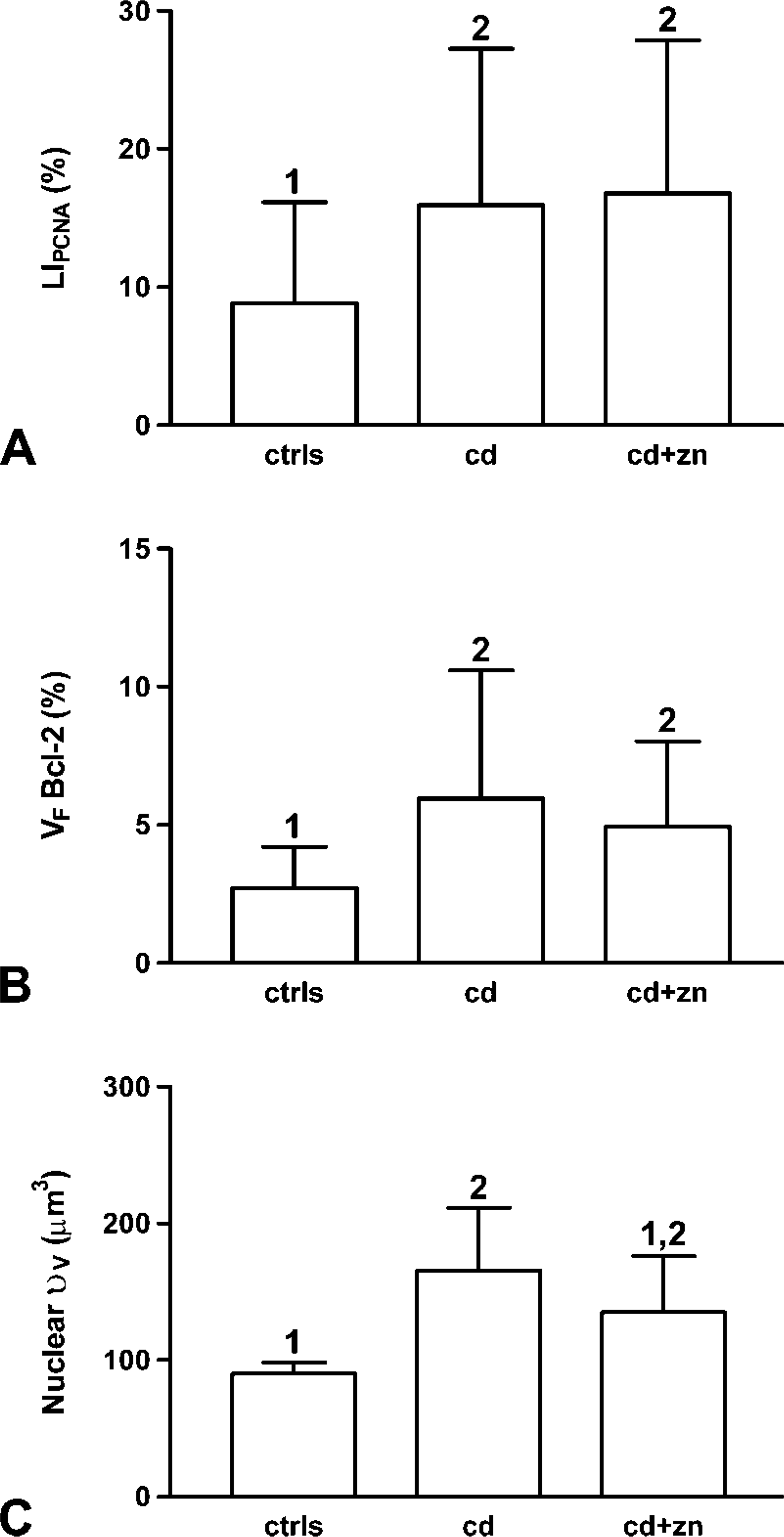
Bar graphs for LIPCNA (
Bar graphs for LIPCNA (
The significant increase of cell proliferation observed in the groups treated >6 months might be due to the growth of dysplasias from 12 months of exposure (Waalkes et al. 1992). No other significant changes have been observed between rats with 6 months of treatment and animals with >6 months of exposure.
According to some authors (King et al. 1999; Waalkes 2000), zinc treatment should reduce or abolish the adverse effects of cadmium, given the similarity in the transport characteristics between both metals. Nevertheless, when all the acini excluding the dysplastic ones were considered, exposure to zinc combined with cadmium had no effect over any of the variables studied. Although LIPCNA and VF Bcl-2 were increased in rats exposed to cadmium or cadmium + zinc in comparison with untreated animals, the differences were not significant, in contrast to those observed in other studies (Martin et al. 2001). According to the low expression of Bcl-2 protein, there were no differences in the single-strand conformation analysis (SSCA)-band pattern of bcl-2 gene in all the animals (data not shown), indicating that the mutation rate is apparently not affected by the treatments assayed. A number of studies have analyzed the p53 expression in human prostate (Downing et al. 2003; Zeng et al. 2004); however, p53 expression is not well documented in rat prostate epithelium. Immunoreactivity to p53 visualized in ventral prostate in this study did not show changes in relation to the different groups of treatment; however, the PCR-SSCA study revealed a new band in one rat treated with cadmium + zinc for 6 months (data not shown). This band appears as a consequence of the conformational change of single strands as suggested by Sheffield et al. (1993), in this case a substitution of adenine by guanine as a normal single nucleotide polymorphism not detected until now at this point of the p53 gene in the rat genome.
Bar graphs for LIPCNA (
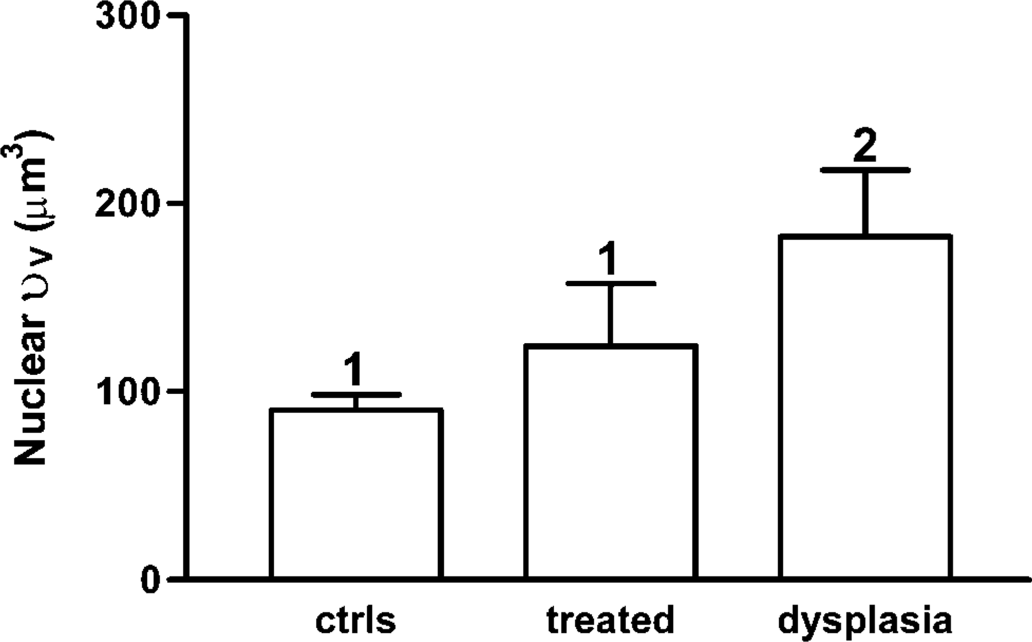
Bar graphs for volume-weighted mean nuclear volume. Measurements correspond to controls (ctrls), non-dysplastic acini from both types of exposed rats (treated), and dysplastic acini from both types of exposed rats (dysplasia). Results are expressed as mean ± SD. Bars showing different numbers at the top of the error bars differ significantly (p<0.05).
Significance of Student's t-test carried out to investigate the influence of exposure time to the treatment (p 6 months, >6 months)
LIPCNA, labeling in ex for PCNA.
VF Bcl-2, volume fraction of Bcl-2.
LIp53, labeling index for p53.
NV caspase-3+ cells, numerical density of caspase-3+ cells (number of caspase-3+ cells/mm3 of epithelial acinar volume).
p<0.05 was considered significant.
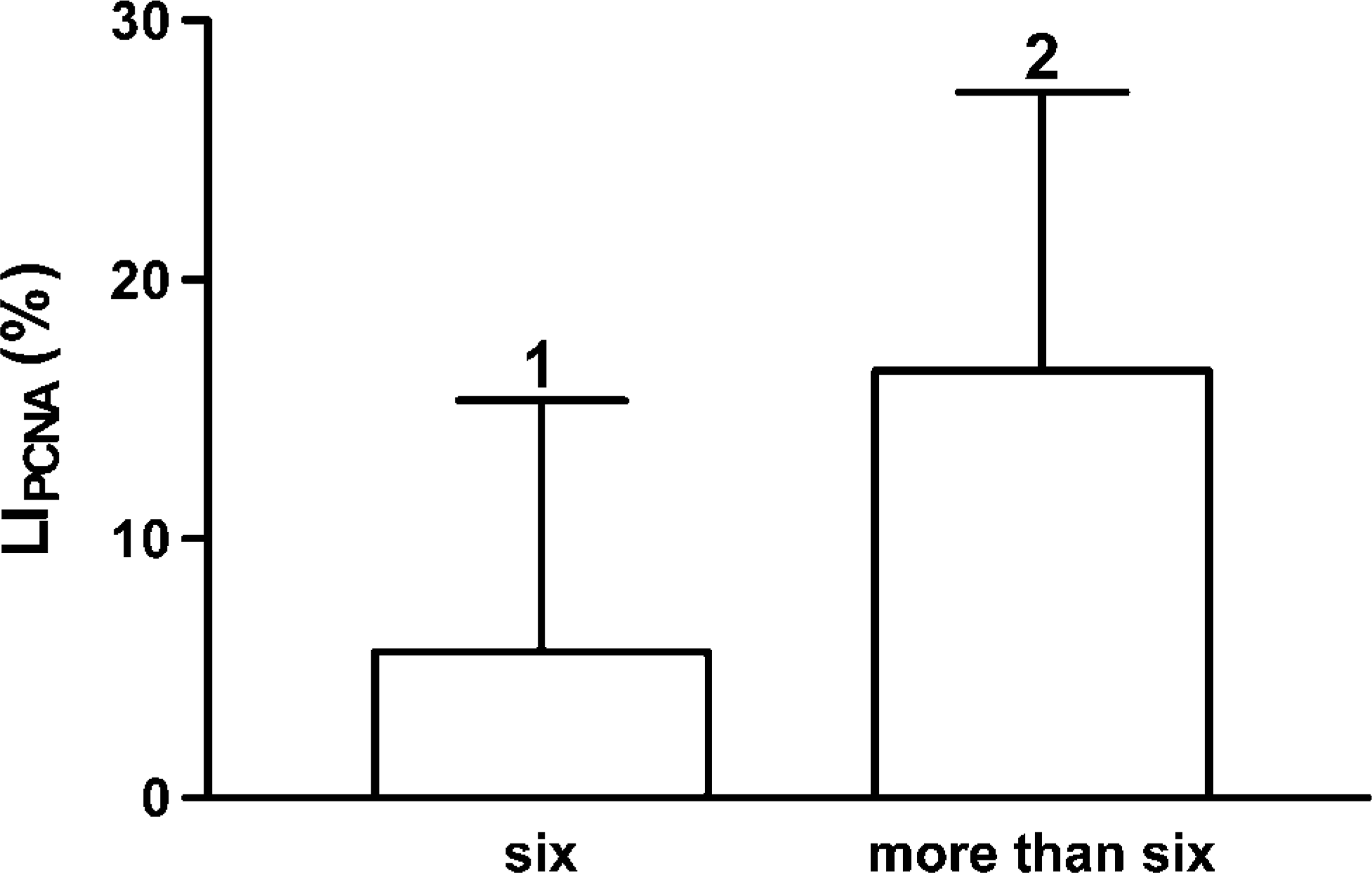
Bar graphs for LIPCNA for specimens of 6 months (six) and >6 months (more than six). Results are expressed as mean ± SD. Bars showing different numbers at the top of the error bars differ significantly (p<0.05).
For the first time, caspase-3 immunoexpression has been detected in the ventral prostate from rats, involving an increase (independent of the zinc intake) of caspase-3 immunoreactive cell density in dysplastic acini. This finding does not agree with that observed in man, where caspase-3 expression is decreased in prostate cancer (O'Neill et al. 2001; Winter et al. 2001).
Cell proliferation was clearly increased in dysplastic acini, independently of the presence of zinc. It seems that, when the dysplastic changes are present in the cadmium-exposed acini, the proliferation rate is equal to that observed in cadmium + zinc-exposed glands, as indicated in a previous study (Arriazu et al. 2005a). LIp53 is dependent on the presence of zinc, which could be related to the binding of zinc by p53 protein, promoting the interaction between DNA and p53 and, consequently, the cell-cycle arrest after DNA damage (Achanzar et al. 2001).
When exposure to metals is prolonged, some focal areas of the epithelium undergo transformation, resulting in upregulation of Bcl-2 expression, which produces an increase in focal resistance to programmed cell death. This increase, together with the proliferation, may induce the progression of these areas toward dysplasia, as has been postulated for human PIN (Martin et al. 2001).
Differences in the expression and localization of PCNA and Bcl-2 between rat and human prostates are remarkably pronounced. In normal human prostate, the proliferation compartment (the basal layer, PCNA, and Bcl-2 positive) is better defined than in rat prostate where the basal compartment is represented by isolated cells and often absent in some portions of the acini. Moreover, proliferative activity in the rat is not confined to the basal cells. It has been reported that the main proliferating cells in rat prostate are the columnar (luminal) cells (Sensibar 1995).
There is an inverse relationship between volume fraction of Bcl-2 and the numerical density of caspase-3 immunoreactive cells in both control and exposed rats. This supports the results of Munshi et al. (2001), who indicated that the increase of Bcl-2 expression caused the decrease of caspase-3 activity. However, the significant increase of LIp53 and NV caspase-3 detected in the dysplastic acini may be related to an increase in apoptotic activity, as described by Montironi et al. (1994) in human PIN.
Several neoplastic conditions in man (Martin et al. 1999) including prostate cancer (Fujikawa et al. 1995; Arai et al. 2001; Fischer et al. 2004) show an increase of nuclear υV that could indicate a more aggressive behavior for these lesions. In the present study, a significant increase of nuclear υV has been detected in rats exposed to cadmium and in the dysplastic tissue, independently of the treatment group. The change in nuclear size of dysplastic lesions may be a good marker of tumoral progression and could be correlated to the molecular and immunohistochemical changes observed in the evolution from normal to dysplastic epithelium (Bostwick 1996; Fischer et al. 2004).
We conclude that (a) a significant increase of cell proliferation occurs in animals treated with cadmium or cadmium + zinc for >6 months, (b) the exposure to zinc combined with cadmium had no effect over any of the variables studied when comparing with normal acini, (c) the increase of apoptosis in the dysplastic lesions was evident by the increment (independent of the zinc intake) of caspase-3 immunoreactive cell density, and (d) the increase of nuclear υV is indicative of a more aggressive behavior for pretumoral lesions.
Footnotes
Acknowledgements
This work was supported in part by grants 9/01 and 9/03 of the Universidad San Pablo-CEU.