
Abstract
The multicolor-banding (mcb) technique is a fluorescence in situ hybridization (FISH)-banding approach, which is based on region-specific microdissection libraries producing changing fluorescence intensity ratios along the chromosomes. The latter are used to assign different pseudocolors to specific chromosomal regions. Here we present the first three available mcb-probe sets for the Mus musculus chromosomes 3, 6, and 18. In the present work, the creation of the microdissection libraries was done for the first time on mouse/human somatic cell hybrids. During creation of the mcb-probes, the latter enabled an unambiguous identification of the, otherwise in GTG-banding, hardly distinguishable murine chromosomes.
A
Here we present for the first time the establishment of murine mcb-probe sets, as well as the establishment of mcb probes from somatic cell hybrids that was not previously reported.
The multicolor-banding technique is based on overlapping region-specific partial chromosome paints generated by glass-needle based microdissection (Chudoba et al. 1999; Liehr et al. 2002b). Each of the probes is based on 15–20 chromosomal fragments and the isolated DNA is amplified by degenerate oligonucleotide-primed polymerase chain reaction (DOP-PCR) (Telenius et al. 1992). Between four and eight micro-dissection libraries were created per mouse chromosome. The chromosomal location of the partial chromosome painting probes (pcps) as shown in Figure 1 was confirmed by reverse painting to normal murine chromosomes. Four to five different fluorochromes are used to label the pcps: SpectrumOrange, Fluorescein, TexasRed, Cyanine 5 (i.e., Cy5 coupled to avidin for detection of biotinylated probes) and diethylaminocoumarine (DEAC). Probe labeling was done by DOP-PCR again. All technical details of MCB/mcb are described in Liehr et al. (2002b).
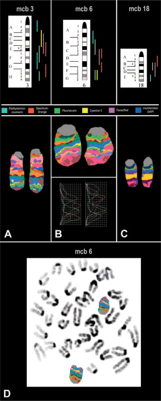
Scheme of the microdissection libraries used for achieving the multicolor banding (mcb) on murine chromosomes 3, 6, and 18 plus the corresponding mcb-pseudocolor banding. For mouse chromosome 6 the fluorochrome profiles are depicted, which are the basis for the mcb pseudocolors. The latter are obtained using the Isis software (MetaSystems; Altlussheim, Germany). (
Probe sets for the murine chromosomes 3, 6, and 18 were established from the cell lines SN11C5–3 sc1.3, N12C1, and SN19C8, respectively. These three mouse/human somatic cell hybrids contain one murine chromosome each, exclusively (Sabile et al. 1997). This enabled the unambiguous recognition of the wanted murine chromosome.
In summary, the creation of mcb probes from somatic cell hybrids is a very elegant approach that could be applied to banding in all species that have cytogenetically hardly distinguishable chromosomes.
Footnotes
Acknowledgements
This work was supported in part by the DFG (436 RUS 17/49/02 and 436 RUS 17/135/03), the INTAS (2143), and the Deutsche Krebshilfe (70–3125-Li1).