
Abstract
An immunocytochemistry (ICC) for the anticancer antibiotic daunomycin (DM) was developed using a combination of anti-DM serum produced against
T
To overcome each of these drawbacks and to develop an easy and generally applicable method revealing a precise localization or uptake of the drug in the cells and tissues, we established the present ICC method. In this study we used an anthracycline antibiotic daunomycin (DM) as a prototype, which has the characteristic of possessing an aliphatic primary amino group in the molecule and the autofluorescence as well. This first characteristic may permit the drug to be covalently fixed in situ with a fixative, and the second characteristic may be used to compare the presently developed ICC methods with the cytofluorescence method.
Materials and Methods
Chemicals
Daunomycin HCl, epirubicin HCl and adriamycin HCl were generously supplied by Meiji Seika Co. Ltd., Tokyo, Japan; Pharmacia Co. Ltd., Tokyo, Japan; and Kyowa Hakko Kogyo Co. Ltd., Tokyo, Japan, respectively. Glutaraldehyde (GA, 25% in water), formalin (37%) and Triton X-100 were obtained from Nacalai Tesque (Kyoto, Japan). Sodium borohydride (NaBH4) and protease (Type XXIV: Bacterial) were from Sigma-Aldrich (St Louis, MO).
Antibody
Anti-DM serum was produced in a rabbit against DM conjugated to bovine serum albumin (BSA) using a heterobifunctional agent
Synthesis of Daunomycin-glutaraldehyde-bovine Serum Albumin Conjugate (DM-GA-BSA)
BSA (10 mg) in 1.0 ml of 1 M sodium acetate and 1.0 ml of 3 mM GA were mixed and incubated at room temperature for 30 sec by stirring. DM (1 mg) in 1.0 ml of 1 M sodium acetate, pH 8.5, was then added to this mixture. This was followed by incubation for 30 min before 1 M monoethanol amine (1 ml) was added to terminate the reaction. The reaction mixture was further incubated for 30 min and was dialyzed against 10 mM Tris-HCl buffer, pH 7.2, for 6 hr at 4C.
EIA method
The wells in microtiter plates (Nunc F Immunoplates I; Nunc, Roskilde, Denmark) were coated by loading 100 μl of daunomycin-glutaraldehyde-bovine serum albumin (DM-GA-BSA) conjugate (10 μg/ml) in 10 mM Tris-HCl buffer, pH 8.5, containing 10 mM NaCl and 10 mM NaN3 and being left for 20 min at 37C. After they were rinsed with 10 mM phosphate buffer, pH 7.4, the plates were incubated with 100 μl of 60 mM phosphate buffer, pH 7.4, containing 1.0% casein and 0.1% NaN3 for 1 hr at 37C to avoid nonspecific adsorption of serum components in the samples. The wells were then incubated overnight at 4C with 100 μl of anti-DM serum, diluted to various degrees with 20 mM phosphate buffer, pH 7.0, containing 0.15 M NaCl, 0.05% Tween 20, and 0.02% BSA, followed by goat anti-rabbit IgG labeled with horseradish peroxidase (HRP, 1:2000) for 1 hr at 25C. The amount of enzyme conjugate bound to each well was measured with
EIA Inhibition Test
Wells in a microtiter plate were coated with 100 μl of the DM-GA-BSA conjugate (10 μg/ml) as described above. The wells were then incubated with 50 μl of a fixed concentration of anti-DM serum (1:5000) and 50 μl of different compounds (DM, adriamycin, epirubicin, bleomycin, pepleomycin, mitomycin C, streptomycin, or gentamycin) at various concentrations overnight at 4C, followed by incubation with goat anti-rabbit IgG (1:2000) for 1 hr at 25C. The bound HRP activity was measured as described above.
Cells
Human melanoma BD cells were grown on 18 × 18-mm coverslips in MEM medium with 10% calf serum (BioWhittaker; Walkersville, MD), penicillin (50 U/ml), and streptomycin (50 μg/ml) in a 95% air/5% CO2 atmosphere, and used 24 hr after plating. DM in aqueous stock solution at 1 mg/ml was then added to the cells to a final concentration (0.03 to 3.0 μg/ml) and further incubated for 2 hr at 37C.
Immunocytochemistry
The DM-uptake BD cells were washed in Mg2+- and Ca2+- free PBS and then immersion fixed in 1.0% to 3.0% GA in 0.1 M sodium phosphate buffer, pH 7.6, at 25C for 30 min. The specimens were treated with a series of 0.05% NaBH4 in TBS (50 mM Tris-HCl buffer, pH 7.4, containing 0.15 M NaCl) for 5 to 20 min, 0.5-4 N HCl for 30-90 min, and 0.001-0.004% protease in TBS for 30 min. During each process of the treatment the specimens were washed three times with TBS. Next, the specimens were blocked with a protein solution containing 10% normal goat serum, 1.0% BSA, and 0.1% saponin in TBS for 1 hr at room temperature and then directly incubated at 4C overnight with anti-DM serum (diluted 1:2,000-10,000 with the above protein solution supplemented with 0.1% Triton X-100). Specimens were washed with TBST (TBS containing 1% Triton X-100) three times, 5 min at a time, and then incubated with a horse-radish peroxidase-labeled goat anti-rabbit-IgG (whole IgG; Cappel, West Chester, PA) 1:500 or IgG/Fab' (MBL; Nagoya, Japan) 1:300 for 1 to 24 hr at 4C. After rinsing with TBS, the site of the antigen-antibody reaction was revealed for 10 min with diaminobenzidine and H2O2 (Fujiwara and Masuyama 1995).
Tissue Materials
Male Wistar rats (250 g body weight) were injected IV with DM at a dose of 4 mg in 0.8 ml of PBS. Twenty-four hr after the injection, the rats were anesthetized with pentobarbital intraperitoneally, perfused transcardially with 100 ml of saline for 2 min, and subsequently fixed by perfusion with 300 ml of 2% GA in 0.1 M phosphate buffer, pH 7.4, for 4 min at room temperature. Specimens of the liver were postfixed in the same fixative overnight at 4C and subsequently routinely embedded in paraffin. Five-μm paraffin sections were deparaffinized and then processed for DM ICC as described above.
Control Experiments
In the DM immunocytochemistry study, the specificity of immunostaining was ascertained by incubating control specimens with the secondary antiserum alone, the anti-mitomycin C serum (Fujiwara et al. 1982), and DM serum preabsorbed with DM-GMBS-BSA conjugate at a concentration of 10 μg/ml.
Fluorescence Microscopy
This was performed essentially according to the method of Rutherford and Willingham (1993). The DM-uptake human melanoma cell line sk-mel-37 cell specimens were either fixed with 3.7% formaldehyde in PBS for 10 min or GA (1-2%) in PBS for 30 min at room temperature, washed with PBS, and examined with an Olympus epi-fluorescence microscope equipped with a 100-W high-pressure mercury lamp light source and filters for excitation wavelength of 490 nm and for emitted light >515 nm.
Results
Antibody Dilution
Anti-DM serum produced against GMBS-conjugated DM (DM-GMBS-BSA conjugate) was characterized by an EIA system employing GA-conjugated DM (DM-GA-BSA conjugate) as the solid phase antigen. This EIA system may be useful as a model of DM ICC, because the ICC is based on the principle that DM in situ is coupled to the tissue proteins with the fixative GA through covalent bonds. As shown in Figure 1, significant binding activity was observed at serial dilutions of anti-DM serum, even at more than 10,000 times dilution. On the other hand, a much lower level of immunoactivity was seen in an EIA system using the solid phase antigen of BSA itself at the same concentration (10 μg/ml) (Figure 1), this showing binding activity of the antibody produced against the carrier BSA molecule of DM-GMBS-BSA conjugate used as the DM antigen. Thus, DM immunoreactivity was indicated as the remainder of their binding activity.
EIA Inhibition Test
This was achieved by the principle of competition between DM and analogs (free in the solution) and a fixed amount of DM-GA-BSA coated on EIA plates for the limited number of binding sites on the anti-DM antibody. Calibration curves were plotted showing the relationship between the concentrations of the analytes and the percentage of bound antibody, giving dose-dependent inhibition curves with DM, adriamycin, and epirubicin in a range between 100 pM and 500 μM. The dose required for 50% inhibition of binding was used as an indication of the strength of inhibition: 0.05 μM with DM, 0.07 μM with adriamycin, and 0.08 μM with epirubicin (Figure 2). No inhibition occurred with other drugs, bleomycin, pepleomycin, mitomycin C, streptomycin, or gentamycin (data not shown).
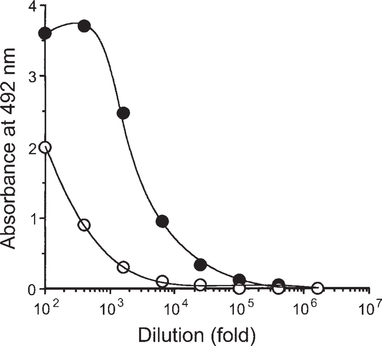
Dilution curves for anti-DM serum: several dilutions of anti-DM were incubated in microtiter wells coated with either DM-GA-BSA conjugate or BSA at a concentration of 10 μg/ml each overnight at 4C, followed by the procedure described for the EIA using HRP-labeled goat anti-rabbit IgG (whole) (diluted 1:2000) as the second antibody. Closed circle, DM-GA-BSA conjugate; open circle, BSA.
Conditions for DM ICC
A variety of experimental conditions necessary for DM ICC were examined using specimens of the human melanoma BD cells cultured on a coverslip, in which DM had been added to give a concentration of 1 μg/ml and then left in a CO2 incubator for 2 hr. The ICC required a series of pretreatments of the cell specimens prior to immunoreaction, such as cell fixation, reduction with NaBH4, treatment with HCl, and then protease treatment. Fixation with 1-3% GA for 30 min at room temperature resulted in intense immunostaining of the BD cells. However, most of the cells that had been fixed with 3.7% formaldehyde peeled off from the slides during the pretreatment with protease, although slight immunostaining occurred in the nuclei of the cells remaining on the slide. No DM was fixed in the cells by the fixation with methanol. Thus, fixation with 2% GA for 30 min was chosen for the present ICC.
The best experimental conditions for DM ICC were reached by determining an optimal concentration of sodium borohydride, HCl, and protease, and an optimal incubation time for their reaction with the cells. After several sets of ICC conditions were tried, it was found that a 5-min incubation with a 0.05% NaBH4 solution, a 30 min-incubation with 1 N HCl solution, and a 30 min incubation with a 0.004% protease solution provided an effective ICC system (Table 1). Also, the present indirect immunoperoxidase procedure for DM ICC needed Triton X-100 at a concentration of 0.1% in an antibody solution for both the first and second immune reaction (Table 1). Furthermore, it was revealed that horseradish peroxidase-labeled goat anti-rabbit IgG/Fab' was preferable to the whole IgG as the second antibody in the ICC (Table 1). On the other hand, when the pretreatment of either HCl or protease was omitted from the ICC protocol, DM immunostaining, especially in the nuclei, was very weak or non-existent (Table 1 and Figure 3b). Also, the elimination of borohydride reduction resulted in nonspecific staining of the cells, and to contrast, the reduction with 0.05% NaBH4 longer than 5 min resulted in weaker immunostaining.
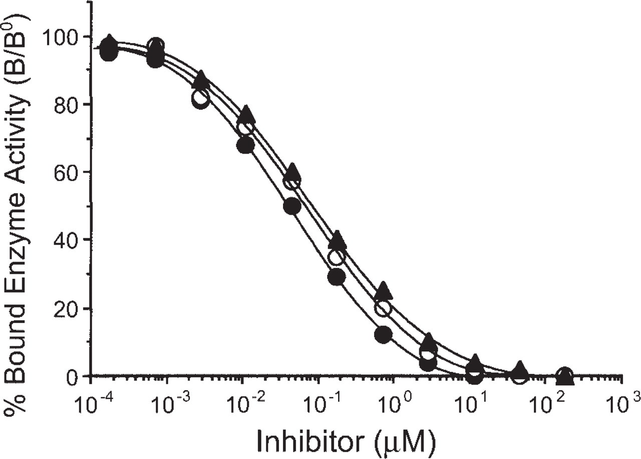
Reactivity of anti-DM serum as measured by its immunoreactivity in the EIA inhibition test. The curves show the amount (percentage) of bound enzyme activity (B) for various doses of DM (closed circles), adriamycin (open circles), or epirubicin (closed triangles) as a ratio of that bound using the HRP-labeled second antibody alone (B°).
Immunocytochemistry (ICC)
Under the presently established ICC conditions, strong immunoreactivity for DM was observed in the nuclei and in the perinuclear Golgi regions in the cytoplasm of the melanoma BD cells (Figure 3a). However, no immunoreactivity was observed in the wider parts of the cytoplasm outside the Golgi regions or in the nucleolei of the cells. Additionally, in the ICC specimens that omitted the hydrochloric acid pretreatment, we failed to detect DM immunoreactivity in the nuclei but were able to do so in the Golgi regions in the cells (Figure 3b). Conventional immunocytochemical staining controls (second level controls) were negative. The absorption controls for anti-DM serum showed that an addition of DM-GMBS-BSA conjugate at a concentration of 10 μg/ml into the serum abolished all staining (Figure 3c).
To compare the results of ICC, the cytofluorometric method was also employed using the melanoma cells pretreated with DM as above. In the cells fixed with 3.7% formaldehyde, the orange-colored fluorescence of a DM signal was clearly seen only in the nuclei of the cells (Figure 3d). Furthermore, in the cells fixed with 1% GA, strong autofluorescence of the green signal, most of which may be due to the autofluorescent mitochondria (Rutherford and Willingham 1993), occurred in the cells, making it difficult to distinguish it from a DM signal: the DM signal was very slight or nonexistent in the nuclei, though it was significant in the Golgi regions of the cells (Figure 3e). However, the green autofluorescence disappeared very rapidly after irradiation by excitation UV light, while some parts of the DM signal were also affected (Figure 3e).
Effects of sodium borohydride reduction, hydrochloric acid treatment, protease digestion, Triton X-100 treatment, and second antibody (goat anti-rabbit IgG/Fab' or whole IgG) on intensity of DM immunostaining a
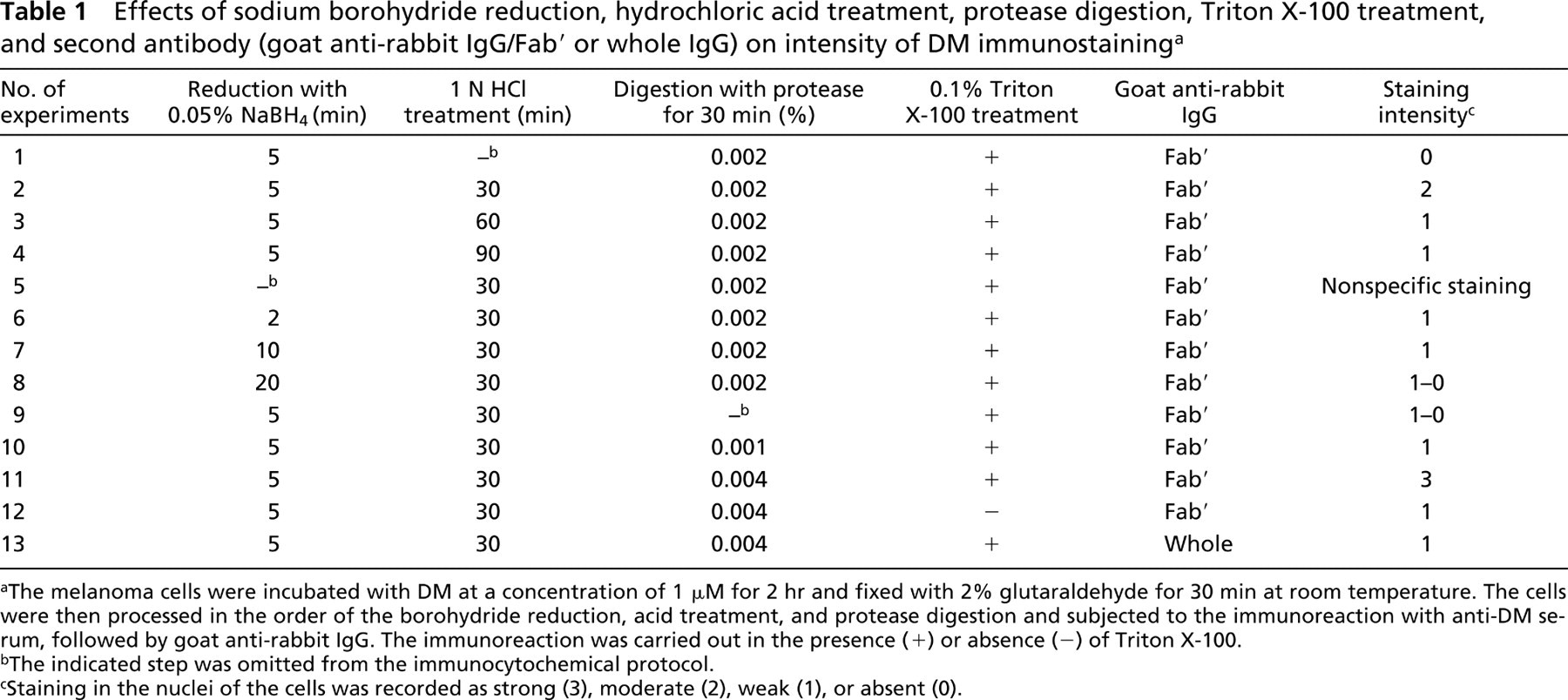
The melanoma cells were incubated with DM at a concentration of 1 μM for 2 hr and fixed with 2% glutaraldehyde for 30 min at room temperature. The cells were then processed in the order of the borohydride reduction, acid treatment, and protease digestion and subjected to the immunoreaction with anti-DM serum, followed by goat anti-rabbit IgG. The immunoreaction was carried out in the presence (+) or absence (−) of Triton X-100.
The indicated step was omitted from the immunocytochemical protocol.
Staining in the nuclei of the cells was recorded as strong (3), moderate (2), weak (1), or absent (0).
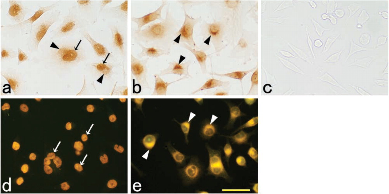
Immunostaining and fluorescence of DM in the DM-uptake human melanoma BD cells. DM immunoreactivity was seen both in the nuclei and paranuclear Golgi regions under the presently established ICC conditions
Specificity and Sensitivity of ICC
Anti-DM serum specifically stained for DM analog adriamycin or epirubicin taken up in the melanoma cells under the same conditions as for DM, the immunostaining pattern of each being completely the same as with DM (data not shown). The DM ICC was sensitive enough to stain for DM in the melanoma cells whose treatment with DM was as little as 30 ng/ml for 2 hr by the imidazole-3,3′-diaminobenzidine tetrahydrochloride procedure. No cross-reactivity was seen with any of the compounds tested (bleomycin, mitomycin C, or streptomycin). Using the cytofluorometric method for the cells fixed with 3.7% formaldehyde, DM could be detected in the cells that had been treated with as little as 100 ng of DM/ml for 2 hr. In the cells fixed with GA, on the other hand, no DM signal was observed in the cells pretreated even to the degree of 300 ng of DM/ml for 2 hr.
Evaluation of Tissue Staining
The optimal conditions for DM ICC established above were applied to ICC for the paraffin sections of the liver of rats 24 hr after an IV injection of DM. Strong immunoreaction for DM was observed in Kupffer cells, bile duct cells, smooth muscle cells surrounding the branch of the hepatic artery, and certain cells in the connective tissues of Glisson's capsule. Less strong staining was seen in the endothelial cells in the branches of the portal vein and hepatic artery (Figure 4a). Only very slight staining was found in the nuclei of the hepatic cells but almost not at all in the cytoplasm (Figure 4a). Conventional immunocytochemical staining controls (second level controls and absorption controls) were all negative (Figure 4b).
Discussion
A variety of quantitative chemical assays have been used for the pharmacological study of drugs using mainly urine, blood, and tissue homogenates from animals (Working and Dayan 1996; Minotti et al. 2004). Also, autoradiography has been an effective and generally applied method that has been employed for studies for the indirect localization of a drug in cells and tissues (Ono et al. 2003; Ke et al. 2004). With the intention of establishing an immunocytochemical method that has the advantage over autoradiography of being able to elucidate more directly, simply, and precisely the localization of drugs in cells and tissues, we have developed an ICC for DM as a prototype method for detecting drugs containing the aliphatic amino group in the molecule. DM is an anthracycline antibiotic that acts against a variety of human cancers (Danesi et al. 2002). However, its therapeutic use is limited by its unique cardiotoxicity, and the pharmacokinetics of the drug in physiological fluids has been extensively studied by specific and sensitive assays (Zagotto et al. 2001).
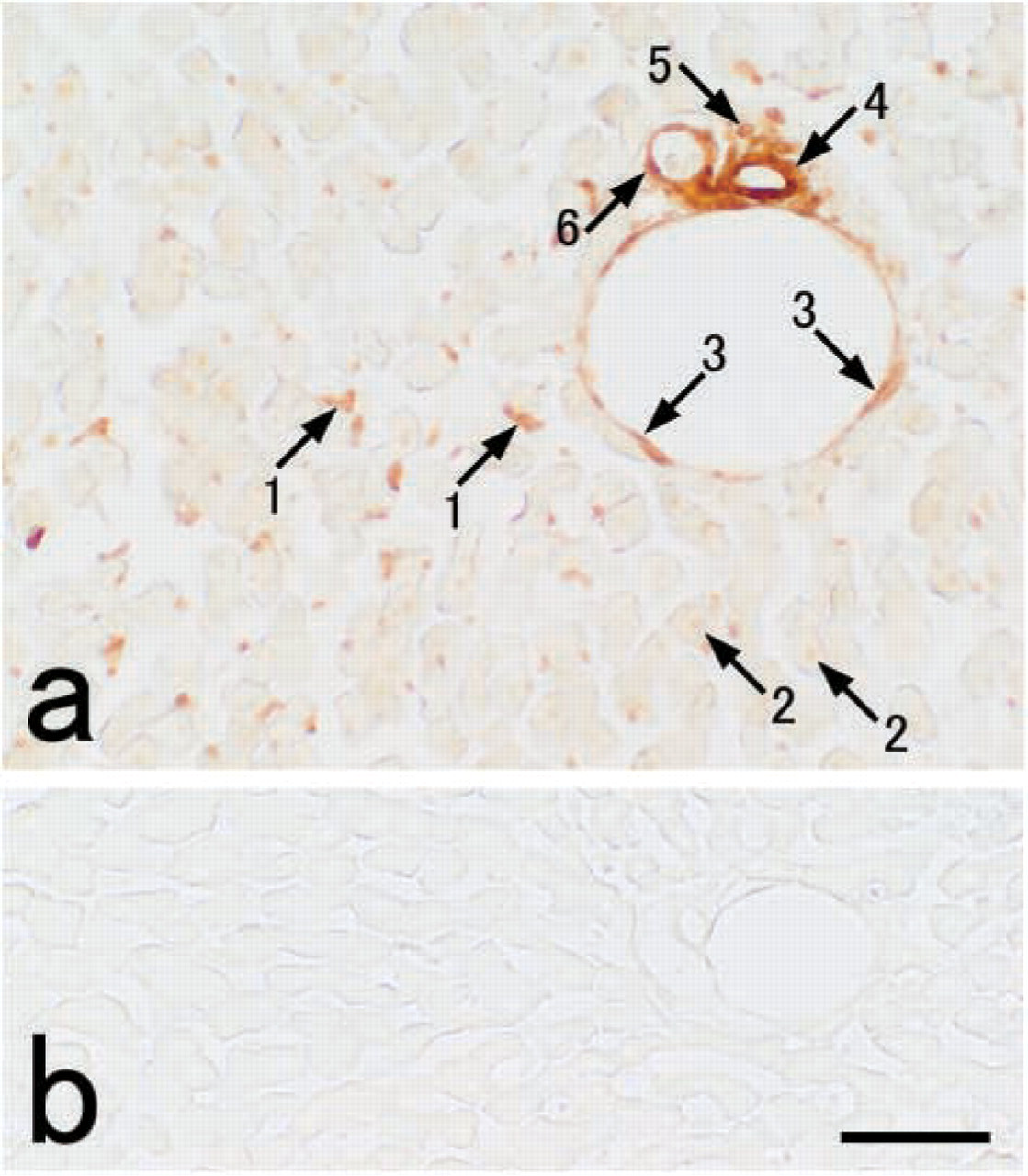
Rat liver stained for DM by immunocytochemistry (indirect peroxidase method). The staining
The detailed conditions for establishing immunocytochemistry for drugs have not yet been elucidated. The anti-DM serum used in the present ICC study was a sample of an antibody produced against GMBS-conjugated DM and, therefore, was tested for binding activity by the EIAs with GA-conjugated DM as the solid phase antigen (Figure 1 and Figure 2). These EIA tests could be useful if they indicate whether or not the antibody reacts with DM in cells and tissues used for the present ICC study using GA as a fixative. The anti-DM serum showed a much higher titered binding activity to DM-GA-BSA conjugate than BSA itself at the same concentration (Figure 1). Furthermore, it was demonstrated by the EIA inhibition test that the antibody binding was inhibited by DM to the highest degree, followed by the structurally related analogs adriamycin and epirubicin with a cross-reactivity of 71% and 62%, respectively (Figure 2). Also, no inhibition was evident with the other antibiotics, mitomycin C, bleomycin, pepleomycin, and actinomycin D (data not shown).
In ICC studies for small-sized drug molecules, a drug that has been specifically distributed according to its characteristic into the cells should be fixed in situ without redistribution during fixation. Thus, the fixation must be rapid and effective. Immersion fixation of the culture cells with 1-2% GA for 30 min fulfilled these requirements, as the immunocytochemical staining revealed a well-localized and specific reaction for DM in the nucleus and perinuclear Golgi region in the cytoplasm of the melanoma BD cells (Figure 3a). However, weaker fixatives often resulted in signs of diffusion artifacts such as weak nuclear staining and diffuse cytoplasmic staining (data not shown). The present immunostaining pattern agrees well with the results of the DM autofluorescence method. Specifically, the DM signal localized only in the nucleus of the cells when fixed with 3.7% formaldehyde, whereas it did so mainly in the perinuclear Golgi region (Figures 3d and 3e) with GA fixation, these being completely consistent with the results that Rutherford and Willingham (1993) reported for DM localization in the culture cells. In other ICC studies, GA has proved useful as a fixative for small molecular compounds with the amino group(s) such as the biogenic endogenous amines (Decavel et al. 1987; Nilsson et al. 1987; Fujiwara et al. 1996, 1998) and amino acids (Hepler et al. 1988; Popratiloff et al. 1996; Spirou and Berrebi, 1997).
Hydrochloric acid treatment (1 N for 30 min) and protease digestion (
Under the optimal conditions established for DM ICC, preliminary experiments were examined using paraffin sections of the liver of rats 24 hr after a single IV injection of DM. It was found that anti-DM serum immunostained for DM in Kupffer cells, the endothelial cells of blood vessels as well as their neighboring cells (smooth muscle cells, bile duct cells, and connective tissue cells) in the Glisson's capsule. This may suggest that Kupffer cells actively endocytize DM injected into the bloodstream. We are now undertaking toxicology studies on DM especially for its specific localization in the kidney and heart, which may help develop a better understanding of the mechanism for the unique renal and cardiac toxicity of DM (Tan et al. 1967; Bachur 1973; Lefrak et al. 1973; Steinherz et al. 1991) (Fujiwara K et al., unpublished data).
In conclusion, DM ICC method was newly developed with an advantage of being able to detect DM in the cells with a higher degree of sensitivity than the previously available cytofluorometric method. In addition, this ICC was demonstrated to be useful for the DM analogs adriamycin and epirubicin. The principle used in the present study for developing a DM ICC method might be applied to other drugs, especially for those containing primary amino group(s) in their molecules. Thus, these ICCs for drugs are direct, precise, and easy new methods that should have potential for pharmacology and toxicology studies of drugs, revealing the localization of the drug in cells and tissues.
Footnotes
Acknowledgments
This study was supported in part by a grant from the Japan Society for the Promotion of Science (15590148).
We are grateful to Dr Masashi Shin of Sojo University for valuable suggestions throughout this study.