
Abstract
To elucidate the behavior of autologously transplanted mesenchymal cells in osteochondral defects, we followed transplanted cells using green fluorescent protein (GFP) transgenic rats, in which all cells express GFP signals in their cytoplasm and nuclei as transplantation donors. Bone marrow-derived mesenchymal cells, which contain mesenchymal stem cells (MSCs), were obtained from transgenic rats. Then, dense mesenchymal cell masses created by hanging-drop culture were transplanted and fixed with fibrin glue into osteochondral defects of wild-type rats. At 24 weeks after surgery, the defects were repaired with hyaline-like cartilage and subchondral bone. GFP positive cells, indicating transplanted mesenchymal-derived cells, were observed in the regenerated tissues for 24 weeks although GFP positive cells decreased in number with time. Because GFP causes no immunological rejection and requires no chemicals for visualization, transplantation between transgenic and wild-type rats can be regarded as a simulation of autologous transplantation, and the survivability of transplanted cells are able to be followed easily and reliably. Thus, the behavior of transplanted mesenchymal cells was able to be elucidated in vivo by this strategy, and the results could be essential in future tissue engineering for the regeneration of osteochondral defects with original hyaline cartilage and subchondral bone.
I
We previously developed a transplantation model using Big Blue transgenic (Stratagene; La Jolla, CA) and Fisher 344 wild-type rats (Watanabe et al. 1999; Oshima et al. 2002). Each cell of these transgenic rats contains multiple copies of a bacteriophage lambda shuttle vector homozygously in the chromosomes, which included bacterial promoter lacI and lacZ genes (Dycaico et al. 1994; Wyborski et al. 1995). Since transgenes express no proteins, this transplantation model can be used as a simulation of autologous transplantation. In situ hybridization (ISH) technique was additionally performed to detect transgenes of the transgenic rat cells. By ISH, their nuclei are stained as a two-dot signal. To elucidate the survival of transplanted mesenchymal cells in osteochondral defects, transgenic rat-derived mesenchymal cells were transplanted into the defects. Then, signal positive cells indicating transplanted-derived cells were detected within both the cartilage and bone layers of the regenerated tissues at 24 weeks after surgery (Oshima et al. 2004). The strategy proved to be effective in following transplanted cells. However, some donor cells may not have stained as staining condition by ISH varies depending on the type of tissue and condition of the sections, for example, matrix type and section thickness. It was difficult to demonstrate, especially within the deeper portion of the defect, the relationship between donor and host. A new strategy displaying all transplanted cells in the section therefore needed to be established.
Green fluorescent protein (GFP) is a 27-kD protein, originally discovered in jellyfish Aequorea victoria. Since GFP is theoretically a non-rejected protein, immunological rejection is eliminated in the transplantation between female GFP transgenic and Sprague-Dawley (SD) wild-type rats, and this model can also be regarded as a simulation of autologous transplantation (Okabe et al. 1997; Ito et al. 2001). Because the fluorescence activity of GFP requires no substrates, cofactors, or additional gene products, cells expressing the intracellular GFP marker can be scored directly by fluorescent microscopy and/or flow cytometry without the requirement for antibody staining or extensive cell manipulation (Persons et al. 1998). Therefore, transgenic rat-derived cells are easily distinguished from wild-type rat-derived cells. Thus, it was hypothesized that the use of GFP transgenic rats as transplantation donors would be helpful in developing autologous transplantation examinations and in comprehending clearly the behavior of transplanted mesenchymal cells during the repair process of the defect. The objectives of this study were to develop a new strategy to follow autologously transplanted cells using GFP transgenic rats, as well as to elucidate the survivability of transplanted mesenchymal cells in osteochondral defect.
Materials and Methods
Animals
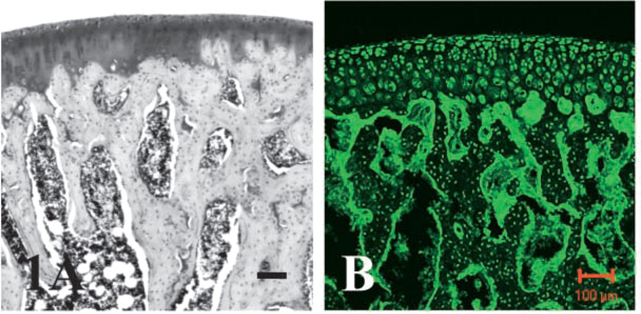
Twelve-week-old female GFP transgenic rats (n=4) (Japan SLC, Inc.; Hamamatsu, Japan) and sd wild-type rats (n = 32), genetically identical to each other except for their transgenes, were used (Okabe et al. 1997; Ito et al. 2001). In GFP transgenic rats, all cells including cartilage, bone, and bone marrow cells express GFP signals in their cytoplasm and nuclei, and the cells divided from GFP positive cells also express GFP signals. Thus, the distribution of cell bodies was visualized by a confocal laser scanning microscope (LSM 510 META; Zeiss, Jena, Germany), while no signal was detected in sections of wild-type rats (Figure 1). All experimental procedures employed in this study have been approved by the Committee for Animal Research at the Kyoto Prefectural University of Medicine, Japan.
Culturing of Mesenchymal Cells
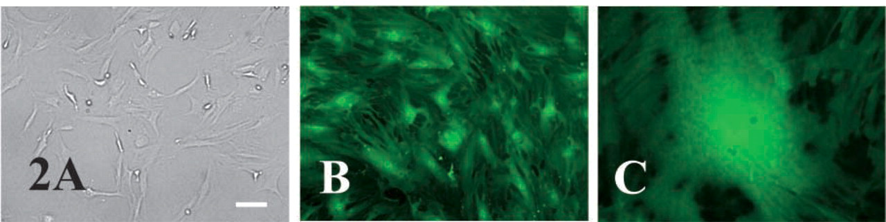
Female transgenic rats (n=4) were anesthetized by an intra-peritoneal injection of sodium pentobarbital (35 mg/kg of body weight) and killed. Bone marrow cells were removed from bilateral femora and tibias of transgenic rats under sterile conditions and then suspended in phosphate-buffered saline (PBS) containing 2% bovine serum albumin (BSA) (Serologicals Proteins Inc.; Kankakee, IL). The cell suspensions were centrifuged (200 × g for 5 min) to remove serum components. The sedimented cells were treated with sterile water for 10 sec to disrupt the erythrocytes. They were then resuspended in PBS containing 2% BSA and again centrifuged (200 × g for 5 min). The number of bone marrow cells was determined using a hemocytometer. Each rat yielded ∼3-5 × 10⁶ cells, including non-adhering ones and erythrocyte cells. The cells were transferred to 100-mm-diameter culture dishes (FALCON; Franklin Lakes, NJ) to which Dulbecco's modified Eagle Medium (DMEM) (GIBCO; Grand Island, NY) containing 10% fetal bovine serum (FBS) (ICN Biomedicals Inc.; Aurora, OH) and 0.06% kanamycin was added. The cultures were incubated at 37C in a humidified atmosphere containing 5% CO2. The medium was first changed 24 hr after seeding and then every 3 days during the 10-14 days of culturing to remove non-adhering cells. At the end of the culturing period, the growing fibroblast-like cells that were considered to be of mesenchymal origin were ∼80% confluent (Figure 2).
The cells were detached from the culture dishes by treatment with 0.05% trypsin and 0.02% ethylenediaminetetraacetic acid (EDTA) for 5 min. They were then counted in a hemocytometer.
Create Cell Masses
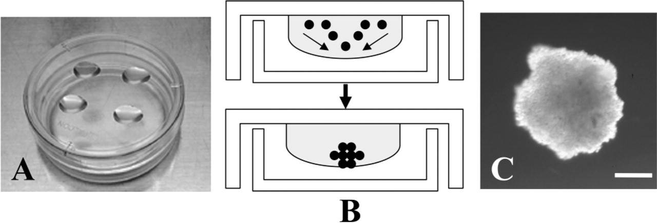
The mesenchymal cells were transferred to DMEM containing 10% FBS and 0.06% kanamycin and maintained in hanging-drop culture in a humidified atmosphere containing 5% CO2 at 37C for 60-72 hr. (Itoh et al. 1988; Iwagami et al. 1994) (Figure 3). Each 40-μl drop initially contained 4 × 10³ cells. Approximately 24-28 cell masses, each ∼0.6 mm in diameter, were obtained from a single rat.
Reverse Transcription-polymerase Chain Reaction (RT-PCR)
To examine whether mesenchymal cell masses express mRNA of differentiation markers for cartilage or bone tissue, RT-PCR was performed. Bilateral lower extremities of 1-day-old female sd rats were used as a positive control.
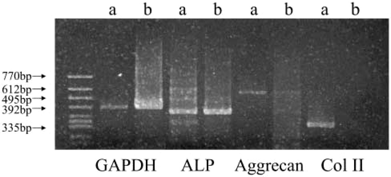
Cultured mesenchymal cell masses or tissues were homogenized and total RNA was isolated by Sepasol-RNA I (Nacalai Tesque, Inc.; Kyoto, Japan). Reverse transcription was carried out using ReverTra Ace (Toyobo Co. Ltd.; Osaka, Japan) to synthesize first-strand cDNA with isolated RNA of mesenchymal cells and whole lower extremities as a positive control. PCR was performed with oligonucleotide primer pairs (Invitrogen Japan K.K.; Tokyo, Japan) based on the sequences of the rat alkaline phosphatase (ALP) cDNA (sense: 5′-AGGCAGGATTGACCACGG-3′, antisense: 5′-TGTAGTTCTGCTCATGGA-3′), aggrecan cDNA (sense: 5′-GGCAACCTCCTGGGTGTAAG-3′, antisense: 5′-TGGGGTCCGTGGGCTCACAA-3′), type-II collagen cDNA (sense: 5′-CTGGCGAGTCTTGCGTCTAC-3′, antisense: 5′-GGCCCTCATCTCCACATCAT-3′), and GAPDH cDNA (sense: 5′-GGTGAAGGTCGGTGTGAACG-3′, antisense: 5′-CAAAGTTGTCATGGATGACC-3′). PCR was carried out with AmpliTaq DNA polymerase (Applied Biosystems; Foster City, CA), for 38 cycles under the following conditions: 94C for 1 min, followed by 55C for 1 min, followed by 72C for 1 min. The amplified PCR products were analyzed on 2% agarose gel electrophoresis and visualized by ethidiumbromide staining. The predicted sizes of PCR-amplified products were 440 bp (ALP mRNA), 702 bp (aggrecan mRNA), 313 bp (type-II collagen mRNA), and 497 bp (GAPDH mRNA) (Hamada et al. 1999; Wang et al. 2001).
Preparation and Treatment of the Osteochondral Defects in Wild-type Rats
Female wild-type rats (n = 32) were anesthetized by an intraperitoneal injection of sodium pentobarbital. An anterior midline incision was made through the skin of the right knee and the articular surface of the femur was then exposed using a medial parapatellar retinacular approach. The knee joint was immobilized in a deeply flexed position, and a cylindrical osteochondral defect, 1.5 mm in diameter and 3 mm in depth, was then created in the central weight-bearing surface of the medial femoral condyle by drilling with a 22-gauge needle. In the experimental group (n = 16), four separate masses of mesenchymal cells were introduced into the defects and affixed therein with fibrin glue (Beriplast, Aventis; Bridgewater, NJ). In the control group (n = 16), the defects were filled with fibrin glue alone. All rats were allowed to move freely after surgery in a temperature-controlled environment with a 12-hr light-dark cycle.
Evaluation of Histological Change and Survivability of Transplanted Cells
Two, 4, 12, and 24 weeks after surgery, the wild-type rats were anesthetized with sodium pentobarbital and then perfused with physiological saline and fixed with 4% paraformaldehyde (PFA) in 0.1 M phosphate buffer (PB). The distal portions of the femora were removed and immersed in the same fixative solution (4% PFA in 0.1 M PB) for 24 hr at 4C. They were then decalcified with 0.5 M EDTA (pH 7.5) for 2 weeks at ambient temperature, this procedure being followed by gradient replacement with 20% sucrose for 24 hr at 4C. The femora were then rapidly frozen cryosectioned in a cryostat (CM3050 S, Leica; Nussloch, Germany). The 14-μm-thick sections were stained either with 0.01% safranin O or with 0.05% toluidine blue in preparation for light microscopy. The following features were assessed: surface regularity, thickness of the cartilage layer, repair of the subchondral bone layer, matrix-staining of the cartilaginous and bony compartments, and integration of the repair tissue with the surrounding host tissues. The extent of the inflammatory response and osteophyte formation were also evaluated.
Macroscopic view (
To examine the survival of transplanted cells, GFP positive cells were detected on the serial sections of 2- to 24-week-models using a confocal microscope. Then, to assess the proportion of the positive cells in the tissues, the number of GFP positive cells and all the cells were calculated in randomly selected areas (0.14 × 0.21 mm) at a magnification of x160 in the merge image of laser and light microscope.
Results
RT-PCR
RT-PCR detecting GAPDH showed that cDNA from mesenchymal cell masses and whole lower extremities was synthesized at similar contents. RT-PCR showed that lower extremities of 1-day-old rat expressed mRNA of ALP, aggrecan, and type II collagen. On the other hand, mesenchymal cell masses expressed mRNA of ALP and faintly expressed aggrecan; however, mRNA of type II collagen was not expressed (Figure 4). Thus, they were not well-differentiated chondrocytes or osteocytes.
Macroscopic Observation
Symptoms of infection, immunological rejection, osteophyte formation and/or osteoarthritis in any of the knees in either group at any time period were not observed.
In the control group, as far as 24 weeks after surgery the margins of the defects were recognizable, and the surfaces were slightly depressed and irregular (Figure 5).
In the experimental group, at 2 weeks after surgery the margins of the defects were recognizable and the surfaces were also recessed, but at 24 weeks the margins were only slightly discernible, the surfaces were mostly smooth and not depressed, and the color of the surfaces was mostly the same as normal cartilage (Figure 6).
Evaluation of Histological Change and Survivability of Transplanted Cells
At 2 weeks after surgery in the control group, the margins of the defects were histologically clearly recognizable, the surfaces were slightly recessed, and the defects were filled with fibrous tissues, unaffected by safranin O or toluidine blue staining. Reconstruction with osseous tissues at the deeper layer was not observed (Figure 7A). At 4 weeks the margins were still clearly recognizable, the surfaces were slightly irregular, and the defects had not repaired with cartilaginous and osseous tissues (Figure 7B). At 12 weeks, there were two cases where a few areas of the defects had repaired themselves with cartilaginous tissues, faintly stained with safranin O; however, the defects were not repaired with cartilageous tissue in the other cases. On the other hand, the deeper layers of the defects were filled with osseous tissues (Figure 7C). At 24 weeks, there were two cases in which the shallow layers of the defects were stained with safranin O; however, the defects were not repaired with cartilageous tissue in the other cases. On the other hand, the deeper layers of the defects were filled with osseous tissues and integration to the surrounding host subchondral bone was observed (Figure 7D).
Results of RT-PCR, (
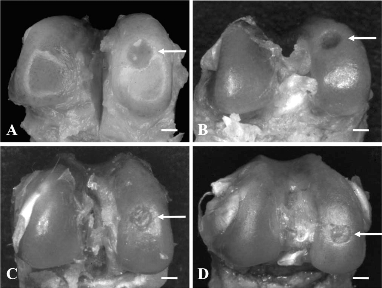
Macroscopic views of the control group of knees 2 weeks (
At 2 weeks after surgery in the experimental group, the margins of the defects were histologically clearly recognizable, the surfaces were irregular and recessed, and the defects were filled with fibrous tissues, unstained by safranin O or toluidine blue. However, the cellularity was very high and almost all the cells throughout the whole area of the regenerated tissue, ∼90-95% of all the cells were GFP positive cells, indicating transplanted mesenchymal-derived cells. On the other hand, invasion of GFP positive cells to surrounding tissue was not evident (Figures 8A-E).
At 4 weeks the margins were still recognizable, but the surfaces were not recessed and comparatively smooth. Regenerated tissues were composed of numerous cells in the whole area of the defects. These cells were deduced as chondrocytes because these cells were surrounded by a lacuna, and these areas were strongly stained with safranin O and showed metachromatic staining with toluidine blue indicating the existence of cartilaginous matrix. They were aligned irregularly and the thickness of regenerated cartilaginous tissue was thicker than normal cartilage. GFP positive cells did not spread uniformly; however, they were observed throughout the whole area and were ∼40-50% of all the cells at a maximum area. Though integration of regenerated tissues to host was not achieved, some GFP positive cells were observed in the surrounding host tissue (Figures 8B-F).
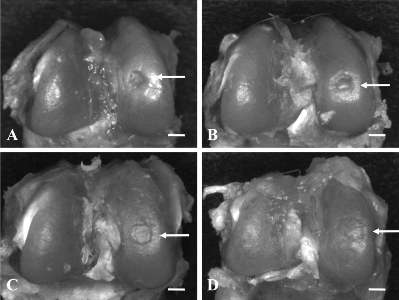
Macroscopic views of the experimental group of knees 2 weeks (
At 12 weeks, the margins were still recognizable; however, the surfaces were mostly regular and were not recessed. The thickness of regenerated cartilaginous tissue was slightly thicker than normal cartilage; however, underlying subchondral bone was remodeled and integration of regenerated tissues to host was found in the deeper layers of the defects. GFP positive cells, ∼20-25% of all the cells at a maximum, were extremely decreased in number, especially in the deeper layer, and were not detected in the surrounding tissues (Figures 8C-8G).
At 24 weeks the surfaces were mostly smooth and the shallow layer of regenerated tissue was stained with safranin O and showed metachromatic staining with toluidine blue. The alignment of chondrocytes was irregular, tidemark was not regenerated and the thickness of the cartilage layer was still slightly thicker than normal cartilage. Subchondral bone was highly remodeled in the deeper layer, and bony contact between the deeper layer of the remodeled subchondral bone and host bone was achieved. GFP positive cells were observed in both hyaline-like cartilage and subchondral bone; however, the number of GFP positive cells, ∼15-20% of all the cells at a maximum, was very small compared with those of 2-weeks model (Figures 8D-8H).
Discussion
Bone marrow-derived mesenchymal cells of GFP transgenic rats were transplanted into osteochondral defects. The defects were repaired with hyaline-like cartilage and subchondral bone, and some of the transplanted-derived cells survived in the regenerated tissues 24 weeks after surgery.
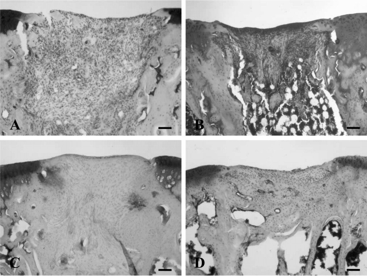
Photomicrographs of tissue sections (stained with safranin O) through the control group of defects 2 weeks (
To regenerate osteochondral defects, tissue transplantations such as periosteum, perichondrium, or osteochondral transplantation were previously performed (Amiel et al. 1985; Yamashita et al. 1985; O'Driscoll et al. 1986; Matsusue et al. 1993). Cell transplantations with chondrocytes or mesenchymal cells, for example, have also been applied, resulting in regeneration with hyaline-like cartilage and improvement in clinical symptoms (Brittberg et al. 1994; Ochi et al. 2002; Wakitani et al. 2002). Growth factor treatment and gene therapy as well have been attempted (Dodds et al. 1994; Flechtenmacher et al. 1996; Mason et al. 2000; Perka et al. 2000; Chubinskaya et al. 2001; Martinek et al. 2003). Nevertheless, regeneration to truly original tissues has not been fully accomplished biochemically and biomechanically, inclusive of tidemark, calcified layer, and underlying subchondral bone.
To improve these trials and obtain the regeneration of articular cartilage, the best resource for repair should be selected first. Compared with tissue transplantations, cell transplantations have been widely performed because it is possible to obtain cells with minor invasive operations and increase their number in vitro. However, the best resource for repair has not yet been determined. Moreover, when cells are used as a transplantation material, it is difficult to fix cells at transplant sites, due to joint motion and weight bearing. Thus, transplantation methods also need to be improved. While these matters are very important, what is foremost is the elucidation of the behavior and role of transplanted cells. Furthermore, the origin of regenerated tissues whether from transplanted cells or host cells has not been clarified. Several tools, such as radioisotopes, gene markers and cell-surface antigens, have been employed to follow the fate of autologously transplanted cells (Brecher et al. 1982; Morse et al. 1987; Lundberg et al. 1996). However, their use may be associated with one of several disadvantages, such as a temporal reduction in signal intensity, immunological rejection or selective marking. Thus, there have been no useful methods for following autologously transplanted cells. Recently, we reported the survival of transplanted mesenchymal-derived cells in both hyaline-like cartilage and subchondral bone layers of the regenerated tissues using Big Blue transgenic rats and ISH as a simulation of autologous transplantation (Oshima et al. 2004). However, it was technically difficult to stain all transgenic rat-derived cells and, thus, the behavior of transplanted cells in osteochondral defect with time has not been clearly demonstrated.
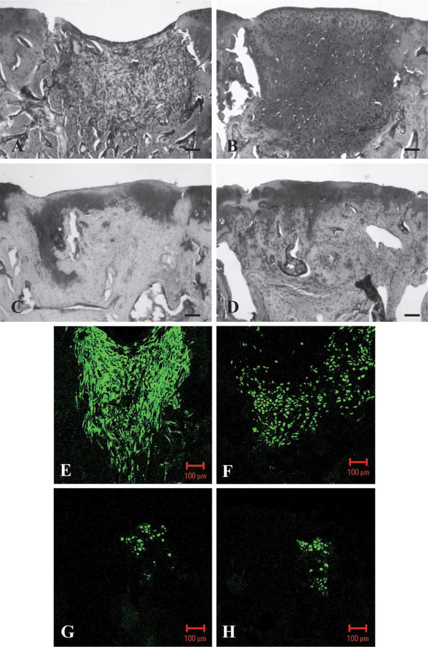
Photomicrographs of tissue sections (stained with safranin O or scanned by confocal laser microscope) through the experimental group of defects 2 weeks (
Mesenchymal cells with MSCs exist in bone marrow and have a capacity to differentiate into multiple mesenchymal-lineage cells, including chondrocytes and osteocytes (Caplan 1991; Lunstrum et al. 1999; Pittenger et al. 1999). Use of bone marrow cells for future clinical trials has an advantage in that they are obtainable with minor invasion. Further, osteochondral defect consists of subchondral bone layer as well as cartilage layer and, thus, we selected multipotent bone marrow-derived mesenchymal cells as a transplanted resource to repair the defects. To harvest MSCs from bone marrow, the density gradient medium and/or fluorescence-activated cell sorter analyses have been performed (Baddoo et al. 2003; Silva et al. 2003). However, these methods are not to select only MSCs, but to remove hematopoietic cells from bone marrow cells because definitive markers of MSCs have not been established. Some investigators only changed the medium of bone marrow cells to remove non-adhering cells for MSCs harvest (Huibregtse et al. 2000; Kotobuki et al. 2004). We used sterile water to rinse bone marrow cells for disrupting the erythrocytes and to change the medium during the monolayer culturing for removing non-adhering cells. For these procedures, the fibroblast-like mesenchymal adherent cells were obtained and we used them as mesenchymal cells, in which MSCs were included.
The results in the control group show that part of the defects were repaired by the invasion of cells from the surrounding tissue; however, the number of cells that have a capacity to repair the defects were few and, thus, the defects were not well repaired compared with the results in the experimental group. These results indicate the necessity for sufficient numbers of cells possessing the capacity to repair the defects. For this reason, we employed hanging-drop culture technique, originally developed for three-dimensional thymus cell culture, to fix high-concentrated transplanted cells at the transplanted sites and fibrin glue as a scaffold (Itoh et al. 1988; Iwagami et al. 1994). This transplantation strategy enables us to transplant dense mesenchymal cell masses into the defect rigidly. This culture method has no remarkable adverse influence on cell survivability; survival rate of mesenchymal cells did not decrease in number after the creation of masses, as evaluated by trypan blue staining.
The in situ hybridization technique used in our previous study to detect transgenic rat-derived cells is a cumbersome technique requiring multiple steps such as staining, washing, protease treatment, blocking and incubation. The technique utilized in this study using confocal laser microscopy alleviated these time-consuming steps making it easier to evaluate cells at multiple time points. We therefore were able to examine the behavior of transplanted mesenchymal cells as a function of time, which has never before been evaluated. Further, we performed RT-PCR of the mesenchymal cells before transplantation to elucidate their phenotype. The result showed that mesenchymal cells were not well-differentiated chondrocytes or osteocytes. Therefore, we suggested that the small number of survived transplanted cells could be MSCs and/or chondrogenic and osteogenic lineage cells, and they promoted the reconstruction of the defects especially at the early stage of the repair process.
In conclusion, the survivability and the behavior of GFP positive transplanted mesenchymal-derived cells were followed in this study. The results showed that mesenchymal cells have the potential to enhance the repair of osteochondral defects, and this strategy was a useful model to elucidate the behavior of transplanted cells in simulating autologous transplantation. Although the regeneration with hyaline cartilage and subchondral bone was not achieved, these results could be a basis in future tissue-engineering strategies for osteochondral repair.
Footnotes
Acknowledgements
This work was partially supported by a grant from the Japanese Ministry of Education, Science, Sports and Culture.