
Abstract
Breast cancers (BCa) with ERBB2 amplification show rapid tumor growth, increased disease progression, and lower survival rate. Deregulated intracellular trafficking and extracellular vesicle (EVs) release are mechanisms that support cancer progression and resistance to treatments. Neratinib (NE) is a Food and Drug Administration–approved pan-ERBB inhibitor employed for the treatment of ERBB2+ BCa that blocks signaling and causes survival inhibition. However, the effects of NE on ERBB2 internalization, its trafficking to multivesicular bodies (MVBs), and the release of EVs that originate from these organelles remain poorly studied. By confocal and electron microscopy, we observed that low nanomolar doses of NE induced a modest ERBB2 internalization along with an increase of clathrin-mediated endocytosis and of the CD63+ MVB compartment in SKBR-3 cells. Furthermore, we showed in the culture supernatant two distinct EV subsets, based on their size and ERBB2 positivity: small (30–100 nm) ERBB2− EVs and large (>100 nm) ERBB2+ EVs. In particular, we found that NE increased the overall release of EVs, which displayed a reduced ERBB2 positivity compared with controls. Taken together, these results provide novel insight into the effects of NE on ERBB2+ BCa cells that may lead to a reduction of ERBB2 potentially transferred to distant target cells by EVs:
Keywords
Introduction
ERBB2/Human epidermal growth factor receptor 2 (HER2) belongs to the ERBB family of receptor tyrosine kinases that includes epidermal growth factor receptor (EGFR)/ERBB1, ERBB2/HER2, ERBB3/HER3, and ERBB4/HER4. In physiological conditions, ERBB2/HER2 plays a major role in the development of the mammary gland, the peripheral nervous system, the heart, and epithelia.1,2 Unlike other receptors of the family, ERBB2 is an orphan receptor with no known ligand and chaperoned by the heat shock protein HSP90. ERBB2 is normally activated through growth factor–dependent heteromerization with other ERBBs. As a coreceptor, ERBB2 functions as a powerful signaling subunit that enhances signaling power and diversity. 1 At the cellular level, ERBB2 localization is restricted at the plasma membrane (PM) and it is considered an endocytosis-resistant receptor. The reasons why ERBB2 is resistant to downregulation have been the subject of intense debate that included the requirement of the chaperone HSP90 for its stability, an active retention mechanism mediated by flotillins, 3 the lack of internalization signals, and the ERBB2 expression itself that inhibits the formation of clathrin-coated pits (CCPs). 4 In contrast, other studies indicated that ERBB2 is internalized but very efficiently recycled. 5
Breast tumors with ERBB2 overexpression are detected in up to 20% of patients and are characterized by rapid tumor growth, increased disease progression, and poor prognosis.6,7 Deregulation of ERBB receptor signaling and intracellular trafficking is a common feature of many cancers, including breast. 1 Cancer cells show altered intracellular and extracellular vesicular trafficking, contributing to tumor progression and therapy resistance. In particular, breast cancer (BCa) cells not only present endocytic pathway abnormalities 8 but also potentiate extracellular vesicle (EV) release to support cancer growth and progression. 9 The development of targeted therapies like the therapeutic monoclonal antibody trastuzumab (Tz) and derivatives as well as tyrosine kinase inhibitors has improved the outcome of ERBB2+ BCa patients. Despite that, approximately one third of women diagnosed worldwide with BCa still die from the disease, mostly from secondary metastasis.10–12 Therefore, there is an urgent need to comprehend the underlying biology of targeted therapies to circumvent resistance. 13
Neratinib (NE, also known as HKI-272) is an irreversible inhibitor of the tyrosine kinase activity of the ERBB family inhibitor members of ERBB2, EGFR/ERBB1, and ERBB4 that was approved in 2017 for extended adjuvant treatment of early-stage ERBB2/HER2-positive BCa based on the ExteNET trial. 14 The final results of this trial demonstrated an improved invasive disease-free survival in patients with ERBB2/HER2-positive BCa. 15 To date, NE is also under clinical evaluation for lung, colorectal, and bladder cancers, as well as diabetes.14,16–21 It has been shown that NE is selectively active in ERBB2+ amplified compared with non-amplified BCa cell lines at very low nanomolar IC50 values; 12 however, many in vitro studies performed on breast and other cancer cell types showed both ERBB2 tyrosine kinase inhibition and its proteolytic degradation at submicromolar doses of NE (from 100 to 500 nM).22–24 As earlier findings, supported by more recent ones, clearly showed that NE targets many additional kinases, especially serine/threonine kinases of the Ste20 family,25,26 we believed that it was therefore essential to minimize possible off-targets effects to correctly interpret specific biological responses to this drug.
It is a common opinion that the aggressiveness of ERBB2 is linked to the its poor endocytic rate and lysosomal degradation, which is at least partially due to the HSP90 chaperone stabilizing activity on the receptor.4,27 ERBB2 resistance to downregulation via endocytosis and the propensity of cancer cells to alter intracellular and extracellular vesicular trafficking are likely contributing factors to tumor progression and therapy resistance. In fact, BCa cells not only present endocytic pathway abnormalities 8 but also release EVs to support cancer growth and progression. 9 EVs are released by many cells in both physiological and pathological conditions in the extracellular environment to mediate intercellular communication.
Based on biogenesis and sizes, three main classes of EVs have been defined: exosomes (30–150 nm), microvesicles (MVs, 200–2000 nm; also termed ectosomes, MVs, or oncosomes), and apoptotic bodies (ApoBDs, 400–2500 nm). Exosomes are membrane vesicles that originate from inward budding within the endosomal/lysosomal pathway, and are released from the cells into the microenvironment upon fusion of multivesicular bodies (MVBs) with the PM. Instead, MVs are formed through direct outward budding of the PM. The formation of ApoBDs is an important process downstream of apoptotic cell death and is considered a hallmark of apoptosis. ApoBDs are released by dying cells, and their size ranges between 500 nm and 2 µm. 28 In the cancer context, EVs collected from body fluids are considered reliable markers for screening, diagnosis, and therapy monitoring of different types of cancer. 29 Exosomes derived from ERBB2+ SKBR-3 and BT474 BCa cells, as well as exosomes derived from serum of ERBB2+ BCa patients, display on their membrane active ERBB2 able to bind Tz reducing the drugs’ effectiveness. 30 Recently, we also showed that Tz modulates 51 proteins associated with organelle organization, cytokinesis, and response to lipids in EVs derived from SKBR-3 cells, suggesting that Tz may influence these cellular processes in distant sites. 31
The types of cargo and signal transmitted by EVs have been associated with their size and morphological features, which therefore were proposed as factors affecting the mechanisms of communications between cells. 32 Thus, a characterization of vesicles derived from ERBB2+ BCa cells is relevant to understand their role in the progression of BCa.
Here we used light and electron microscopy (EM) techniques in ERBB2+ BCa cells to investigate the effects of low nanomolar doses of NE, used to minimize off-targeting, on the cellular compartments involved in ERBB2 trafficking and EV release, and to characterize the morphology and ERBB2 content of the two subpopulations of EVs derived from ERBB2+ SKBR-3 cells.
Materials and Methods
Cell Culture and NE Treatment
ERBB2+ overexpressing SKBR-3 cells and ERBB2-negative MFC7 cells were obtained from Banca Biologica and Cell Factory in IRCCS Ospedale Policlinico San Martino belonging to the European Culture Collections’ Organization. SKBR-3 and MFC7 cells were cultured in complete medium (DMEM high glucose supplemented with 10% heat-inactivated fetal bovine serum, 1% glutamine, penicillin, and streptomycin [Euroclone S.p.A.; Milan, Italy]). NE (Nerlynx; Puma Biotechnology, Inc., Los Angeles, CA) was purchased from Sigma-Aldrich (CAS Number: 698387-09-6; St. Luis, MO) and dissolved in DMSO at 50 µM. A final concentration of 6-nM NE was used to treat SKBR-3 and MFC7 cells for 72 hr. This treatment effectively inhibits cell survival at 50% compared with control condition (0.012% DMSO) (not shown). Higher concentrations of NE were employed when indicated.
EV Isolation
The purification of the EVs from cell culture medium was carried as follows. The EVs were isolated by sequential centrifugation starting from 95% confluent SKBR-3 plates. Culture media was collected and subjected to a first centrifugation at 300 × g for 10 min at 4C. The supernatants thus obtained were centrifuged again at 2000 × g for 30 min at 4C to eliminate debris and any cells in suspension. Then, the supernatant was filtered (0.22-µm mesh) and transferred into ultracentrifuge tubes (Beckman) to be ultracentrifuged by Optima XPN-100 Beckman Coulter Ultracentrifuge at 100,000 × g for 2 hr at 4C (rotor SW28; Beckman Coulter Inc, Brea, CA). At the end of this phase, the supernatant was removed and the pellet resuspended in PBS filtered and subjected to a second ultracentrifugation at 100,000 × g for 1 hr and 30 min at 4C (SW55ti rotor). Finally, the EVs were resuspended in filtered Dulbecco phosphate-buffered saline (DPBS), fixed in 3.7% paraformaldehyde (PFA) in 2% sucrose in DPBS and stored at 4C if used for EM or resuspended in filtered DPBS 1× and stored at −80C if dedicated to Western blot analysis.
Immunofluorescence Imaging
For immunofluorescence analysis, SKBR-3 cells were treated for 72 hr with 6-nM NE (single-dose treatment), fixed in 3% PFA in PBS pH 7.4 for 20 min and then quenched with 30-mM NH4Cl. Cell permeabilization was performed with 0.2% saponin in 0.1% BSA for ERBB2 (9G6, sc-08; Santa Cruz Biotechnology, Dallas, TX), CD63 (H5C6; Developmental Studies Hybridoma Bank, Iowa City, IA), and LAMP1 (H4A3, Developmental studies Hybridoma Bank) staining. Cells were washed 3× with PBS and incubated with secondary antibodies conjugated to Alexa Fluor 546 or 488 (Thermo Fisher Scientific; Waltham, MA) for 20 min at room temperature. Coverslips were washed 3× in PBS and mounted using a mounting media containing 4′,6-diamidino-2-phenylindole (DAPI) (ProLong Gold with DAPI, Thermo Fisher Scientific) to visualize nuclei. Image acquisition and deconvolution were performed using an Axio Imager A2M microscope equipped with an Apotome module (Carl Zeiss; Jena, Germany). For endocytic assays, acetylated LDL (Ac-LDL) conjugated with Alexa488 (Thermo Fisher Scientific) was diluted into culture medium to 10 µg/ml and incubated for 2 hr at 37C. Cells were then processed for immunofluorescence and labeled as described. Endocytic assay analysis has been carried out using Icy software (version 2.1.4.0; Institut Pasteur, Paris, France; http://icy.bioimageanalysis.com). Ac-LDL mean spot intensity and number of spots per cell were automatically analyzed using the spot detector tool within manually drawn region of interests (ROIs) for each cell. The results were plotted as histograms (mean ± SEM).
Transmission EM
SKBR-3 cells were incubated with 5-nm BSA-gold (5 OD) (Cell Microscopy Core; Utrecht, The Netherlands) for 2 hr at 37C, then washed out twice in 0.1-M cacodylate buffer, and fixed in 0.1-M cacodylate buffer containing 2.5% glutaraldehyde (Electron Microscopy Science; Hatfield, PA) for 1 hr at room temperature. The cells were postfixed in 1% osmium tetroxide for 10 min and 1% aqueous uranyl acetate for 1 hr. Subsequently, samples were dehydrated through a graded ethanol series and flat embedded in epoxy resin (Poly-Bed; Polysciences, Inc., Warrington, PA) for 24 hr at 60C. Ultrathin sections (50 nm) were cut parallel to the cell monolayer and counterstained with 5% uranyl acetate in 50% ethanol. Electron micrographs were acquired using a Hitachi 7800 120-kV electron microscope (Hitachi; Tokyo, Japan) equipped with a Megaview G3 digital camera and Radius software (EMSIS; Muenster, Germany).
For EM morphometry, we defined as CCPs/clathrin-coated vesicles (CCVs) all the CCPs attached to the PM and as putative vesicles, those located within 200 nm from the PM. Because NE did not affect the size of the cells (not shown), it was not necessary to normalize the number of CCP/V counted for the linear length of the PM. The cell surface was compared using ImageJ software. Ten cell profiles for each condition were analyzed in an unbiased manner.
Negative Staining of EVs
Electron microscopic analysis on isolated vesicle preparations was performed as follows. The EV preparations were fixed by adding 2% PFA in 0.1-mol/L phosphate buffer (pH 7.4) in the same volume of EV resuspension buffer. Five µl drops of EVs were then adsorbed for 10 min on formwar-carbon-coated copper grids. Subsequently, grids were rinsed in PBS and negatively stained with 2% uranyl acetate for 5 min at room temperature. Stained grids were finally embedded in 2.5% methylcellulose for improving preservation and air-dried before examination. Electron micrographs were acquired using a Hitachi 7800 120-kV electron microscope (Hitachi) equipped with a Megaview G3 digital camera and Radius software (EMSIS). Size was calculated using the arbitrary line function embedded in the measurement dialog box of Radius software (EMSIS).
Immunolabeling of EVs
Five µl of concentrated solution of EVs was dropped on formwar-coated copper grids and incubated for 20 min at room temperature. After washing in PBS for 5 min, EVs were fixed in 1% glutaraldehyde for 5 min. A second wash in PBS and two passages in 0.2% glycine in PBS for 5 min each were then performed to saturate the nonspecific reactive sites. We then blocked grids with 1% BSA in PBS for 5 min and then proceeded with an incubation incubated with the mouse monoclonal anti-ERBB2 antibody (9G6, sc-08, Santa Cruz) for 30 min at room temperature. After four washes in 1% BSA in PBS of 2 min each, grids were incubated with 10-nm protein A gold (PAG) for 20 min. After the incubation with the PAG, grids were washed for 4× in 1% BSA in PBS of 2 min each and then postfixed in 1% glutaraldehyde for 5 min. Grids were then further washed in PBS and in distilled water for 5 min. At this point, the samples were counterstained with 2% uranyl acetate in 0.15-M oxalic acid for 5 min at room temperature. The procedure was completed with a passage in 2.5% methylcellulose in uranyl acetate 4% for 5 min at room temperature, which allows to confer further contrast and to form a beam protective layer on the grids.
Immunoblot Analysis
For Western blotting, SKBR-3 cells were lysed using lysis buffer (Hepes pH 7.4 20 mM, NaCl 150 mM, 10% glycerol, 1% Triton X-100) supplemented with protease inhibitors cocktail cOmplete (Roche Applied Science; Penzberg, Germany) and sodium orthovanadate and PhosStop (Roche). Proteins were resolved on SDS-PAGE (Thermo Fisher Scientific) and blotted on nitrocellulose (Amersham, GE Healthcare Life Science, Chicago, IL) or polyvinylidene fluoride (Merck Millipore; Darmstadt, Germany) membranes. Detection was performed using an enhanced chemiluminescent (ECL) Detection Reagent (BIORAD, Hercules, CA) according to manufacturer’s protocol. ECL signals were detected, recorded, and measured with the Uvitec Cambridge gel doc system and software (Cambridge, UK). ERBB2 was detected with antibodies anti-ERBB2 N-terminal (Ab-20, Thermo Fisher Scientific) and anti-ERBB2 C-terminal (C-18, sc-284, Santa Cruz Biotechnology). Antivinculin antibody (V9131, Sigma) was used as loading control.
Statistical Analysis
All parameters measured are presented as mean ± SEM and were analyzed with two-tailed Student’s t-test or the non-parametric Mann–Whitney U test. Values of p<0.05 were considered statistically significant.
Results
ERBB2 is Trafficked to Late Endocytic Compartments Under NE Treatment
It has been reported that ERBB2 is poorly internalized in basal conditions but can be internalized and degraded in lysosomes upon pharmacological inhibition. 5 NE triggers rapid ERBB2 internalization and lysosomal degradation at high concentrations (500 nM) in BCa cells. 22 To minimize possible off-target effects of the drug, we first determined the IC50 dose of NE during 72 hr of treatment which was found to be 6 nM (not shown). We then investigated the effects of NE on ERBB2 internalization, endocytic trafficking, and EV release in SKBR-3 cells using this concentration. We performed double immunofluorescence studies to localize ERBB2 with early (EEA1+) and late (CD63+) endosomal compartment markers after 72 hr of NE treatment. Results showed that ERBB2 localized mostly on the PM and at a lesser extent with EEA1-positive early endosomes (EEs) and CD63-positive compartments, suggesting that only a fraction of ERBB2 is trafficked to MVBs, which represent the organelle in which biogenesis of exosomes occurs (Fig. 1A). Accordingly, immunoblot analysis showed a slight decrease of ERBB2 protein levels occurring within 2–6 hr of NE treatment that returned to control levels within 72 hr (Fig. 1B). In contrast, a high dose of NE (500 nM) induced a substantial internalization of ERBB2 and a dramatic decrease of the 185-kDa full-length ERBB2 protein, strongly suggestive of protein degradation (Supplementary Fig. 1A and B).
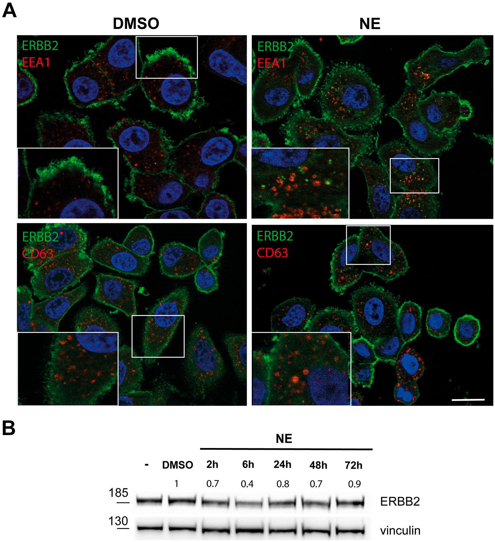
NE modulates ERBB2 trafficking in SKBR-3 cells. (A) Representative confocal microscopy images showing SKBR-3 treated for 72 hr with DMSO or 6-nM NE stained with antibodies against the ERBB2 extracellular domain (green signal), EEA1 (red signal), and CD63 (red signal). Nuclei were stained with DAPI (blue signal). Colocalization is shown as a merged yellow signal. Insets show signal colocalization at a higher magnification. Scale bars: 20 μm. (B) Cell lysates from SKBR-3 cells treated with DMSO or 6 nM for 2, 6, 24, and 72 hr and subjected to immunoblot analysis with antibodies against ERBB2. Vinculin was used as loading control. On the left side of the panel, the migration of protein molecular mass standards expressed in kDa is shown. The blot shown is representative of three independent experiments. Abbreviations: NE, neratinib; DAPI, 4′,6-diamidino-2-phenylindole.
NE Impacts on CME Endocytosis
A body of literature suggests that eukaryotic cells maintain up to five distinct, constitutive pinocytic pathways. These are clathrin-mediated endocytosis (CME), caveolae, and three non-caveolar clathrin-independent endocytic routes. 33 As the CME route is the most represented endocytic pathway in SKBR-3 cells and ERBB2 entry in these cells follows this pathway under pharmacological inhibition, 4 we sought to investigate whether NE treatment may impact this pathway. To this aim, we performed a morphometric analysis by EM of PM profiles from 10 randomly chosen SKBR-3 cells to score CCP/V in DMSO- and NE-treated cells. Interestingly, we found that NE significantly increased the assembly of CCP/V at the cell surface (Fig. 2A and B). This result prompted us to verify whether CME endocytosis was modulated by NE. We performed an endocytic assay using Alexa488-conjugated Ac-LDL as a specific endocytic tracer for CME. As shown in Fig. 2C and D, SKBR-3 cells showed a significant increase of internalized Ac-LDL after 72 hr of NE treatment, suggesting that NE impacts CME endocytosis. In contrast, no differences in endocytosis of Alexa488-conjugated Ac-LDL were found in ERBB2− MFC7 cells, suggesting that NE endocytic modulation depends upon ERBB2 and is not an off-target effect of the drug (Fig. 2E and F).
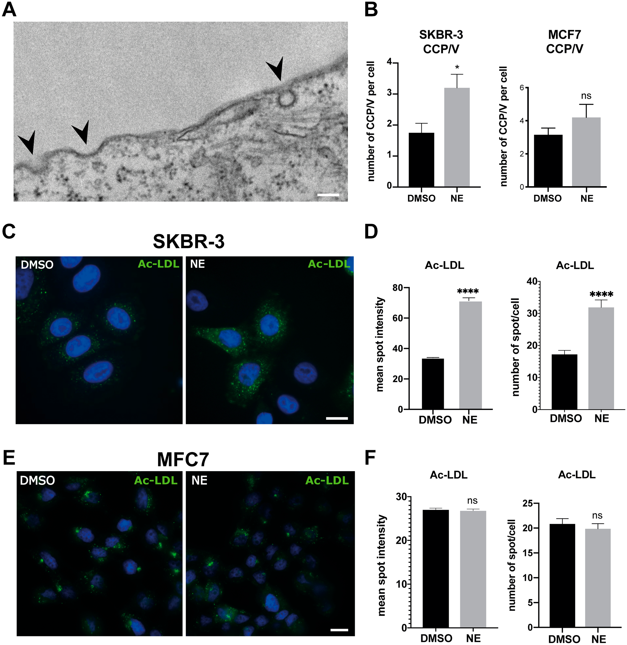
(A) Electron micrograph showing the cell surface of SKBR-3 cells. Black arrowheads point to clathrin-coated pits (CCPs) and clathrin-coated vesicles (CCVs). (B) Morphometry of CCP/Vs. Plasma membranes of 10 randomly chosen SKBR-3 cells were screened to count CCPs and CCVs. We considered as plasma membrane-derived CCVs those vesicles detected at the cell surface and within 200 nm from it. Data are expressed as mean ± SEM. Unpaired t-test, *p=0.028. Scale bar: 100 nm. (C) Representative immunofluorescence images of SKBR-3 cells treated with NE or DMSO and then incubated with 10-µg/ml Alexa488 Ac-LDL for 2 hr at 37C. Cells were then fixed and costained with DAPI to detect nuclei. Scale bar: 10 μm. (D). Graphs showing quantification analysis of Ac-LDL dots intensity in NE- versus DMSO-treated cells. Data are expressed as mean ± SEM from two independent experiments in which 50 cells were analyzed for each condition. Unpaired t-test, ****p<0.0001. (E) Representative immunofluorescence images of MFC7 cells treated with NE or DMSO and then incubated with 10-µg/ml Alexa488 Ac-LDL for 2 hr at 37C. Cells were then fixed and costained with DAPI to detect nuclei. Scale bar: 10 μm. (F). Graphs showing quantification analysis of Ac-LDL mean dots intensity and number of spots/cells in NE- versus DMSO-treated cells. Data are expressed as mean ± SEM from two independent experiments in which 100 cells were analyzed for each condition. Unpaired t-test, not significant. Abbreviations: SEM, standard error of the mean; NE, neratinib; Ac-LDL, acetylated LDL; DAPI, 4′,6-diamidino-2-phenylindole.
NE Expanded CD63+ Late Endosomal Compartments
To test whether the observed increase of CME capacity reflected a parallel expansion of the endocytic compartments, we performed immunofluorescence analysis using EEA1, a well-known marker for EEs, and the tetraspanin protein CD63, which is a marker for late endosomes/MVBs. In particular, EEA1+ EEs represent a sorting station in which endocytosed cargoes are routed to recycling or to degradation, whereas CD63 is a key factor in EV production and endosomal cargo sorting. CD63 is predominantly localized to the intraluminal vesicles (ILVs) of late endosomes and MVBs and is thus enriched on exosomes. After 72 hr of NE treatment, SKBR-3 cells showed a significant increase in the number of CD63+ endosomes but not of EE, suggesting that NE influences MVB/late endosomal maturation and/or biogenesis (Fig. 3A). To better characterize this phenotype, after 72 hr of NE treatment, SKBR-3 cells were further probed for 2 hr at 37C with 5-nm gold conjugated BSA, a widely used endocytic tracer to visualize endocytic compartments, and then processed for EM. Upon morphometric analysis, we confirmed that NE significantly increased the number of MVBs/late endosomes (Fig. 3B).
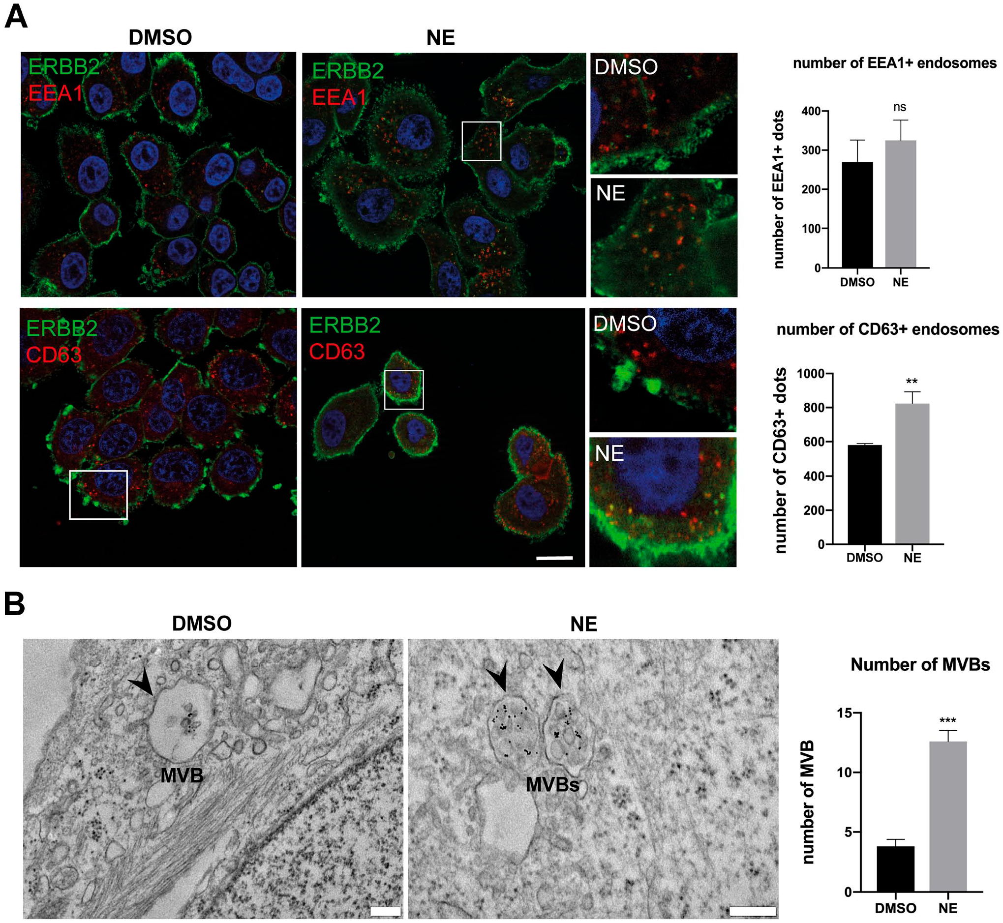
(A) Left, representative confocal images of SKBR-3 cells treated with DMSO or NE and labeled for EEA1 (upper panel, red signal), CD63 (lower panel, red signal), and ERBB2 (green signal). Nuclei were stained with DAPI (blue signal). Scale bar: 20 μm. Insets show signal colocalization at a higher magnification. Upper right, quantification of the number and size of EEA1+ dots and per cell in NE- versus DMSO-treated cells are depicted and expressed as mean ± SEM from n=3 experiments; 50 cells were analyzed for each experiment. Insets show signal colocalization at a higher magnification. Lower right, quantification of the number and size of CD63+ dots and per cell in NE- versus DMSO-treated cells are depicted and expressed as mean ± SEM from n=3, 50 cells were analyzed for each experiment. **p=0.038. (B) Left, ultrastructural analysis of multivesicular bodies (MVBs) (arrowheads) in SKBR-3 cells treated with DMSO or NE. After 72 hr of treatment, SKBR-3 cells were further probed with BSA 5-nm gold for 2 hr at 37C and processed for flat embedding in epoxy resin. Ultrathin sections show BSA-gold labeling (black dots) in MVBs in DMSO- and NE-treated cells. Right, quantification showing a significant increase in the number of MVBs in NE-treated cells estimated over 30 randomly chosen cell profiles. Scale bars: 200 nm. Abbreviations: NE, neratinib; DAPI, 4′,6-diamidino-2-phenylindole; SEM, standard error of the mean.
NE Promotes the Release of EVs
As NE promoted the increase of CD63+ MVBs, which are the precursors of EVs, and because a fraction of ERBB2 is found colocalizing with these compartments, we investigated whether NE modulated EV release. By performing EM negative staining, exosomes appear as vesicles of 30–130 nm in size and have a characteristic round or cup-shaped morphology,34,35 while MVs are approximately 100 nm to 1 μm, and are composed of a round-shaped population. For EV separation, medium from SKBR-3 cell cultures treated with NE or with DMSO (control) was collected and subjected to differential ultracentrifugation. Isolated EVs were then either processed for negative staining and/or immuno-EM. As shown in Fig. 4A and B, the number of EVs isolated from NE-treated SKBR-3 cells was significantly increased compared with that obtained from control cells, but only a subpopulation of both EVs was labeled with an anti-ERBB2 antibody. In addition, the size of ERBB2+ EVs was larger than that of ERBB2− EVs (mean 150 vs 100 nm, respectively) in both control and NE-treated samples (Fig. 4C).
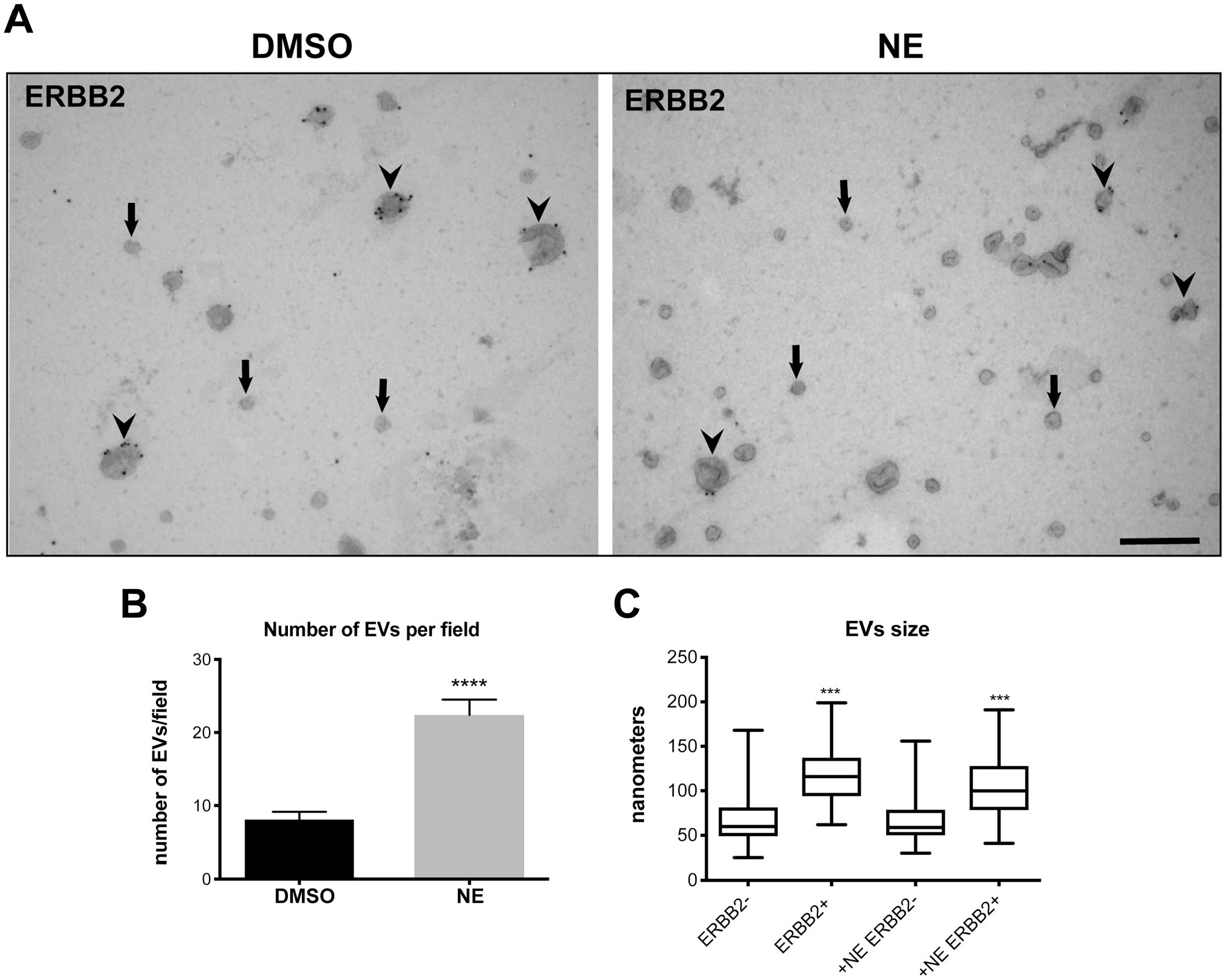
(A) Representative TEM images of immuno-labeled EVs indicating the presence of both ERBB2− small EVs (arrows) and ERBB2+ larger EVs (arrowheads). Scale bar: 200 nm. (B) Morphometric analysis of EV number. Results are plotted as mean ± SEM from N=3 independent preparations. Unpaired t-test, ****p<0.0001. (C) TEM analysis of EV size. Data are visualized as box plots showing EV size of ERBB2− versus ERBB2+ EVs. The plot shows the median value as a horizontal bar, and the interquartile range as a box; the whiskers are the minimum and maximum values. EVs were counted and measured on 30 randomly taken micrographs for each condition from n=3 independent experiments. Abbreviations: EV, extracellular vesicle; TEM, transmission electron microscopy; SEM, standard error of the mean; NE, neratinib.
NE Decreases ERBB2 Content on EVs
As cancer cell–derived EVs contain oncogenic proteins which can be transported to surrounding cancer cells to elicit phenotypic changes, or may transfer drug resistance to promote tumor progression, we sought to evaluate the effects of NE on the content of ERBB2 at the single EV level by immuno-EM. This was performed using an anti-ERBB2 antibody (9G6, N-terminal domain of ERBB2) and 10-nm PAG. As shown in Fig. 5A and B, NE dramatically decreased the number of ERBB2 gold particles associated with each individual EV, suggestive of treatment efficacy in reducing the amount of ERBB2 that could be transferred to other cells.
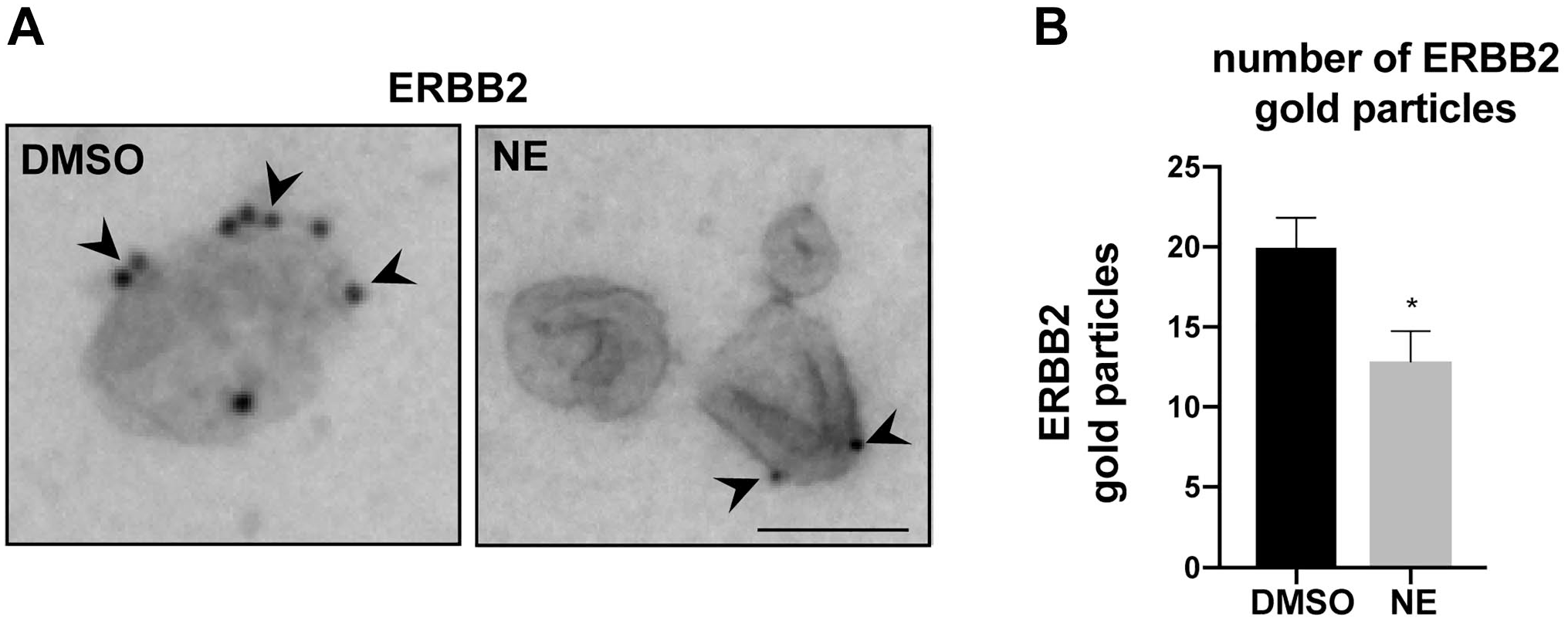
(A) High magnification images of EVs isolated from DMSO- and NE-treated SKBR-3 cells immuno-labeled for ERBB2 (9G6 antibody, 10-nm protein A gold), indicating the presence of ERBB2 (arrowheads) on EVs. Scale bar: 100 nm. (B) Morphometric TEM analysis showing that the total number of gold particles associated with EVs is significantly decreased in NE-treated cells. ERBB2 gold particles detected on EVs were counted on 30 randomly taken micrographs for each condition (n=3 independent experiments). Unpaired t-test, *p=0.011. Abbreviations: EV, extracellular vesicle; NE, neratinib; TEM, transmission electron microscopy.
Discussion
ERBB2+ BCa represents an aggressive subtype of breast tumors that develop in roughly 20–30% of patients. In 2017, NE has been approved by the Food and Drug Administration as an adjuvant therapy alongside the anti-ERBB2 antibody Tz in ERBB2+ BCa, with improved disease-free survival in the subgroup of ERBB2+/hormone receptor+. 15 To date, NE is considered the most effective ERBB2-targeted tyrosine kinase inhibitor against ERBB2-amplified, ERBB2-mutant, and EGFR-mutant cell lines. 12 Recent work showed that NE not only inhibits ERBB1/2/4 catalytic activity but also triggers their rapid internalization and subsequent proteolytic degradation. 22 To make the picture more complex, NE also equally targets and inhibits multiple Ste20 family kinases like MST3, MST4, MAP4K5, and MAP4K3 involved in the regulation of cell polarity, adhesion, and cell migration.25,26 However, all these studies relied on submicromolar concentrations of NE (from 50 to 500 nM) in human BCa cell lines 18 or were performed with NE in combination with other antineoplastic drugs in different cancer models.36,37 This study explored the effects of NE, as a single agent, on endocytic trafficking, as well as on the release of EVs in SKBR-3 cells, a widely used ERBB2+ overexpressing cell line. To do that, we treated SKBR-3 cells with NE at the IC50 dose of 6 nM for 72 hr, which is remarkably close to the IC50 used in a previous study. 12 In our opinion, employing these low nanomolar concentrations lessen possible off-target effects of the drug.
We found by confocal microscopy analysis that 6-nM NE increased the capacity of CME and expanded late endocytic compartments. However, simultaneously, it induced only a modest internalization of ERBB2 compared with the fraction retained at the PM, whereas a high-dose of NE (500 nM) induced a substantial internalization and degradation of ERBB2 (Supplementary Fig. 1). CME is a major route for the entry of diverse cargo in the cellular endocytic pathway. ERBB2 downregulation via endocytosis has been a subject of debate for long time. It is thought that, in SKBR-3 cells, ERBB2 is either retained on the cell surface where it modulates intracellular signaling or is constitutively internalized and recycled via CME with slow kinetics. 5 Indeed, our previous work showed that ERBB2 silencing in SKBR-3 cells caused the increase of the internalization rate of transferrin and cholera toxin B subunit, two CME-internalized cargoes, and of the number of CCP/Vs per cell, suggesting that ERBB2 overexpression is sufficient to interfere with the overall CME capacity involved in both constitutively and regulated receptor internalization. Here, we showed that NE increases CME capacity without inducing ERBB2 downregulation, suggesting that the inhibition of kinase/s signaling was playing a major role in the regulation of CME. 4 In addition, we report that CME endocytosis was not affected in ERBB2− MFC7 cells, supporting that the unlocking effect of NE on CME endocytosis depends on the presence of ERBB2. Our results also show an increase in the number of late CD63+ MVBs after NE treatment, supporting the increased capacity of the endocytic system. As ERBB2 appears neither to be degraded nor to be secreted at low nanomolar concentrations of NE, we might speculate that the fraction of internalized ERBB2 is most likely recycled to the PM. EVs, including exosomes and MVs, are secreted in large quantities by cancer cells into the local microenvironment and are important mediators of multiple biological functions such as metastasis, immunosuppression, and resistance to therapy.29,30,38 The observed expansion of the MVB compartment, where the exosome biogenesis takes place, prompted us to investigate whether NE promoted EV release. 39 In particular, we took advantage of immuno-EM techniques, to effectively reveal the inner structure of EVs, and evaluate EV ERBB2 content by using immunogold labeling. As reported in other studies, the typical cup-shaped morphology that we observed in our EVs preparations is likely due to dehydration artifacts, while the appearance of membranes inside the EVs has been also described with cryo-EM techniques, revealing a wide diversity in EV morphology. 35 We found that, while NE increased the total amount of EVs released, immuno-labeling of purified EVs examined at a single level showed a significant decrease in their ERBB2 content. In addition, we identified at least two subpopulations of EVs based on their size and ERBB2 positivity: ERBB2+ EVs, which ranged between 100 and 150 nm, and ERBB2− EVs, which appeared smaller, ranging between 50 and 100 nm. However, we cannot completely exclude that part of the increased size of ERBB2+ EVs originated also from EV aggregation and collapse that occur after fixation even in fresh preparations. As EVs mediate intercellular communication by shuttling functional signaling molecules as cargo, the decrease in ERBB2 content may reflect an additional unreported antitumor activity of NE.
To the best of our knowledge, this is the first study that reports that a low nanomolar dose of NE, effective in determining a 50% reduction of cell survival, increases CME and release of ERBB2+ EVs but has a limited impact on ERBB2 endocytic trafficking. ERBB2 is the primary target of NE, and the low nanomolar doses used in this study should have minimized off-target effects. Indeed, the impact of NE on CME endocytosis appears to be an ERBB2-dependent process. All these processes are deeply connected and influence several cellular processes linked to cancer progression at distant sites; however, we cannot rule out that the extensive modulation of intracellular and extracellular trafficking might depend on concomitant inhibition of other kinases expressed in SKBR-3 cells, like EGFR/ERBB1 or others. In conclusion, these results warrant further studies to better evaluate to what extent the observed effects depend exclusively on the specific inhibition of ERBB2.
Supplemental Material
sj-pdf-1-jhc-10.1369_00221554211026297 – Supplemental material for Imaging of Endocytic Trafficking and Extracellular Vesicles Released Under Neratinib Treatment in ERBB2+ Breast Cancer Cells
Supplemental material, sj-pdf-1-jhc-10.1369_00221554211026297 for Imaging of Endocytic Trafficking and Extracellular Vesicles Released Under Neratinib Treatment in ERBB2+ Breast Cancer Cells by Sara Santamaria, Maria Cristina Gagliani, Grazia Bellese, Silvia Marconi, Anastasia Lechiara, Martina Dameri, Cinzia Aiello, Erica Tagliatti, Patrizio Castagnola and Katia Cortese in Journal of Histochemistry & Cytochemistry
Footnotes
Competing Interests
The author(s) declared no potential conflicts of interest with respect to the research, authorship, and/or publication of this article.
Author Contributions
KC and PC conceived of and designed the study. SS, MCG, GB, SM, AL, MD, and CA performed experiments, microscopy, biochemistry, and data analysis, and drafted the article. ET critically revised the work and edited the article. KC and PC wrote the article with input from all other authors. All authors read and approved the final article.
Funding
The author(s) disclosed receipt of the following financial support for the research, authorship, and/or publication of this article: This work was supported by grants from the Italian Ministry of Health (Ricerca Corrente) to PC and by the University of Genova research grant funding (FRA, Fondi Ricerca Ateneo, 100008-2020-EC-FRA_001_FRA2020 and 100008-2019-EC-FRA_002—PROGETTO FRA 2019) to KC.
References
Supplementary Material
Please find the following supplemental material available below.
For Open Access articles published under a Creative Commons License, all supplemental material carries the same license as the article it is associated with.
For non-Open Access articles published, all supplemental material carries a non-exclusive license, and permission requests for re-use of supplemental material or any part of supplemental material shall be sent directly to the copyright owner as specified in the copyright notice associated with the article.